Thursday, June 30, 2016

Saudi MOH On KKUH MERS Outbreak & Increased Asymptomatic Detections

Credit MOH/CCC


#11,512


We've been following the outbreak at KKUH (King Khalid University Hospital) in Riyadh for nearly 3 weeks, and what has set this outbreak apart from others we've seen are the number of asymptomatic cases being reported by the Saudis.

While asymptomatic (or perhaps very mild) cases have been reported previously by the Saudi MOH, their incidence was roughly 1 in 10.  

Most of those were detected through RT-PCR testing of close contacts of symptomatic patients, although until late last summer, the Saudi MOH didn't appear to be aggressively looking for asymptomatic cases.


This came to a head very publicly last September with the WHO Statement On The 10th Meeting Of the IHR Emergency Committee On MERS, which rebuked the Saudis for their handling of asymptomatic cases (among other issues).

Since then, the Saudis have been far more diligent in seeking out, and reporting, asymptomatic cases.  But this does make it difficult to compare rates today to rates reported in previous years.

Still, over the past 20 days the Saudi's have reported 28 cases from the KKUH outbreak, and of those, 21 are listed as being asymptomatic.  An unusually high ratio by any standard.


While they don't provide answers, the MOH offers several possible explanations why this may have occurred, including.  
  • Better and more diligent testing of asymptomatic contacts
  • One or more `superspreaders' 
  • Increased transmissibility of the virus
  • Or (optimistically), the virus has lost some of its virulence, resulting in fewer serious infections


While more mild cases sounds like an improvement, this could make the virus harder to contain, and promote greater community spread.


The following comes from the most recent MOH/CCC Weekly monitor report.


MERS Outbreak at KKUH
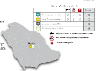
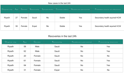
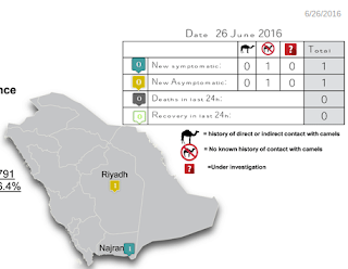
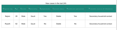
During the period between 14 and 26 June, 2016 a total of 28 cases of MERS eere reported from King Khalid University Hospital (KKUH), Riyadh. Out of the total cases, 7 (25%) were symptomatic.
There has been a recent increase in reports of asymptomatic or mild MERS-CoV infection. Data from Ministry of Health (MoH) showed that the proportion of asymptomatic infections increased from 10% (2012- July ,2015), to 57% by the end of 2015.

The increased proportion of asymptomatic infections could be attributed to increased number and improved criteria for detection of contacts and/or improved swabbing techniques. 


According to MoH guidelines all exposed HCWs should be screened for the virus. It is difficult to rule out presence of a super spreader and increased infectiousness of the virus. The increased proportion of asymptomatic MERS may indicate that the virus has lost some of its virulence. 

However, it is hard to estimate the proportion asymptomatic patients among infected individuals because there is no perfect method for defining the exposed contacts. In addition, lack of serological testing could have resulted in missing some other asymptomatic infections.

There was delay in diagnosing the primary case of MERS who happened to be a female admitted for management of a diabetic septic foot. Conceivably, the diagnosis of MERS was least expected. 


Nevertheless, the large number of secondary infections within a short period of time in a well-defined health institution points out that there is a clear gap in infection prevention and control in that institution.


Credit MOH/CCC

Brazil's MOH To Re-Evaluate Criteria For Zika Congenital Syndrome


#11,511


In light of the study, published yesterday in the The Lancet (see The Lancet: Two Congenital Zika Virus Studies), finding that Zika Congenital Syndrome may exist without overt microcephaly, the Brazilian MOH has announced plans to re-evaluate their surveillance criteria.

As a side note, we usually get Brazil's weekly Microcephaly  report on Tuesday or Wednesday, but this week's has yet to be posted.


(Translation)
Registration Date: 29/06/2016 19:06:45 the amended 06/29/2016 19:06:45 the 


Largest study ever conducted on the Zika indicates that signs and symptoms of neurological disorders are included as criteria for trigam babies, regardless of the presence of microcephaly

The microcephaly and rash history during pregnancy are insufficient criteria to identify the consequences of infection Zika virus in newborns. The finding is part of a study commissioned by the Ministry of Health, published on Wednesday (29) by the British journal, The Lancet. The study results suggest that the signs and symptoms of neurological disorders are included as criteria for screening of babies, regardless of the presence or absence of microcephaly.

The study was done in partnership with the Federal University of Pelotas (UFPel), the Brazilian Society of Medical Genetics, the Latin American Center for Perinatology of the Pan American Health Organization (PAHO / WHO) and Harvard University, US. 1,501 live births were analyzed, which had already been investigated by state and local health departments, which is the largest study ever done on the subject in the world. Before, the largest study had analyzed 104 children.

Research shows that for every five children with congenital infection Zika virus - confirmed or probable - a non had microcephaly, indicating that 80% of children were taken through investigation using the criteria of microcephaly and rash. "We are adapting our protocols to those found to expand research and improve our surveillance system. At this time, Brazil and the world have accumulated more knowledge about the disease and can, with this learning, improve the monitoring of the consequences of congenital infection Zika virus, "said the general coordinator of Surveillance and Response to Emergencies in Public Health, the Ministry of Health, Wanderson Oliveira.

At the beginning of the epidemic investigation in November 2015, the Ministry of Health adopted a more sensitive case definition to identify the microcephaly, with the criterion of 33 cm head circumference for newborns of both sexes. This definition was subsequently changed to 32 cm. The adoption of these more sensitive definitions identified that 20% of children with a condition of clinical importance not had microcephaly. So this is an important aspect being considered to adjust the microcephaly surveillance model for Congenital changes.
"Using this cutoff point more sensitive at the beginning of the investigation allowed us to identify those children who had not microcephaly, but who had other disorders of the central nervous system, possibly related to infection by Zika virus," he stressed.

The article was based on information from the Event Log in Public Health (RESP) and Live Birth Information System (SINASC) of the Ministry of Health, with the support of all states. Based on this finding, the Ministry of Health is now studying the adequacy of current surveillance protocol and Response to Occurrence of microcephaly and / or central nervous system changes. It is noteworthy that the investigation of other causes is already held by the Ministry of Health in conjunction with states and municipalities, through hearing tests and vision.

Saudi MOH: 2 New Primary MERS Cases (Al-Baha & Dammam)


#11,510


The Saudi MOH reports two new Primary MERS Cases (1 with camel exposure), one who is listed in critical condition, and the other who has already expired. 
 
Posthumous announcements are always worth noting since  they may indicate delayed detection and isolation, raising the chances of seeing additional infections.

While camel contact is frequently cited in primary cases, for the vast majority of community acquired cases, the source of their infection is never known. 


The Lancet: Two Congenital Zika Virus Studies


#11,509


Although microcephaly has been the most obvious birth defect linked to maternal Zika infection, for months we've been warned by Brazilian MOH, the CDC, NIH, and WHO that Zika may cause other congenital defects that may not become apparent until the child is much older.


Cranial development is generally advanced enough by the 30th week of gestation that a 3rd trimester exposure would be unlikely to seriously impact head circumference, yet it could still cause neurological damage. 

So, while investigators have discarded 2/3rds of all suspected Zika related microcephaly cases as not meeting their criteria (see last week's chart below), it may still turn out that some of these kids have serious neurological deficits. 


Shedding new light on the effects of maternal Zika carriage for the developing fetus, we a pair of Lancet studies, published late yesterday.   


Below you'll find links, excepts from the abstracts, and some snippets from the press releases.


Congenital Zika virus syndrome in Brazil: a case series of the first 1501 livebirths with complete investigation
Giovanny V A França, PhD, Prof Lavinia Schuler-Faccini, PhD, Wanderson K Oliveira, MSc, Claudio M P Henriques, MD, Eduardo H Carmo, PhD, Vaneide D Pedi, MSc, Marília L Nunes, DVM, Marcia C Castro, PhD, Suzanne Serruya, PhD, Mariângela F Silveira, MD, Prof Fernando C Barros, MD, Prof Cesar G Victora, MD

Published online: June 29, 2016
Open Access

(Excerpt)

Background

In November, 2015, an epidemic of microcephaly was reported in Brazil, which was later attributed to congenital Zika virus infection. 7830 suspected cases had been reported to the Brazilian Ministry of Health by June 4, 2016, but little is known about their characteristics. We aimed to describe these newborn babies in terms of clinical findings, anthropometry, and survival.

Methods

We reviewed all 1501 liveborn infants for whom investigation by medical teams at State level had been completed as of Feb 27, 2016, and classified suspected cases into five categories based on neuroimaging and laboratory results for Zika virus and other relevant infections. Definite cases had laboratory evidence of Zika virus infection; highly probable cases presented specific neuroimaging findings, and negative laboratory results for other congenital infections; moderately probable cases had specific imaging findings but other infections could not be ruled out; somewhat probable cases had imaging findings, but these were not reported in detail by the local teams; all other newborn babies were classified as discarded cases. Head circumference by gestational age was assessed with InterGrowth standards. First week mortality and history of rash were provided by the State medical teams.    

Findings

Between Nov 19, 2015, and Feb 27, 2015, investigations were completed for 1501 suspected cases reported to the Brazilian Ministry of Health, of whom 899 were discarded. Of the remainder 602 cases, 76 were definite, 54 highly probable, 181 moderately probable, and 291 somewhat probable of congenital Zika virus syndrome. Clinical, anthropometric, and survival differences were small among the four groups. 
Compared with these four groups, the 899 discarded cases had larger head circumferences (mean Z scores −1·54 vs −3·13, difference 1·58 [95% CI 1·45–1·72]); lower first-week mortality (14 per 1000 vs 51 per 1000; rate ratio 0·28 [95% CI 0·14–0·56]); and were less likely to have a history of rash during pregnancy (20·7% vs 61·4%, ratio 0·34 [95% CI 0·27–0·42]). Rashes in the third trimester of pregnancy were associated with brain abnormalities despite normal sized heads. One in five definite or probable cases presented head circumferences in the normal range (above −2 SD below the median of the InterGrowth standard) and for one third of definite and probable cases there was no history of a rash during pregnancy. The peak of the epidemic occurred in late November, 2015.   

Interpretation
  
Zika virus congenital syndrome is a new teratogenic disease. Because many definite or probable cases present normal head circumference values and their mothers do not report having a rash, screening criteria must be revised in order to detect all affected newborn babies.


You'll find a nice summation in the Journal's press release: The Lancet: Microcephaly screening alone won't detect all cases of Zika virus in newborns, study suggests 


The second study finds evidence of Zika virus infection in the brain tissue of a deceased 2-month-old microcephalic baby, in two newborns who died shortly after delivery, and in the placental tissue of two fetuses that spontaneously aborted.

While a small sample, all five mothers reported Zika-like symptoms (fever and rash) during their first trimester.  Of the three live-births, all three showed signs of microcephaly.

You'll find a summary of their findings in The Lancet: Zika virus identified in brain and placenta tissue, strengthening link to birth defects, or you can follow the link below to read the full full report (warning: PDF contains graphic autopsy photos).

Pathology of congenital Zika syndrome in Brazil: a case series

Roosecelis Brasil Martines, MD†, Julu Bhatnagar, PhD†, Prof Ana Maria de Oliveira Ramos, MD, Helaine Pompeia Freire Davi, MD, Silvia D'Andretta Iglezias, MD, Cristina Takami Kanamura, MSc, M Kelly Keating, DVM, Gillian Hale, MD, Luciana Silva-Flannery, PhD, Atis Muehlenbachs, MD, Jana Ritter, DVM, Joy Gary, DVM, Dominique Rollin, MD, Cynthia S Goldsmith, MGS, Sarah Reagan-Steiner, MD, Yokabed Ermias, MPH, Tadaki Suzuki, MD, Prof Kleber G Luz, MD, Wanderson Kleber de Oliveira, MSc, Robert Lanciotti, PhD, Amy Lambert, PhD, Wun-Ju Shieh, MD, Dr Sherif R Zaki, MDcorrespondenceemail

Published Online: 29 June 2016
Article has an altmetric score of 6
DOI: http://dx.doi.org/10.1016/S0140-6736(16)30883-2 |
(Excerpt)

Methods

In this case series, formalin-fixed paraffin-embedded tissue samples from five cases, including two newborn babies with microcephaly and severe arthrogryposis who died shortly after birth, one 2-month-old baby, and two placentas from spontaneous abortions, from Brazil were submitted to the Infectious Diseases Pathology Branch at the US Centers for Disease Control and Prevention (Atlanta, GA, USA) between December, 2015, and March, 2016. Specimens were assessed by histopathological examination, immunohistochemical assays using a mouse anti-Zika virus antibody, and RT-PCR assays targeting the NS5 and envelope genes. Amplicons of RT-PCR positive cases were sequenced for characterisation of strains.

Findings

Viral antigens were localised to glial cells and neurons and associated with microcalcifications in all three fatal cases with microcephaly. Antigens were also seen in chorionic villi of one of the first trimester placentas. Tissues from all five cases were positive for Zika virus RNA by RT-PCR, and sequence analyses showed highest identities with Zika virus strains isolated from Brazil during 2015.

Interpretation

These findings provide strong evidence of a link between Zika virus infection and different congenital central nervous system malformations, including microcephaly as well as arthrogryposis and spontaneous abortions.


Wednesday, June 29, 2016

Riyadh: Two More (Asymptomatic) MERS Cases


#11,508


Despite declaring the MERS outbreak `controlled and ended' at King Khalid Hospital last week (see Arab News MoH rejects WHO charges of MERS negligence), we continue to get sporadic reports of new nosocomial infections out of Riyadh.

Today two more asymptomatic Health Care Workers have been identified as being infected, bringing this outbreak's total to roughly 30 since mid-June. 

On the plus side, the detection of so many asymptomatic cases (> 20) in this outbreak is a hopeful sign that the Saudis are being more diligent about testing asymptomatic contacts for the virus, and are taking their infections more seriously (see Saudi MOH CCC: Asymptomatic MERS-CoV Infections).


Meanwhile, 6 cases from this outbreak have recovered (i.e. no longer test positive for the virus).


Upcoming COCA Call: Identification and Care of Patients with Hantavirus Disease

Credit CDC


#11,507


Hantavirus  is a collective term for a group of viruses in the Bunyaviridae family – hosted by various types of rodents - that vary in distribution, symptomology, and severity around the world.

The clinical symptoms of Hantavirus were first recognized by western medicine back in the early 1950s during the Korean war, when 3,000 UN troops stationed there were infected with a mysterious viral illness. 

The mortality rate was 10%-15%, with patients experiencing fever, hypotension, renal failure, and internal bleeding (disseminated intravascular coagulation).

Originally called Korean Hemorrhagic Fever (later dubbed Hantavirus after the Hantaan River of Korea), we now know it by a variety of names today, including the American Sin Nombre Virus,  Europe's milder Puumala Virus, and  the Andes Virus in South America.


As summer is the time when we typically see the most cases of Hantavirus, tomorrow the CDC will hold a COCA call for healthcare providers on:
   
Identification and Care of Patients with Hantavirus Disease


Date:Thursday, June 30, 2016

Time:2:00 - 3:00 pm (Eastern Time)

Participate by Phone:

    800-779-5346 (U.S. Callers)
    517-308-9340 (International Callers)

Passcode:2718213

Participate by Webinar: https://www.mymeetings.com/nc/join.php?i=PW8830943&p=2718213&t=c
Presenter(s)

Barbara Knust, DVM, MPH, DCAVPM
Epidemiologist
Office of Infectious Diseases
National Center for Emerging and Zoonotic Infectious Diseases
Centers for Disease Control and Prevention

Gregory Mertz, MD
Professor Emeritus
Department of Internal Medicine
University of New Mexico

Michelle Harkins, MD
Associate Professor of Medicine
Division Chief, Pulmonary, Critical Care, and Sleep Medicine
Department of Internal Medicine
University of New Mexico

Overview

Hantavirus infection in the United States can cause severe and life-threatening illness, requiring rapid assessment, presumptive diagnosis, and high-level supportive care of respiratory and cardiac functions. Hantavirus infection causes a cardiopulmonary syndrome, which includes rapid accumulation of pulmonary edema following a flu-like prodrome. 

Approximately 37% of cases end in death. Although hantavirus disease is rare, clinicians should be aware of the risk factors, clinical picture, and essential care elements. During this COCA call, clinicians will learn about the epidemiology, diagnosis, and clinical care of patients with hantavirus disease in the United States.

Objectives

At the conclusion of the session, the participant will be able to accomplish the following:

  • Describe the risk factors, endemic areas, and incubation period of hantavirus infection
  • Identify the clinical presentation and methods to identify a patient with hantavirus in the clinical setting
  • Understand the parameters of clinical management and critical care for patients with hantavirus

Additional Resources


I confess to having a bit of of a personal interest in the Hantavirus story because my ex-wife’s cousin (Dr. Ron Voorhees) was one of the original investigators of the famous 4 corner’s outbreak of Hantavirus in 1993 when he worked as an epidemiologist for the state of New Mexico. 

It was during this outbreak that the New World `Sin Nombre’ (Spanish for `No Name’) Hantavirus was identified, and found to be widely prevalent in its natural host, the deer mouse.

If you would like to learn more about the history of that outbreak, and the epidemiological work done to identify the pathogen, I can direct you to an excellent account at:

Tracking a Mystery Disease:
The Detailed Story of Hantavirus Pulmonary Syndrome


Hubei Zoo: African Lions Infected With Avian H5N1

Credit Wikipedia


#11,506


We've another report of large cats, kept in a Chinese Zoo, infected by the H5N1 virus.  This time - instead of Asian tigers - the victims are African lions.


Despite reporting only 15 human H5N1 infections (and 6 deaths) over the past 5 years, and with H7N9 and H5N6 making bigger a splash in recent years, we continue to see evidence that HPAI H5N1 is alive and well on the Chinese Mainland.

Last May, in Fatal H5N1 Infection In Tigers By Different Reassortant Viruses - China, we looked at 5 recent tiger infections and/or deaths from H5N1 over 2014-2015.   

Three of these infections occurred in Yunnan Province over an 18 month period, and remarkably, all three viruses represented different clades of H5N1.
  • A/tiger /Yunnan /tig1404 /2014(H5N1)  subclade 2.3.4.4e  reassortant with six internal genes from avian influenza A(H5N2) virus
  • A/tiger /Yunnan /tig1412 /2014(H5N1) subclade 2.3.2.1b
  • A/tiger /Yunnan /tig1508 /2015(H5N1) clade 2.3.2.1c reassortant with three internal genes from the A(H9N2) avian virus.
A study found molecular markers associated with increased virulence and transmission in mammals in all three isolates. After describing multiple findings (see text for details) the authors wrote:
In summary, all of mutations/substitutions of the gene segments of the tiger originated viruses could contribute to the enhancement of virulence or the increase of the H5N1 virus binding to the α2-6 receptor.

The authors also documented replication and pathogenicity in mice, and a loss of effectiveness of China's RE-6 vaccine against the most recent (Aug 2015) tiger isolate


As we've noted previously (see Subclinical Highly Pathogenic Avian Influenza Virus Infection among Vaccinated Chickens, China) - poultry vaccines don’t always prevent infection – sometimes they only mask the symptoms, allowing the virus to spread and evolve under the radar.

This `healthy facade' likely explains why so many zoo animals have been infected with H5N1, as raw poultry is often used to feed carnivorous animals in captivity.

While the following government report lacks some crucial details (the event was described only as `recent'), it does provide a pretty good narrative of the incident.


Hubei successful handling of the H5N1 avian flu African lion

Hubei Provincial People's Government portal www.hubei.gov.cn 


2016-06-27 15:28:00
 

Source: Hubei Emergency Management website 

Recently, the provincial wild animal epidemic sources and disease monitoring center has successfully disposed of together Ezhou African lion H5N1 infection anomalies.

Provincial wildlife epidemic sources and disease monitoring center Ezhou monitoring stations, the city zoo SEG two African lions (1 male 1 female) high fever, dying, sudden abnormal situation, the provincial wild animal epidemic sources and disease monitoring immediate establishment of a panel by the center of Changchun military Medical veterinary hospital, Huazhong Agricultural University and other emergency experts, arrived in the afternoon Ezhou, two African lions to carry out isolation and treatment, to identify the cause and related investigations. Male African lion condition suddenly deteriorated because he died soon afterwards. The other a female African lion, the careful treatment of the Expert Group has been recovered, and other park animals not see the death of the new phenomenon.

Group of Experts on the African lion was death autopsy, found it impossible to exclude the possibility of death from communicable diseases, immediately took samples back to the laboratory. Provincial wildlife epidemic sources and disease monitoring center immediately to the State Forestry Protection Department, Monitoring Station, the Provincial Forestry Department and the provincial joint prevention and control office to report in a timely manner to Ezhou health planning, forestry and animal husbandry and veterinary department informed, and requested the zoo, take Feng Yuan, disinfection and other treatment, the African lion death was burned deep disposal, the park of birds have been vaccinated. Meanwhile, the province also organized to go to other zoos technology, domestication and breeding places to carry out sample testing, tracing investigation, as of now, the province found no abnormalities other wildlife.

African lion samples Conservancy Military Medical Veterinary Hospital detect H5N1 influenza virus gene sequence homology with the white tiger isolates 2015 Nanning death up to 99%. Experts believe that a comprehensive analysis, African lions since the H5N1 flu virus and bacterial mixed infection caused by lung, kidney and other organ damage and death.

To further strengthen the province's wild animal epidemic sources and disease monitoring and prevention and control work, the provincial Forestry Department issued a document requiring the forestry sector at all levels, in accordance with the requirements of the focus period, to strengthen prevention and control of wild animal epidemic sources and disease monitoring, so early detection, early reporting, early disposal; further implement the ban order of birds, wildlife and out Closure transport permits, each zoo prohibited person close contact with wild animals, wild animals in captivity breeding strengthening workplace health supervision. (Provincial Forestry Department)

As we discussed last year in HPAI H5: Catch As Cats Can,
felines (and canines) are susceptible to HPAI H5 viruses, including H5N1.


In 2006, virologist C. A. Nidom of the Institute of Tropical Disease, Airlangga University demonstrated that of 500 cats he tested in and around Jakarta, 20% had antibodies for the H5N1 bird flu virus.  Findings that prompted the FAO in 2007 to warn that: Avian influenza in cats should be closely monitored.

More recently, in 2012 the OIE reported on Cats Infected With H5N1 in Israel, while in early 2015 we saw Guangxi Zoo Reports 2 Tiger Deaths Due To H5N1.  

Just over a year ago we saw reports of Fatal H5N6 Avian Influenza Virus Infection in a Domestic Cat and Wild Birds in China While last May, in Report: Skunks and Rabbits Can Catch And Shed Avian Flu, we saw evidence that other peridomestic animals are susceptible as well.


Although reports of H5N1 have declined out of Asia over the past few years (while surging in the Middle East and Africa), reports like the one today are a reminder that it remains a player in China's diverse avian flu ecology.

Tuesday, June 28, 2016

Saudi MOH: 2 More Primary MERS Cases


#11,505


While reports from our two recent outbreaks (Riyadh & Najran) take a break, we've two new primary (community acquired) cases, one with camel contact and the other without a known risk exposure.


Additionally, one death of a previously reported case from Hail was reported.


Is Something New Killing Poultry In Egypt?


#11,504

Since it first arrived in the Middle East just over a decade ago, avian H5N1 has devastated Egypt's poultry sector, and has produced hundreds of human infections and more than 115 deaths.

The chronic use of older, often ineffectual vaccines (see 2012's A Paltry Poultry Vaccine) helped to spread  H5N1, and likely contributed to the Emergence Of A Novel Cluster of H5N1 Clade 2.2.1.2 in 2014.  

In addition to H5N1, LPAI H9N2 is also endemic in Egypt's poultry, and while Egypt hasn't reported it to the OIE - earlier this month in PLoS One: Serological Evidence Of Human Infection with Avian H7 in Egyptian Poultry Growers - we saw circumstantial evidence of avian H7 in Egypt's poultry.

Testing, surveillance, and reporting out of Egypt has been seriously lacking in recent years, and so it is very difficult to know exactly what the situation is there. 

But earlier this month, in HPAI H5 Worries From Egypt's Poultry Sector we saw reports of greatly increased poultry deaths, and concerns that HPAI H5N8 might have arrived in Egypt as well.

After launching an investigation, on June 9th Egypt's Ministry of Agriculture announced Initial Tests were Negative For H5N8, but the cause of the excess mortality remained unknown. 

Reports of unusual poultry losses have continued, and in the past few days Egyptian media have begun reporting on a new syndrome - الالتهاب الارتعاشى -  which translates to Inflammation tremens, that is reportedly spreading rapidly.
 
The Egyptian Ministry of Agriculture's website - which is only rarely updated - has been unreachable for me the past few days, so I've not found any official statement on this syndrome.

We do have one of the many syntax-challenged Arabic media reports on this crisis posted this morning by Sharon Sanders of Flutrackers called Three crises besieging the poultry industry.


While it briefly describes the new `epidemic' it spends most of its time quoting an industry representative's criticisms of the  MOA for a long list of shortcomings, including failing to provide effective vaccines.

A few excerpts follow:

(EXCERPTS)
The poultry sector faces severe crises since the beginning chapters since the emergence of the bird flu virus in 2006 caused the retreat despite industry achieved before it jumps the productivity, where production exceeds $ 2 million birds a day.


(SNIP)


This year a new epidemic is severe tremens a disease do not have veterinary services special vaccines vaccines to meet it, leading to massive deaths of poultry farms.
(SNIP)
Dr. Abdulaziz Al Sayed, head of the massacres and poultry Cairo Chamber of Commerce Division, that the current crisis is the most powerful crises experienced by the sector since the avian flu crisis in virus emerges in 2006, pointing out that the fundamental problem facing the sector is not the emergence of the virus and to reverse and return production to normal visits but other lies in the working methods in the sector, whether producers or the Ministry of Agriculture or government decisions, which resulted in many errors that made this sector groaning
(SNIP)

He referred to the Ministry of Agriculture failed to work serums strong and vaccines that can cope with bird flu even though the settlers of the virus in Egypt for more than 9 years, they resorted to import vaccines and vaccines from abroad, which it increased both the emergence of inflammation tremens is a new virus does not have the body serum is able to confront what led to the deaths of large numbers of more than 40% of production
(Continue . . . )


Given the vague nature of these reports, and the lack of comment by the MOA, it is impossible to know exactly what is going on with Egypt's poultry sector, beyond the fact they are dealing with a crisis.


But with Egypt's history of avian flu expansion and evolution, hopefully we'll get some clarification on all of this sooner rather than later.


DRC's Plan To Vaccinate 11.6 Million Against Yellow Fever


#11,503


Just over 10 days ago, in WHO: Lower Doses Of Yellow Fever Vaccine Could Be Used In Emergencies - in view of Africa's ongoing Yellow Fever outbreak and a shortfall in global vaccine supplies - we saw their SAGE (Strategic Advisory Group of Experts) on immunization approve the use of `fractional dose' vaccination in an emergency.

While fractional dosing greatly extends the supply of available vaccine, the downside is recipients would be unlikely to gain lifetime immunity (more likely 12 months or so), and would not receive a standard yellow fever vaccination certificate. 

The decision announced on June 17th was described as an emergency fall-back plan, with the WHO stating the situation didn't currently call for its implementation, but if the situation in Africa worsened substantially . . . .

Fast forward a little over a week and the Health Minister of the DRC has announced plans to vaccinate the entire population (over the age of 9 mos) of the capital -  Kinshasa -  and target populations in Kwango, Lualaba and Kasai provinces. 

With reportedly only 1.6 million doses of vaccine available in the DRC - even if diluted to 1/5th strength - it isn't clear how they will obtain enough vaccine to provide the estimated 11.6 million doses required.

Six days ago the WHO announced the Launch of emergency vaccination campaigns on the DR Congo and Angola border (see below), but this new plan would seem to far exceed it in the DRC.

    Brazzaville, 22 June 2016 – As the yellow fever outbreak in Angola and Democratic Republic of the Congo continues, the World Health Organization will launch emergency pre-emptive vaccination campaigns on the DR Congo, Angola border and the city of Kinshasa in the DR Congo to halt the epidemic and prevent the risk of further international spread.
    The initial phase of the campaign which begins in July will focus on districts where there is high movement of people and intense trade activities, particularly the northern border districts of Angola and targeted border districts in neighbouring countries. Specifically, within a 75-100km belt spanning the border between Angola and DR Congo and targeted health zones/communes at risk in Kinshasa city in the DR Congo. This will create an immune buffer to prevent further international spread.

    So far, I haven't seen any official comment from the WHO on the DRC's more aggressive plan.  The most complete coverage of the DRC's decision comes from VOA NEWS.

    Congo Launches Vaccination Campaign Against Yellow Fever
     

    VOA News

    June 27, 2016 8:15 PM

    The Democratic Republic of Congo says it will begin a campaign next month to vaccinate 11.6 million people against yellow fever after an epidemic was declared in the capital.

    Health Minister Felix Kabange said the campaign will begin July 20 and will aim to vaccinate everyone in the capital of Kinshasa except children under nine months, and will also target populations in the provinces of Kwango, Lualaba and Kasai.

    Last week, Congo's government announced an epidemic in Kinshasa and two other provinces after reporting 67 confirmed cases of yellow fever and more than 1,000 other suspected cases. An outbreak in neighboring Angola has led to the deaths of about 345 people.

    Kabange did not say how health authorities would acquire enough vaccination doses for the campaign. The vaccine is in short supply around the world, and takes about a year to make.

    The World Health Organization recommended this month that the vaccine be diluted up to a fifth of the standard dose to deal with the current emergency.

    The WHO says the lower dosage will protect people for at least a year, but will likely not give lifelong immunity.

    (Continue . . . )

    Monday, June 27, 2016

    Nature Sci Rpts: Continued Evolution Of The 2009 H1N1 Virus

    Credit NIAID


    #11,502


    In August of 2010, roughly 16 months after the new H1N1 virus first emerged, the WHO finally declared the end of the 2009 Pandemic.

    For most people the switch over to seasonal status signaled the pH1N1 threat was over.

    But of course, seasonal influenza kills hundreds of thousands of people each year, and as we've seen, some seasons are much worse than others.


    In the United States, the 2011-2012 and the 2015-2016 flu seasons were unusually mild (see chart above), while the years in-between were moderately severe.


    The severity of flu can also be regional, as we've seen severe flu outbreaks reported in India, Eastern Europe, and the Middle East at the same time there was relatively little flu in North America (see WHO: Update On Ukraine's Flu Season).

    While some of the reasons behind this variance in flu severity remain murky, two factors - the waxing and waning levels of community immunity and antigenic (and other) changes in the virus - appear to be major factors.

    Although the hope is always that the longer a virus circulates in a host population, the less severe it will become, the other side of the coin is the longer it circulates, the more evolutionary changes it may acquire.

    And that could not only make it more transmissible, it could make it more virulent as well.

    The most recent ECDC Influenza Virus Characterization report describes pH1N1's evolution:
    Since 2009, the HA genes have evolved, and nine clades have been designated. For well over a year, viruses in clade 6, represented by A/St Petersburg/27/2011 and carrying amino acid substitutions of D97N, S185T and S203T in HA1 and E47K and S124N in HA2 compared with A/California/7/2009, have predominated worldwide with a number of subclades emerging.

    In other words, the 2009 H1N1 viruses of today are markedly different from those that emerged in the spring of 2009, and they continue to evolve and diversify.


    Some mutations in pH1N1 - including the D225 Variant linked to more severe deep lung infection and the H275Y mutation conferring antiviral resistance -  are worrisome, but are seen in fewer than 2% of samples and are not yet viewed as major public health concerns. 

    But today we've a study, published in Nature's Science Reports, that identifies 6 genetic changes (PA-L581M, NP-V100I, NP-I373T, HA-S202T, NA-N248D and NS1-I123V) that appeared shortly after the virus jumped from pigs to humans, and that are still seen in 99% of the pH1N1 viruses circulating today. 

    These changes appear to to be the result of host adaptation, are believed to have made the pH1N1 a more `humanized' virus, conferring greater transmissibility and improved viral fitness. 

    This study has significance beyond just pH1N1, as it suggests the longer a poorly adapted virus influenza circulates in humans (think: H7N9, H5N1, H1N1v, H3N2v, etc.), the better its chances are of adapting to human hosts.  

    It's a long, fascinating article, and the abstract only scratches the surface, so you'll want to follow the link to read it in its entirety.


    I'll have a bit more when you return. 


    Evolution of 2009 H1N1 influenza viruses during the pandemic correlates with increased viral pathogenicity and transmissibility in the ferret model.


    Abstract

    There is increasing evidence that 2009 pandemic H1N1 influenza viruses have evolved after pandemic onset giving rise to severe epidemics in subsequent waves.However, it still remains unclear which viral determinants might have contributed to disease severity after pandemic initiation.

    Here, we show that distinct mutations in the 2009 pandemic H1N1 virus genome have occurred with increased frequency after pandemic declaration. Among those, a mutation in the viral hemagglutinin was identified that increases 2009 pandemic H1N1 virus binding to human-like α2,6-linked sialic acids. 

    Moreover, these mutations conferred increased viral replication in the respiratory tract and elevated respiratory droplet transmission between ferrets. 

    Thus, our data show that 2009 H1N1 influenza viruses have evolved after pandemic onset giving rise to novel virus variants that enhance viral replicative fitness and respiratory droplet transmission in a mammalian animal model. 

    These findings might help to improve surveillance efforts to assess the pandemic risk by emerging influenza viruses.
    (BIG SNIP)

    In summary, our findings here suggest that increased vigilance in viral surveillance is required even after pandemic onset since IAV seem to harbour the potential to further evolve causing severe subsequent epidemics in the human population.


    In 1957, after almost 40 years where H1N1 had dominated the global flu world, a new H2N2 virus appeared, and sparked a fresh pandemic.

    The Asian flu was less severe than 1918, but more severe than 1968 and 2009, and probably killed around 4 million people.


    As the chart below illustrates, intermittent severe outbreaks of that virus continued beyond the pandemic period, with H2N2 returning every few years with renewed vigor.


    We saw similar spikes in H1N1 in the decade following the 1918 pandemic, with peaks reported in 1923, 1926 and 1929 (see The Pandemic Influenza Enigma).

    While we are always on watch for a novel flu to appear, sparking the next pandemic, history has proven time and again that sometimes even an old flu can learn new tricks.

    So we watch for changes in both of our seasonal influenza A viruses, always  mindful that either of them could produce a particularly nasty flu season down the road.  

    UK PHE Stands By The Nasal Spray Flu Vaccine


    #11,501


    It's a case of `PHE said - CDC said', but there's a sharp division between the two public health agencies over the effectiveness of the LAIV (live attenuated influenza vaccine) called FluMist Quadrivalent in the US market and Fluenz Tetra in the UK and EU market.

    Both products are manufactured by AstraZeneca - Medimmune, and while sold under different banners, are pharmaceutically identical. 

    Last week, in CDC Statement On ACIP Recommendation Against Use Of Inhaled (LAIV) Flu Vaccine, we looked at the surprising (and as yet, unexplained) drop in VE (vaccine effectiveness) of the inhaled flu vaccine reported by the CDC over the past three flu seasons.


    After years of posting superior VE numbers, the first signs of trouble appeared in the fall of 2014 when the CDC announced they could find no measurable effectiveness against the H1N1 strain among children who received the 2013-2014 vaccine.

    The LAIV posted another poor showing (against both H1N1 and a `drifted' H3N2) the following year (see CIDRAP ACIP drops preference for nasal-spray flu vaccine in kids).

    And just last week, after reviewing yet another disappointing set of VE numbers from last year's flu season, ACIP voted against recommending the nasal spray LAIV vaccine for the upcoming flu season.

    Citing their own studies, and a study from Finland, the UK's PHE finds the performance of the LAIV (57%) in children to be lower than commonly cited in previous years, but still acceptable and on par with the flu jab in adults. 

    Why there would be such a stark difference between the US and UK VE studies is a mystery.   In any event, the UK continues to support the use of the LAIV vaccine for the upcoming flu season.  

    This from the PHE.

    Child flu vaccine plays important role in annual flu programme


    Provisional figures show that the nasal spray flu vaccine has been effective in the UK.


    Public Health England (PHE), the Department of Health and NHS England remain confident that the children’s nasal spray flu vaccine plays an important role in protecting children, their families and others in the community from flu during the winter.

    Provisional figures released by PHE show that the childhood nasal spray flu vaccine has been effective in the UK, both in protecting the children themselves and their communities from flu. Reports from the US have suggested a possible lower vaccine effectiveness, unlike the findings in the UK.

    The National Institute for Health and Welfare in Finland has confirmed that they saw similar effectiveness levels to the UK in 2015 to 2016 (46% against laboratory confirmed disease), and have confirmed the nasal spray flu vaccine will continue to be used in Finland for the forthcoming winter.

    From October 2016, the vaccine will be extended to healthy children in school year 3 in England. Once again, children aged 2, 3 and 4, and in school years 1 and 2 will also be eligible to receive the free vaccine which is quick, effective and painless.

    Dr Richard Pebody, head of flu surveillance for PHE said:

    We estimate that overall, the vaccine was 57.6% (95% confidence interval: 25.1, 76) effective in preventing influenza infection amongst children in 2015 to 2016. These findings are encouraging and in line with what we also typically see for the adult flu vaccine.
    Prior to offering vaccination to all our youngest primary school aged children this season, school age pilots took place in a number of areas across England in 2014 to 2015. In areas where flu vaccine was piloted amongst primary school age children, there was a 94% reduction in GP influenza like illness consultation rates, 74% reduction in A&E respiratory attendances and 93% reduction in hospital admissions due to confirmed influenza in primary school children. In the same pilot areas, GP ‘influenza like illness’ consultation rates for adults were 59% lower compared to non-pilot areas.

    Flu vaccine is the best protection we have against an unpredictable virus which can cause severe illness and deaths each year not only amongst children but also amongst at-risk groups, including older people, pregnant women and those with an underlying health condition.
    Based on intelligence to date, there is no reason to change current recommendations regarding use of the children’s nasal spray vaccine in the UK. We’re delighted that the UK leads the way in offering this vaccine to children and we remain confident that the vaccines used in the Annual Flu Vaccine programme are the most effective that are currently available in protecting both those vaccinated and in reducing transmission of the flu virus in our communities. We will continue to keep the vaccines used in our programmes under review and to take advice from our independent expert scientific committee, the Joint Committee on Vaccination and Immunisations (JCVI).
    PHE will publish a complete report in late summer 2016.
    PHE recently confirmed the use of the children’s nasal spray for the 2016 to 2017 flu vaccination programme through its annual flu plan and letter.

    ECDC Policy Briefing On Zika Virus

    Credit ECDC


    #11,500


    Although we've seen some signs suggesting Zika outbreaks may be less severe in communities without a prior history of Dengue transmission (see Nature Immunology: Previous Dengue Infection May Make Zika Infection Worse), we really don't know what impact this Asian Zika strain will have on an immunologically naive population - or for that matter - the number of people in Europe with previous Dengue exposure.

    It's a big unknown, and while North America and Europe seem highly unlikely to see widespread outbreaks, there are too many ways this could go badly to ignore the threat. 

    So for the past 6 months the ECDC has been rolling out a series of guidance documents, Rapid Risk Assessments, and (today) a policy briefing on Zika's threat to the EU.


    Preparing for Zika in the EU – Policy briefing


    27 Jun 2016

    ​To assist policymakers in preparing for possible local transmission of Zika in the EU, ECDC has produced a policy briefing which highlights preparedness measures to minimise the risk of Zika virus spreading in continental Europe. This is primarily to protect pregnant women and women who wish to become pregnant, considering the evidence of the association between Zika virus and congenital malformations of the brain of the developing foetus.


    Locally acquired cases of Zika virus infection are possible in the EU this summer in countries which have a large Aedes albopictus mosquito population (a mosquito capable of transmitting Zika), and where the ecological and climatic factors favour transmission. In the Autonomous Region of Madeira (Portugal), there is a higher probability of locally acquired cases of Zika than in continental Europe as the main mosquito capable of transmitting Zika, Aedes aegypti, is present there.


    Imported cases of Zika virus are already being seen in Europe as well as sexual transmission of Zika through travellers returning from affected areas, and this can be expected to continue given the high number of people travelling between the most affected regions and Europe.


    Failure to adequately prepare for Zika in the EU could lead to the disease spreading more widely, resulting in greater costs for mosquito control and care for affected people, and greater concern among the general public. The briefing suggests policymakers focus on operational plans for response measures, including the capability to detect and diagnose cases early and perform surveillance, and the provision of adequate resources to sustain enhanced mosquito control.
      

    Sunday, June 26, 2016

    Saudi MOH Announces 2 New MERS Cases

    http://www.moh.gov.sa/en/CCC/PressReleases/Pages/statistics-2016-06-26-001.aspx 


    #11,499


    Two Saudi MERS clusters continue today, with household contacts in both Riyadh and Najran testing positive.

    The Riyadh cluster, which began with a primary case and 3 asymptomatic household contacts 2 weeks ago - and then spilled over into a major nosocomial outbreak at King Khalid Hospital - adds another asymptomatic case.

    Somewhat confusingly, in Najran - where two primary cases were reported a week ago - a secondary household contact is reported in stable condition, although this patient is also listed as a Health Care Worker.  


    While it is possible to be both a HCW and a house mate, we'll probably have to wait for a WHO update to clarify this case's details.


    Differences In Poultry Exposure Between Human H7N9 and H5N1 Infection


    #11,498


    While H7N9 remains very much an avian influenza virus - and in 2014 a Chinese case-control study pegged exposure to birds at live bird markets as the biggest risk factor (see CDC: Risk Factors Involved With H7N9 Infection) - there have been subtle hints that other routes of transmission might be at work as well.


    During the first two winter epidemics (spring of 2013 & 2014), the Chinese government won deserved praise for providing a good deal of detail on each and every case.

    Daily updates were provided with the patient's age, gender, location, date of onset, likely exposures, condition, and even the number of close contacts being followed up on. 

    That welcomed openness ended abruptly very early in 2015, and since then the timeliness, and level of detail, of China's avian flu reporting has diminished significantly (see H7N9: No News Is . . . . Curious).  


    While we don't know the reasons behind this shift in reporting policy, we were starting to see subtle signs of clustering - and fewer direct links with poultry being cited -  around the time their detailed reporting ended.


    A couple of examples:
    First the EID Journal: Nosocomial Co-Transmission Of H7N9 & H1N1pdm09 which involved 2 patients at a hospital in Zhejiang Province in 2014, and then in NEJM: Probable Hospital Cluster of H7N9 - China, 2015, we saw a cluster involving a patient and two doctors, again in Zhejiang Province.

    And then there were the studies (see Lancet: Clinical Severity Of Human H7N9 Infection) that estimated the actual number of symptomatic H7N9 cases in China were likely anywhere from 10 to 200 times greater than had been officially reported.


    Although both are endemic in China's poultry, in the 3+ years since H7N9 emerged, China has officially reported more than 780 H7N9 cases, but they've only reported 10 H5N1 cases.

    Even if one assumes both viruses are seriously under reported in the human population, the ratio of 78 to 1 strongly suggests H7N9 is better adapted to infecting humans than is H5N1.

    And that raises some interesting questions about how the transmission dynamics of the H7N9 virus in China might differ from H5N1.
     

    Which brings us to a analysis recently published in Epidemiology & Infection that finds (among admittedly sparse data) that poultry contact was far more commonly reported with Chinese H5N1 infection than with H7N9.


    Epidemiol Infect. 2016 Jun 7:1-8. [Epub ahead of print]
     

    Quantified degree of poultry exposure differs for human cases of avian influenza H5N1 and H7N9.

    Bethmont A1, Bui CM1, Gardner L2, Sarkar S3, Chughtai AA1, Macintyre CR1.
        
        Abstract

    Preliminary evidence suggests that direct poultry contact may play a lesser role in transmission of avian influenza A(H7N9) than A(H5N1) to humans.

    To better understand differences in risk factors, we quantified the degree of poultry contact reported by H5N1 and H7N9 World Health Organization-confirmed cases. We used publicly available data to classify cases by their degree of poultry contact, including direct and indirect. To account for potential data limitations, we used two methods: (1) case population method in which all cases were classified using a range of sources; and (2) case subset method in which only cases with detailed contact information from published research literature were classified.

    In the case population, detailed exposure information was unavailable for a large proportion of cases (H5N1, 54%; H7N9, 86%). In the case subset, direct contact proportions were higher in H5N1 cases (70·3%) than H7N9 cases (40·0%) (χ 2 = 18·5, P < 0·001), and indirect contact proportions were higher in H7N9 cases (44·6%) than H5N1 cases (19·4%) (χ 2 = 15·5, P < 0·001).

    Together with emerging evidence, our descriptive analysis suggests direct poultry contact is a clearer risk factor for H5N1 than for H7N9, and that other risk factors should also be considered for H7N9.


    The H7N9 virus is constantly changing, and has evolved into at least 48 genotypes in China (see Nature: Dissemination, Divergence & Establishment of H7N9 In China).


    Over the past 18 months we've seen several studies that have documented changes in the behavior of H7N9 since it first emerged.


    In 2015's EID Journal: The Transmission Potential Of A(H7N9) In China, the authors found that while no evidence of sustained transmission was detected, they noted:


    • `evidence of a small but significant amount of transmission between humans in the first and second waves’
    • `evidence of increased transmission potential in the second wave
     
    While last month, in EID Journal: Human Infection With H7N9 During 3 Epidemic Waves - China, researchers found patients hospitalized in the 2nd and 3rd wave with severe H7N9 tended to be younger, and from more rural areas, than those from the 1st wave.

    They also found that the risk of death among hospitalized patients was greater in the second and third waves, although that varied between provinces.

    Exactly why the demographics of the later outbreaks have changed is a much tougher question to answer, although the authors suggest:

    The increased risk in waves 2 and 3 might imply a changing pathogenesis associated with genetic clades of H7N9 virus that appeared in later epidemic waves or differences in clinical management in different provinces, although case ascertainment bias could not be ruled out.

    Although the case-control study performed in 2013 citing live bird exposure as the #1 risk factor was probably valid, the H7N9 viruses circulating in China today have had 3+ years to evolve, adapt, and diversify.

    And the H7N9 viruses that turn up next fall may well have learned some new tricks over the summer.  So it is important to track any changes in their behavior.
     
    While the recent reduction in H7N9 cases reported over the few months merits some guarded optimism, the lack of detailed reporting out of China - along with the co-circulation of so many dangerous HPAI viruses (H5N1, H5N6, H10N8, etc.) - continues to give pause.