Tuesday, July 31, 2018

UK: PHE Updated Risk Assessment On MERS-CoV

Credit ECDC


#13,426


It has now been 11 weeks since the Saudi MOH last publicly reported any MERS-CoV cases, and dismantled months worth of existing data from their surveillance website.  Since mid-May we've been limited to monthly summaries from WHO EMRO, and a single WHO report.
The Saudi press has been equally silent these past couple of months, and mention of  كورونا  - aka the  `Corona' virus - has been eerily absent from Arabic social media. 
During the month of June, which is historically the most active month for MERS cases, only 4 cases were reported to the WHO. While the number of human infections may have truly fallen off a cliff, the recent lack of public reporting doesn't exactly instill confidence.

With the Hajj now less than 3 weeks away, when international visitors to the Holy sites in the Kingdom will surge, public health authorities around the world are preparing for the possible importation of MERS cases.
Overnight, Korean officials announced they were testing a nurse - recently returned from working at a Saudi Arabian hospital - for suspected MERS-CoV after she developed mild respiratory symptoms (see Korean Times Suspected MERS patient tests negative, awaits 2nd test).
While this appears to be a low probability case, South Korea's traumatic 2015 outbreak - which was sparked by a single imported case but exploded into 186 patients across 16 hospitals left an indelible mark on their national psyche  (see Study: Burnout & PTSD Among Nurses Working During A Large MERS-CoV Outbreak - Korea, 2015)

Since MERS can present with a wide array of symptoms - ranging from mild fever to severe pneumonia - anyone who develops respiratory symptoms or fever within two weeks of returning from the Mid-East is immediately suspect.

This complicates MERS detection greatly, as a 2012 study published in  Clinical Infectious Diseases (co-authored by former Saudi Deputy Minister of Health, Ziad Memish), called  Unmasking Masks in Makkah: Preventing Influenza at Hajj  found:.
Each year more than 2 million people from all over the world attend the Hajj pilgrimage to Saudi Arabia. At least 60% of them develop respiratory symptoms there or during outward or homebound transit [1, 2]
While things appear relatively quiet on the MERS front, the UK's PHE (Public Health England) has published an updated Risk Assessment, including travel advice or this year's Hajj, that emphasizes the need for vigilance, stating:
travellers developing fever and a cough within 14 days of travel from the Middle East with should seek medical advice and must report their travel history so that appropriate clinical assessment, infection control measures and testing can be undertaken
All of which means - even if no MERS cases are exported - that public health entities around the world will be extra busy towards the end of August and well into September screening suspected cases.  The full guidance follows:

Guidance
PHE risk assessment of MERS-CoV

Updated 31 July 2018

Contents
1. Epidemiological update
2. MERS-CoV in Saudi Arabia
3. Risk assessment
4. Travel advice
5. The Hajj

1. Epidemiological update

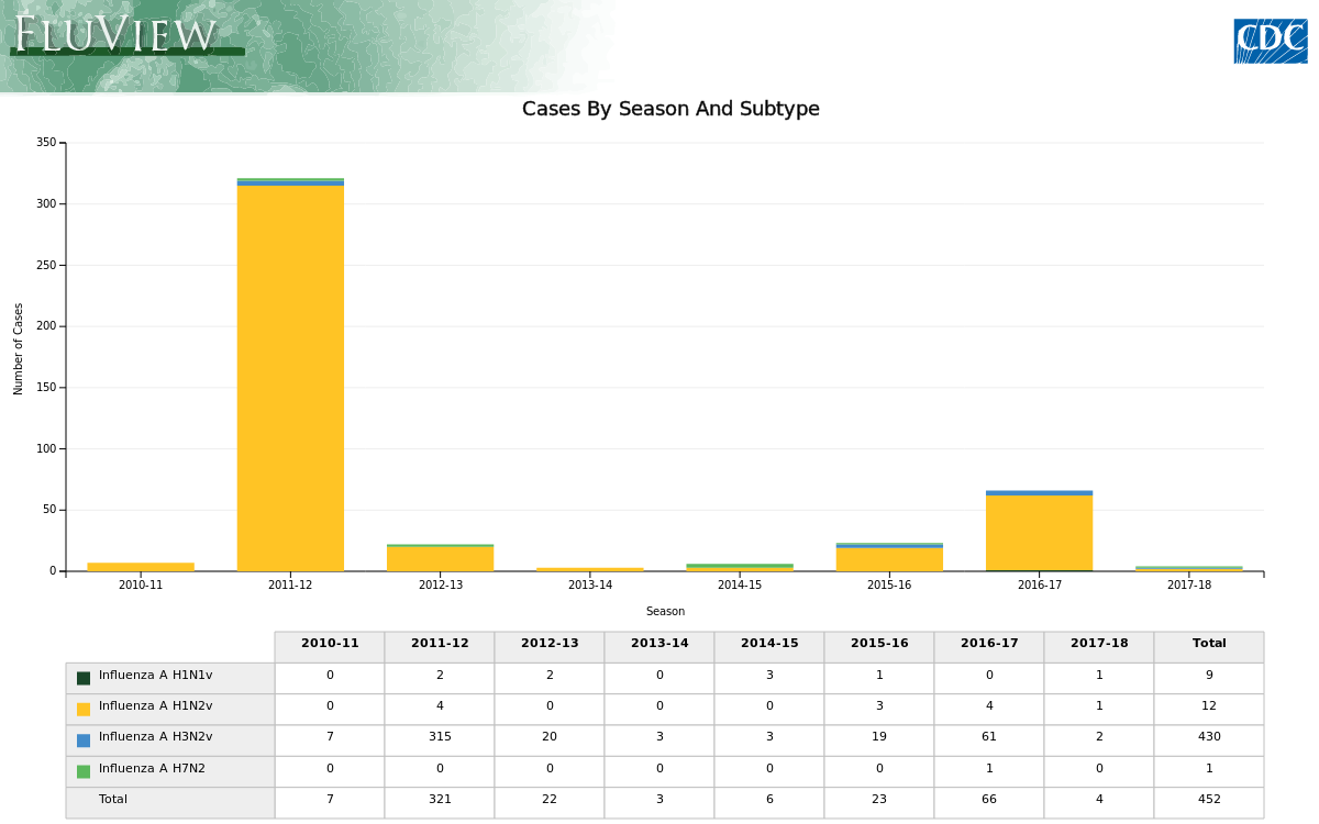
As of 24 July 2018, 2,229 cases of Middle East Respiratory Syndrome (MERS-CoV) have been reported to WHO with at least 791 related deaths. The majority of MERS-CoV cases have been reported from the Arabian Peninsula, with one large outbreak outside this region involving 186 cases in the Republic of Korea (RoK) in 2015. Cases have been exported to countries outside of the Middle East, with the most recent being identified in Malaysia in January 2018. This case was a Malaysian Umrah pilgrim who had visited a camel farm while in Saudi Arabia.

Excluding the outbreak in RoK, over 90% of all reported cases of MERS-CoV have been from the Kingdom of Saudi Arabia (KSA). Dromedary camels in the Middle East are an identified host and the likely source of infection in sporadic human cases. There is currently no evidence of sustained community transmission; human-to-human transmission is most likely to occur in healthcare facilities and household clusters. Large outbreaks linked to healthcare facilities are a feature of MERS-CoV and have occurred both within the Middle East and RoK. This underlines the significance of exposure to healthcare facilities in affected countries as a risk factor for infection, but also the importance of effective and rapid implementation of infection prevention and control practices for possible cases to limit the potential for onward transmission.
2. MERS-CoV in Saudi Arabia

In KSA, cases of MERS-CoV occur throughout the year, with occasional peaks which are a result of large hospital outbreaks; for example, 3 hospital outbreaks were reported from Riyadh in June 2017 leading to a peak in cases. Outside of these hospital outbreaks, the majority of cases are sporadic and usually occur in individuals with a history of contact with camels or camel products such as consumption of raw camel milk. PHE will continue to monitor the situation in KSA.
3. Risk assessment

There continues to be a risk of imported cases to the UK, and health professionals should remain vigilant. Early identification and rapid implementation of appropriate infection control measures for suspected cases is crucial.
The risk of infection with MERS-CoV to UK residents in the UK remains very low.

The risk of infection with MERS-CoV to UK residents travelling to the Middle East is very low but may be higher in those with exposure to specific risk factors within the region, such as camels (or camel products) or the local health care system.

Although the risk of MERS-CoV in individuals who meet the case definition for a possible case in the UK following travel to/from the Middle East is low, testing for MERS-CoV is warranted together with rapid implementation of appropriate infection control measures while awaiting results of testing.

The probability that a cluster in the UK of cases of severe acute respiratory infection of unexplained aetiology requiring intensive care admission is due to MERS-CoV remains very low but warrants investigation and testing. A history of travel to the Middle East would increase the likelihood of MERS-CoV.

The majority of outbreaks of MERS-CoV in the Middle East have been linked to healthcare settings. A previous WHO mission to Saudi Arabia concluded that gaps in infection control measures have most likely contributed to these outbreaks; this reinforced the importance of strict adherence to recommended infection control measures in healthcare facilities.

Where UK infection control procedures have been followed, the probability that a case of acute respiratory infection in a healthcare worker caring for a MERS-CoV case or a case of severe acute respiratory infection of unknown aetiology in ICU, is due to MERS-CoV is very low, but warrants testing; any healthcare worker who had recent contact with a confirmed case developing an acute respiratory illness would be tested for MERS-CoV. The risk will be higher for healthcare workers exposed to MERS-CoV who have not adhered to UK infection control procedures such as use of adequate personal protective equipment.

The risk to contacts of confirmed cases of MERS-CoV infection is low but contacts should be followed up for 14 days following last exposure and any new febrile or respiratory illness investigated urgently for MERS-CoV.

Further information and guidance on MERS-CoV is available on online
4. Travel advice

All travellers to the Middle East are advised to avoid contact with camels as much as possible:
  • travellers should practice good general hygiene measures, such as regular hand washing with soap and water at all times, but especially before and after visiting farms, barns or market areas
  • travellers are advised to avoid raw camel milk and/or camel products from the Middle East
  • more generally, travellers are also advised to avoid consumption of any type of raw milk, raw milk products and any food that may be contaminated with animal secretions unless peeled and cleaned and/or thoroughly cooked
  • travellers should follow the advice of local health authorities; there are currently no travel restrictions in place
  • travellers developing fever and a cough within 14 days of travel from the Middle East with should seek medical advice and must report their travel history so that appropriate clinical assessment, infection control measures and testing can be undertaken
  • people who are acutely ill with an infectious disease are advised not to travel but to seek health advice immediately
5. The Hajj

The most recent annual Muslim pilgrimage to Mecca in Saudi Arabia, known as the Hajj, took place between 30 August and 4 September 2017. This took place with no reported increase in travel related cases. However, cases of MERS-CoV have been imported to countries outside of Saudi Arabia following return from Umrah, a minor pilgrimage, as illustrated by the most recent Malaysian case. The Foreign and Commonwealth Office has stated that the 2018 Hajj is expected to take place between 19 August and 24 August 2018.

PHE remains vigilant and closely monitors developments in the Middle East and in the rest of the world where new cases have emerged and continues to liaise with international colleagues to assess whether our recommendations need to change.
PHE has published infographics for people travelling to or returning from the Middle East. These are available in a range of languages online.

NaTHNaC publishes travel health advice for Hajj and Umrah

Health professionals can access the possible MERS case definition online

While 99.999% of all returning travelers from the Middle East with respiratory symptoms will undoubtedly have something far less exotic or dangerous than MERS, the 2015 outbreak in South Korea is proof that it only takes one getting through to spark a national crisis.

For more on the Korean experience, and how France handled their clinical management of suspected MERS cases in Paris between 2013 and 2016, you may wish to revisit last week's BMC Inf. Dis.: Clinical Management Of Suspected MERS-CoV Cases.

Monday, July 30, 2018

BMC Vet.: Novel Reassortant H1N2 & H3N2 Swine Influenza A Viruses - Chile


#13,425


As mentioned in yesterday's blog (see Michigan: Pigs At Fowlerville Family Fair Test Positive For Swine Flu) - while surveillance in most of the world is marginal at best - we know that many different variants of swine influenza circulate in pigs around the globe.
Nearly all of these viruses contain genetic contributions from swine, avian, and human flu viruses - aka are `triple reassortants' - and as they circulate and co-mingle in swine, they continue to evolve down numerous evolutionary pathways.
This was the process by which the last H1N1 pandemic virus emerged in 2009, and there is little reason to doubt it could happen again someday.  For this reason we pay particular attention whenever a swine flu virus shows an ability to jump to humans.

In the fall of 2011 a new strain of swine H3N2 appeared in American pigs. What set this virus apart from earlier swine H3N2 viruses was that it was a triple reassortant swine virus which had picked up (via reassortment) the matrix (M) gene from the 2009 H1N1 pandemic virus.

The CDC has speculated that:
`This M gene may confer increased transmissibility to and among humans, compared to other variant influenza viruses.’CDC HAN 2012
Since then, swine viruses carrying the 2009 H1N1 M gene have become more common around the world, and we've seen this M gene turn up in other novel flu viruses as well (see Canine H3N2 Reassortant With pH1N1 Matrix Gene).

Increasingly, we're seeing novel swine flu viruses in the wild being described as `human-like', which arguably brings them closer to becoming a species jumping strain. From 2015's J. Virol: Novel Reassortant Human-like H3N2 & H3N1 Influenza A Viruses In Pigs.
The authors described both of these novel subtypes as “. . . virulent and can sustain onward transmission in pigs, and the naturally occurring mutations in the HA were associated with antigenic divergence from H3 IAV from human and swine’ and goes on to warn that  ``. . . the potential risk of these emerging swine IAV to humans should be considered”.
Also in 2015, in EID Journal: Influenza A Viruses of Human Origin in Swine, Brazil, we looked at surveillance in Brazil that uncovered multiple new lineages of swine influenza viruses that were related to seasonal influenza viruses that had circulated in humans more than a decade ago.
 
Three years ago, in Novel Human-like Influenza A Viruses Circulate in Swine in Mexico and Chile - published in PloS Currents - researchers reported finding:

Results: Our analysis identified multiple IAV-S lineages that appear to have been circulating undetected in swine for decades, including four novel IAV-S lineages of human seasonal virus origin that have not been previously identified in any swine populations globally.

We also found evidence of repeated introductions of pandemic H1N1 viruses from humans into swine in Mexico and Chile since 2009, and incursions of H1 and H3 viruses from North American swine into Mexico.
As the lines between human and swine flu viruses continue to blur, we have a new report - published last week in BMC Vet Research - that examines how these recently discovered human-like swine viruses isolated in Chile replicate in the guinea pig model.
Infection of novel reassortant H1N2 and H3N2 swine influenza A viruses in the guinea pig model

Rodrigo Tapia, Victoria García, Juan Mena, Sergio Bucarey, Rafael A. Medina and  Víctor Neira

Veterinary Research201849:73

https://doi.org/10.1186/s13567-018-0572-4

© The Author(s) 2018

Novel H1N2 and H3N2 swine influenza A viruses (IAVs) were identified in commercial farms in Chile. These viruses contained H1, H3 and N2 sequences, genetically divergent from IAVs described worldwide, associated with pandemic internal genes. 


Guinea pigs were used as human surrogate to evaluate the infection dynamics of these reassortant viruses, compared with a pandemic H1N1 virus. All viruses replicated and were shed in the upper respiratory tract without prior adaptation although H1N2 viruses showed the highest shedding titers. This could have public health importance, emphasizing the need to carry out further studies to evaluate the zoonotic potential of these viruses. 


Influenza A virus (IAV) has economic and public health relevance, being considered ubiquitous in the swine industry worldwide [1, 2]. There was limited information about IAVs circulating in swine population around the world, but the pandemic H1N1 (pdmH1N1) lineage that emerged in 2009 underscored the need to increase the surveillance and research of IAV in swine (IAV-S) [3].


In Chile, novel IAVs-S of the H1 and H3 subtypes have been recently identified in commercial swine farms, which are genetically divergent from IAVs described in other countries. These novel IAV-S lineages were most closely related to human seasonal H1N1 and H3N2 viruses from the late 1980s and early 1990s and were named: Chile H1 human I, Chile H1 human II, Chile H3 human I, and Chile H3 human II [4]. 
To date, phylogenetic analyses have been based on hemagglutinin (HA) sequences only, highlighting the need for additional studies to fully characterize these viruses, including the evaluation of their zoonotic potential.
        (SNIP)
These results demonstrate that novel reassortant H1N2 and H3N2 IAVs-S with pandemic internal genes were able to infect, replicate and be shed efficiently in the upper respiratory tract of guinea pigs without prior adaptation. These viruses were compared with a pdmH1N1 virus, lineage that has been well studied in the guinea pig model since its emergence in 2009 [7, 8, 20]. 

Overall, all viruses had similar shedding kinetics except that both H1N2 viruses showed higher shedding titers than pdmH1N1 and H3N2 viruses. On the other hand, only animals inoculated with the H3N2 virus showed titers at 7 dpi. Using guinea pig model, Sun et al. [21] reported similar shedding kinetics for H1N2 and H3N2 IAVs-S isolated in 2006 in China and a pdmH1N1 virus isolated from human in 2009, where H3N2 IAVs-S took a longer time to clear from the nasopharyngeal cavity than pdmH1N1 and seasonal human viruses.

This is of importance, since the respiratory system of guinea pigs has anatomical and physiological characteristics comparable to those of humans [11] along with a similar IAV receptor distribution [21]. Hence, these results suggest that these novel reassortant IAVs-S might be capable of replicating efficiently in other mammal hosts, such as humans.
        (Continue . . . )

Admittedly, the primary thrust of this short report revolves around the advantages of using guinea pigs - instead of mice and ferrets - for characterizing novel flu swine flu viruses.

But it also provides us with a useful update on the 2015 Chilean swine flu report, and reminds us of the tremendous diversity of novel flu viruses that continue to circulate- and evolve - in swine herds around the globe.

For more on the swine-origin flu viruses, you may wish to revisit:
J. Virology: Pathogenesis & Transmission of H3N2v Viruses Isolated in the United States, 2011-2016
JVI: Divergent Human Origin influenza Viruses Detected In Australian Swine Populations
Emerg. Infect. & Microbes: Novel Triple-Reassortant influenza Viruses In Pigs, Guangxi, China

Sunday, July 29, 2018

Michigan: Pigs At Fowlerville Family Fair Test Positive For Swine Flu


#13,424


Although we've only seen one reported human infection with a swine variant virus this summer (see June 30th's Indiana Reports H3N2v Infection In Fairgoer), this is the time of year when we generally start seeing more cases.
Some years, only one or two are identified.  Other year's - such as we saw in both 2017 and 2012 -  dozens or even scores have been reported (see chart below).
 

The CDC describes Swine Variant viruses in their Key Facts FAQ.
What is a variant influenza virus?
When an influenza virus that normally circulates in swine (but not people) is detected in a person, it is called a “variant influenza virus.” For example, if a swine origin influenza A H3N2 virus is detected in a person, that virus will be called an “H3N2 variant” virus or “H3N2v” virus.
Most (but not all) swine variant infections are mild or moderate, and are clinically indistinguishable from regular seasonal influenza. Which is why it is believed many swine variant infections go undetected (see CID Journal: Estimates Of Human Infection From H3N2v (Jul 2011-Apr 2012).

Yesterday the Livingston County (Michigan) Health Department (LCHD) announced the detection of swine flu among pigs at the Fowlerville Family Fair - which ran from July 23rd-July 28th.

So far, I've not found any indication of the swine flu subtype. While outliers are very occasionally reported (see USDA IAV-S Surveillance Program Detects Novel H3N1 In US SwineNorth American pigs normally carry H1N1, H1N2, or H3N2 swine viruses.
More than 90% of all human swine variant infections have been with H3N2v (see J. Virology: Pathogenesis & Transmission of H3N2v Viruses Isolated in the United States, 2011-2016).
While no human infections have been reported from Livingston County, local public health officials are asking swine exhibitors, their families, and anyone who visited the swine barn to notify them of possible exposure to infected pigs.

The LCHD is also asking local healthcare providers to be alert for any cases presenting with respiratory symptoms who had recent exposure to swine or who visited the fair.

From the LCHD press statement:

FOR IMMEDIATE RELEASE
Swine Flu Confirmed in Pigs at Fowlerville Family Fair

HOWELL, Michigan. – (July 27, 2018) The Michigan Department of Agriculture and Rural Development (MDARD) identified pigs at the Fowlerville Family Fair that tested positive for swine flu (influenza A).


The Fowlerville Fair Board has taken steps to isolate infected pigs to prevent additional exposure. Infected pigs began showing symptoms in the evening of Thursday, July 26 and laboratory results were confirmed late Friday afternoon. The fair is scheduled from July 23-July 28. At this time, there are no reported human illnesses.


The Livingston County Health Department (LCHD), in coordination with the Fowlerville Fair Board, are reaching out to swine exhibitors, their families, and those attendees who visited the swine barn at the Fowlerville Family Fair to notify them of possible exposure to infected pigs. The LCHD is also instructing healthcare providers in the area to watch for patients presenting with respiratory symptoms who report exposure to swine or who visited the fair.


Swine flu can spread quickly between pigs and while rare, can pass to humans through droplets in the air when sick pigs cough or sneeze. Human symptoms of swine flu are similar to those of seasonal flu and can include fever, cough, runny nose, and sometimes body aches, nausea, vomiting, or diarrhea. Symptoms usually appear within three days of exposure but can occur up to 10 days. 


Sometimes swine flu causes severe disease even in healthy people, such as pneumonia which may require hospitalization, and sometimes death. People who are at high risk of developing complications if they get swine flu include children younger than five years of age, people 65 years of age and older, pregnant women, and people with certain chronic health disease, such as asthma, diabetes, heart disease, weakened immune systems, and neurological conditions.

(Continue . . . )

While the pigs at this fair were symptomatic - thus sparking the alert -  pigs can often carry swine influenza with little or no signs of illness. Last August's  EID Journal: Transmission Of Swine H3N2 To Humans At Agricultural Exhibits - Michigan & Ohio 2016 examined the risks of novel flu transmission at these types of venues, including from healthy-looking pigs.
Of particular interest, while widespread illness in pigs was only rarely reported, surveillance revealed an average prevalence of influenza A in fair pigs of 77.5%. The authors cautioned that this suggests `that subclinical influenza A infections in pigs remain a threat to public health (3).'
Despite the current low risk of infection, there are some things you should consider doing to reduce your chances of getting sick, particularly if you are at `high risk' of flu complications.  This from the CDC.
CDC Recommendations For People At High Risk:
  • If you are at high risk of serious flu complications and are going to a fair where pigs will be present, avoid pigs and swine barns at the fair. This includes children younger than 5 years, people 65 years and older, pregnant women, and people with certain long-term health conditions (like asthma, diabetes, heart disease, weakened immune systems, and neurological or neurodevelopmental conditions).
If you are not at high risk, take these precautions:
  • Don’t take food or drink into pig areas; don’t eat, drink or put anything in your mouth in pig areas.
  • Don’t take toys, pacifiers, cups, baby bottles, strollers, or similar items into pig areas.
  • Wash your hands often with soap and running water before and after exposure to pigs. If soap and water are not available, use an alcohol-based hand rub.
  • Avoid close contact with pigs that look or act ill.
  • Take protective measures if you must come in contact with pigs that are known or suspected to be sick. This includes wearing personal protective equipment like protective clothing, gloves and masks that cover your mouth and nose when contact is required.
  • To further reduce the risk of infection, minimize contact with pigs in the pig barn and arenas.

While not expected to be as severe as some of the other novel viruses we are watching, immunity to these swine variant viruses is likely very low, and so the CDC's IRAT Influenza Risk Assessment Tool lists H3N2v as having moderate pandemic potential.

Swine flu viruses aren't just a North American concern, as they circulate all over the world. Surveillance in many places, however, is either close to nonexistent or marginal at best. 
How many human infections occur, and how many different variants there are in circulation, is unknown.
A few of of these viruses, such as the EAH1N1 virus in China, are viewed as having significant pandemic potential.  A few of my recent blogs on swine variant influenza outside the United States include:

PNAS: The Pandemic Potential Of Eurasian Avian-like H1N1 (EAH1N1) Swine Influenza
Trans. & Emerg. Dis.: Appearance Of Reassortant European Avian‐origin H1 influenza A viruses in Swine - Vietnam
I&ORV: Triple-Reassortant Novel H3 Virus of Human/Swine 
Origin Established In Danish Pigs Influenza A(H1N2) Virus Variant, Brazil


Friday, July 27, 2018

Frontiers Public Health: Live Poultry Trading Drives China's H7N9 Viral Evolution


Credit Frontiers Public Health


#13,423


As we've discussed many times in the past (see here, here, and here for just a few), the `secret sauce' common across most of China's growing array of avian flu threats has been the donation of internal genes (via reassortment) from an endemic low path poultry virus - H9N2 - which is widely dispersed across Asia and making inroads in the Middle East and Africa.

Of the four avian flu viruses we currently watch with the most concern – H5N1, H7N9, H10N8 and H5N6 – all share several important features (see Study: Sequence & Phylogenetic Analysis Of Emerging H9N2 influenza Viruses In China):
    • They all first appeared in  Mainland China
    • They all  have come about through viral reassortment in poultry
    • And most telling of all, while their HA and NA genes may differ - they all carry some or all of the internal genes from the avian H9N2 virus
While H9N2's involvement can be traced back two decades to the emergence of H5N1, since 2013 we've seen a sudden surge in the number of new avian reassortants appearing in China - nearly all carrying the incriminating fingerprints of H9N2.
This increase in reassortant avian viruses appears to coincide with recent evolutionary changes in H9N2, which include increased mammalian adaptation.
In 2014, in PLoS Path: Genetics, Receptor Binding, and Transmissibility Of Avian H9N2 researchers found evidence of Chinese H9N2 viruses binding preferentially to alpha 2,6 receptor cells -  the type commonly found in the human upper respiratory tract - rather than to alpha 2,3 receptor cells which are found in the gastrointestinal tract of birds.
H9N2's reach extends beyond just avian flu subtypes, as we've also seen evidence of it reassorting with other influenza viruses, including A Canine H3N2 Virus With PA Gene From Avian H9N2 - Korea and PNAS: Reassortment Of H1N1 And H9N2 Avian viruses.
Aiding and abetting H9N2 in the creation of new viable avian reassortant viruses has been the common practice of housing together many different species of birds and poultry at live bird markets (LBMs), where viruses that might never meet up normally in the wild are provided an ideal environment to reassort. 

Add in transport and trade of poultry between markets (LPTs), and what might have remained local, isolated, outbreaks of a novel avian virus are provided additional opportunities to become regional - or even international - threats. 
We've a new study and analysis published yesterday in Frontiers of Public Health of the impact of both H9N2's genetic contributions, and the influence of LBMs and the poultry trade, on the evolution and geographic spread of H7N9 in China.
It's a long, and at times, technical read.  But well worth following the link to read in its entirety.  I've only posted a few excerpts below.

Live Poultry Trading Drives China's H7N9 Viral Evolution and Geographical Network Propagation
Ruiyun Li1†, Tao Zhang2, Yuqi Bai2, Haochuan Li3, Yong Wang4, Yuhai Bi5, Jianyu Chang6* and Bing Xu1,2*

The on-going reassortment, human-adapted mutations, and spillover events of novel A(H7N9) avian influenza viruses pose a significant challenge to public health in China and globally. However, our understanding of the factors that disseminate the viruses and drive their geographic distributions is limited.
We applied phylogenic analysis to examine the inter-subtype interactions between H7N9 viruses and the closest H9N2 lineages in China during 2010–2014. We reconstructed and compared the inter-provincial live poultry trading and viral propagation network via phylogeographic approach and network similarity technique. 

The substitution rates of the isolated viruses in live poultry markets and the characteristics of localized viral evolution were also evaluated. We discovered that viral propagation was geographically-structured and followed the live poultry trading network in China, with distinct north-to-east paths of spread and circular transmission between eastern and southern regions.
The epicenter of H7N9 has moved from the Shanghai–Zhejiang region to Guangdong Province was also identified. Besides, higher substitution rate was observed among isolates sampled from live poultry markets, especially for those H7N9 viruses. 
Live poultry trading in China may have driven the network-structured expansion of the novel H7N9 viruses. From this perspective, long-distance geographic expansion of H7N9 were dominated by live poultry movements, while at local scales, diffusion was facilitated by live poultry markets with highly-evolved viruses.
(SNIP)
Recent analyses demonstrated that the novel H7N9 virus was a reassortant, with surface and internal gene segments originating from wild birds and the H9N2 lineage in poultry, respectively.
This indicates that wild birds were the most likely source of infection, introducing the virus into domestic ducks and chickens through sequential reassortment events, with a consequent spillover to humans by means of live poultry exposure (29).
The resulting multiple H7N9 lineages and genotypes suggest that the evolution of H9N2 has facilitated the genesis of the internal segments of this novel reassortant (4, 10, 11).
It has therefore exhibited greater genetic diversity compared with the surface genes (4, 12). As our preliminary researches have pointed out, this interaction at the wild birds–poultry–humans interface was common in the spread of infectious diseases on various scales (1317).
(SNIP)

In the perspective of live poultry trading network in the spatially-structured spread of AIVs presented here, additional interventions should be jointly implemented to restrict viral expansion along LPTs paths and targeted at live poultry workers.
Despite the effectiveness in reducing the daily number and growth rate of new human cases (21, 22) and the amount and detection rate of viable viruses (37) the mandatory closure of LBMs alone was unlikely to eliminate the zoonotic threat (38) accounting for the LPTs as the pathway with the highest likelihood of viral spread2. It is also reported that humans who engaged in the transportation work of live chickens and ducks was particularly susceptible to infections from AIVs (39).
These facts ascertained the role of live poultry trading in the spillover to humans at avian–human interface and the occurrence of H7N9 human cases. Therefore, a multi-sector, cost-effective approach and even international collaboration will be essential for the substantial reduction in the risk of disease spread and the build of a safer trade in animals3.

(Continue . . . )


While H7N9 activity has been greatly suppressed over the past year due to a combination of the introduction and nationwide deployment of a new H5+H7 poultry vaccine - and greater restrictions on live bird markets - the avian flu threat has not gone away. 
H5 and H7 viruses continue to circulate (along with H9N2) in wild bird and poultry, and earlier this year we saw the emergence of a novel H7N4 virus in Jiangsu Province (see UK PHE Guidance & Risk Assessment On Human H7N4 In China).
And despite government interventions, live bird markets continue to operate across much of China, often outside the law.  Add in the transport of live poultry  - identified as a significant threat by this study - and this respite in China's avian flu activity may prove temporary.

But the greater truth is, avian influenza viruses (AIVs) can reassort into human threats anywhere in the world.  The next big avian threat could easily come from Egypt, Indonesia, West Africa, India, or even the United States or Europe.


https://www.cdc.gov/flu/pdf/avianflu/avian-flu-transmission.pdf


Because Nature's laboratory is open 24/7, and never takes a break.

Thursday, July 26, 2018

EID Journal: Elizabethkingia & Association with Tap Water & Handwashing, Singapore

Credit CDC


#13,422


Ubiquitous in nature, and often multi-drug resistant, Elizabethkingia is a gram negative aerobic bacillus that only rarely causes opportunistic infections in (generally immunocompromised) humans, usually in hospital settings.
The genus Elizabethkingia includes not only E. meningoseptica, but E. miricola, E. endophytica, and E. anophelis - which in 2011 was initially found in the gut of an Anopheles gambiae mosquito (cite).
Despite its name E. anophelis has since been detected in the environment (both water and soil) and in health care settings (cite), and there's no solid evidence of transmission via mosquitoes.

While community acquired infections are rare, Elizabethkingia is viewed as an emerging nosocomial pathogen.  A few past outbreaks include:

A little over two years ago we were following an unusual event; for more than two months we watched an unprecedented 3-state Elizabethkingia anophelis outbreak unfold in America's Midwest (see WHO Takes Notice Of U.S. Elizabethkingia Outbreak).
Unlike the examples listed above - this was a community outbreak - and despite  intense multi-agency investigations, no epidemiological link has been found to explain the mysterious spike in the number of infected.
Had this outbreak occurred during the summer a mosquito vector might have been considered, but the first cases in this outbreak were detected in December and January, a time when mosquitoes aren't active in the upper Midwest. 
After 65 cases and 20 deaths were reported across 3 states - but mostly in Wisconsin - this outbreak ended as mysteriously as it began by early summer.

A paper published in Nature Comms the following year called Evolutionary dynamics and genomic features of the Elizabethkingia anophelis 2015 to 2016 Wisconsin outbreak strain found the Wisconsin isolates had demonstrated remarkable evolution over a short period of time.

They wrote:
Unlike other E. anophelis , the outbreak strain had a disrupted DNA repair mutYgene caused by insertion of an integrative and conjugative element. This genomic change probably contributed to the high evolutionary rate of the outbreak strain and may have increased its adaptability, as many mutations in protein-coding genes occurred during the outbreak. This unique discovery of an outbreak caused by a naturally occurring mutator bacterial pathogen provides a dramatic example of the potential impact of pathogen evolutionary dynamics on infectious disease epidemiology.
Two years later this  outbreak remains under investigation, and state health officials continue to monitor for any signs of a recurrence. 
Singapore, in the meantime, experienced a relatively small (3 case) nosocomial outbreak last year in a children’s intensive care unit.
Yesterday the EID Journal published a Dispatch describing the epidemiological investigation which found (as seen before) that hand washing stations were the likely source of the infection, which ironically, was spread to patients by HCWs after washing their hands.

The full article is worth reading, including their recommendation prioritizing the use of ABHR (Alcohol Based Hand Rubs) over hand washing in clinical care settings unless hands are visibly soiled.

Volume 24, Number 9—September 2018
Dispatch
Elizabethkingia anophelis and Association with Tap Water and Handwashing, Singapore

Chee-Fu Yung , Matthias Maiwald, Liat H. Loo, Han Y. Soong, Chin B. Tan, Phaik K. Lim, Ling Li, Natalie WH Tan, Chia-Yin Chong, Nancy Tee, Koh C. Thoon, and Yoke H. Chan

Author affiliations: KK Women’s and Children’s Hospital, Singapore.
Abstract

We report an Elizabethkingia anophelis case cluster associated with contaminated aerators and tap water in a children’s intensive care unit in Singapore in 2017. We demonstrate a likely transmission route for E. anophelis to patients through acquisition of the bacteria on hands of healthcare workers via handwashing.

Elizabethkingia anophelis is an emergent pathogen first described from midgut specimens of the Anopheles gambiae mosquito (1). To date, there have been 2 reported confirmed E. anophelis outbreaks in humans. One occurred in an adult critical care unit in Singapore; the second was a large community outbreak in the United States (Wisconsin, Michigan, and Illinois) (25).

Water sources have been identified to harbor members of the genus Elizabethkingia, but the source of the community outbreak in the United States remains unknown (3,6). Effective interventions for outbreak control and transmission routes of E. anophelis remain unclear (3).

KK Women’s and Children’s Hospital (KKH) is the single largest public tertiary-care specialist women’s and children’s hospital in Singapore. The Children’s Intensive Care Unit (CICU) is a 16-bed unit that provides advanced monitoring and therapeutic technologies for critical pediatric cases.

On May 30, 2017, an alert was triggered due to the detection of 3 patients with Elizabethkingia spp. within 13 days in the unit. The incidence rate of the cluster, 2.87/1,000 bed-days, was ≈4 times higher than the average rate in the previous 5 years, 0.63/1,000 bed-days (2012 through 2016).

Initially, the strains were reported as E. meningoseptica, but subsequent testing confirmed the cluster to be associated with E. anophelis. We conducted an epidemiologic investigation to identify the source of the cluster. We also conducted a pragmatic experiment to test our hypothesis that E. anophelis could be transmitted by healthcare workers during handwashing with water contaminated with E. anophelis.

        (SNIP)
We showed how handwashing, despite the use of chlorhexidine soap, is a possible vehicle of transmission for E. anophelis from an affected tap outlet via the hands of healthcare workers to patients. Perinatal transmission of E. anophelis was previously documented to have occurred from a mother with chorioamnionitis to her neonate (11).
We confirmed that hand hygiene using ABHR was effective in removing E. anophelis from hands of healthcare workers, which has implications for infection control. Although current hand hygiene guidelines prioritize ABHR over handwashing when hands are not visibly soiled, there is no requirement to perform ABHR in addition to handwashing (12).
Therefore, most staff consider handwashing as complying with hand hygiene requirements. Our findings support using ABHR as the primary hand-hygiene method in clinical care, especially in critical care units and in outbreak situations involving waterborne organisms such as E. anophelis.
        (Continue . . . .)

Alcohol rub has some weaknesses as well; the CDC states that soap and water are more effective than hand sanitizers at removing or inactivating certain kinds of germs, like Cryptosporidium, norovirus, and Clostridium difficile 11-15.
All of which suggests that a traditional soap and water hand scrub followed by a healthy dollop of ABHR - particularly in certain clinical settings - may be the better overall solution. 
Achieving a high level of compliance, however (see APIC: The Impact Of The Hawthorne Effect On Hand Hygiene Audits), is another matter entirely.

Wednesday, July 25, 2018

More Avian Flu Dispatches From Western Russia

Rostov Region


#13,421


Official reports, either from Russia's Rosselkhoznador or posted by the OIE, have been slow in coming (see Russia: Rosselkhoznador Statement On Prevention & Control Of Avian Influenza), but Russian language media sources continue to report new outbreaks on a daily basis. 
What we do know, officially, is that up until last week (OIE Update #5) more than 50 HPAI H5 outbreaks have been reported since early June, across at least 9 oblasts.
While HPAI H5N8 is strongly suspected, media reports and the official OIE filings only describe these outbreaks as due to HPAI H5.
This isn't Russia's first summer avian epizootic.
Last year - starting in April - Russia fought numerous outbreaks of HPAI H5N8 which began in a major turkey owned by Evrodon, which eventually spread across much of Western Russia (see Russia: Rosselkhoznadzor Reports Bird Flu Outbreak In Rostov Region).

Tons of contaminated poultry products managed to get into the retail chain (see Rosselkhoznador : HPAI Contaminated Poultry Shipped To At Least 9 Regions Of Russia) prompting nationwide recalls. 
Two weeks ago we saw OIE Notification Of Avian Flu Outbreaks In Rostov, Oryol, Kursk and Nizhegorod Oblasts, with the largest outbreak (600,000+ birds at risk) at JSC the "Poultry Factory Belokalitvinskaya", Belokalitvinsky, Rostov Region.
Today, via the Rostov government website, we learn of a second large outbreak, affecting one of the large turkey facilities owned by Evrodon.

In two areas of the Rostov region held events to eliminate flu outbreak birds July 25, 2018

Two weeks ago at the JSC "Poultry Belokalitvenskyi" recorded outbreak of avian influenza. The diagnosis was confirmed by the regional veterinary laboratory and the laboratory of the Federal Center for Animal Health in Vladimir. Epizootic has been recognized as a hotbed of the entire territory of the poultry farm, which contained about 616,000 chickens.
- Since the detection of the virus set of preventive measures to prevent the introduction and spread of avian influenza in the Rostov region is carried out in accordance with approved plans and the veterinary rules for the fight against bird flu - said the head of the department of veterinary medicine of the Rostov region Vladimir Zhilin.
In accordance with current regulations, the number in the hearth had to be liquidated. 
- In addition to the birds, feed the remains subject to liquidation, eggs and meat stored at the factory, - said Vladimir Zhilin.- incineration production activities will be completed within a few days. On their completion, as well as after the disinfection of premises, the factory will be able to get to work as usual.
In addition, birds flu foci in July were recorded in the two areas of cultivation of "Evrodon" (October district). Order of the Governor in the territory as quarantine restrictions were introduced. Subject to destruction poultry rearing № portion 5 of "Evrodon".

In the 10-kilometer zone around the poultry farms veterinarians bypass private farms citizens visiting vaccinated and vaccinated birds newcomers. Total to date, examined more than 14 thousand birds. Since the owners are encouraged to talk about the need to comply with veterinary regulations zoohygienic poultry.
Information Policy Department of the Rostov Region Government

Numerous other reports appearing in Russian media this morning suggest new outbreaks of avian flu continue to be reported across much of Western Russia, including:


In Ulyanovsk region detected bird flu center

Published: 07/25/2018 16:53

In Sura district of the Ulyanovsk region found hotbed of avian influenza. It is reported by the press service of the regional veterinary agency.

The hearth was discovered in the village Vypolzovo. It turned out that the source of introduction of the disease is delivery of poultry from Batyrevsky business district of the Chuvash Republic.

Already prepared a draft decree of the governor to impose a quarantine.

July 24
Bird flu found in another region of Tatarstan

The bird flu virus found in another region of Tatarstan - Buinsk. According to the newspaper " The Banner ", the chief of the district epizootologist vetobedineniya Ranis Nigmatullin confirmed deaths from the virus in chickens and ducks Stone Ford and Lower Naratbashe (one pair), willingly pitched in two households.
Samples of three other private enterprises of the Free Mill, one - of Buinsk sent to the Republican veterinary laboratory. Tatarstan President Rustam Minnikhanov signed a decree imposed a quarantine in these localities.

And lastly, a (translated) cautionary statement from the Veterinary Committee of Moscow, issued this morning (local time).

The situation for avian influenza remains tense

July 25, 2018, 14:18

Owners of private farms, as well as citizens, containing birds, it is necessary to take measures to prevent bird flu disease.
Veterinary Committee of Moscow informs that the Russian Federation is maintained tense situation for highly pathogenic avian influenza. As of July 16 in nine subjects recorded 60 foci. Avian influenza is registered in the private farms, and in large poultry farms.

Avian influenza - an acute infectious viral disease characterized by lesions of the digestive, respiratory and high mortality in domestic and wild birds of different species. The source of infection with avian influenza is sick birds and products of its life.

Owners of private farms, citizens, containing a bird, you must take the following measures to prevent the introduction of avian influenza virus:

  • The identification and ensure poultry, excluding its contact with wild and synanthropic bird.
  • Used for feeding birds only feed, feed mixtures, coming from areas free from avian influenza, and the presence of accompanying veterinary documents.
  • At the request of gosvetsluzhby experts to carry out laboratory monitoring of the circulation of the avian influenza virus.
  • Ensure separation of feeding and watering of different animal species.
  • Prevent access to unauthorized persons bird except gosvetsluzhby specialists for preventive and anti-epizootic measures.
  • Do not buy the bird and animal feed in places unauthorized trade.
  • Regularly mechanical cleaning and disinfection of cells and places for poultry.
  • Feathers, products of slaughter of birds disposed of (destroyed), in accordance with veterinary and sanitary rules of collection, recycling and disposal of biological waste.
  • the bird care should be carried out only in special clothes and footwear, which should be regularly washed and cleaned.
All cases of contagious poultry disease or suspicion of their origin, as well as bird deaths in the city, to immediately report to the territorial division of the State Veterinary Service of the City of Moscow GBU "Mosvetobedinenie" (contacts in the "Clinic" site mosobvet.ru) or clock telephone "hot line" SBD "Mosvetstantsiya": 8 (495) 612-12-12


Although I'm seeing some reports of successful eradication in some oblasts hit last month, this summer's epizootic seems far from over.


Stay tuned.

CDC Update: Candida Auris - July 2018

https://www.cdc.gov/fungal/diseases/candidiasis/pdf/Candida_auris_508.pdf


#13,420


Just over two years ago (June 2016) the CDC issued a Clinical Alert to U.S. Health care facilities about the Global Emergence of Invasive Infections Caused by the Multidrug-Resistant Yeast Candida auris.
C. auris is an emerging fungal pathogen that was first isolated in Japan in 2009. It was initially found in the discharge from a patient's external ear (hence the name `auris').  Retrospective analysis has traced this fungal infection back over 20 years.
Since then the CDC and public health entities have been monitoring an increasing number of cases (and hospital clusters) in the United States and abroad, generally involving bloodstream infections, wound infections or otitis.


As the CDC explains in their fact sheet:

Why is Candida auris a problem?

  • It causes serious infections. C. auris can cause bloodstream infections and even death, particularly in hospital and nursing home patients with serious medical problems. More than 1 in 3 patients with invasive C. auris infection (for example, an infection that affects the blood, heart, or brain) die.
  • It’s often resistant to medicines. Antifungal medicines commonly used to treat Candida infections often don’t work for Candida auris. Some C. auris infections have been resistant to all three types of antifungal medicines.
  • It’s becoming more common. Although C. auris was just discovered in 2009, it has spread quickly and caused infections in more than a dozen countries.
  • It’s difficult to identify. C. auris can be misidentified as other types of fungi unless specialized laboratory technology is used. This misidentification might lead to a patient getting the wrong treatment.
  • It can spread in hospitals and nursing homes. C. auris has caused outbreaks in healthcare facilities and can spread through contact with affected patients and contaminated surfaces or equipment. Good hand hygiene and cleaning in healthcare facilities is important because C. auris can live on surfaces for several weeks.
Yesterday the CDC updated their C. Auris surveillance page, where they show - as of June 30th  - 340 confirmed cases and 29 probable cases, across 11 states.

https://www.cdc.gov/fungal/candida-auris/tracking-c-auris.html


Additionally, based on targeted screening in four states reporting clinical cases, the CDC reports an additional 643 patients have been discovered to be asymptomatically colonized with C. auris
An increase of more than 13% over the previous month.
While the numbers remain relatively small, they are undoubtedly significantly under reported, both here in the United States, and around the world.  From the latest CDC update:

https://www.cdc.gov/fungal/candida-auris/tracking-c-auris.html

  • Single cases of C. auris have been reported from Australia, Austria, Belgium, Malaysia, the Netherlands, Norway, Russia, Singapore, Switzerland, and the United Arab Emirates.
  • Multiple cases of C. auris have been reported from Canada, China, Colombia, France, Germany, India, Israel, Japan, Kenya, Kuwait, Oman, Pakistan, Panama, Saudi Arabia, South Africa, South Korea, Spain, the United Kingdom, the United States (primarily from the New York City area, New Jersey, and the Chicago area) and Venezuela; in some of these countries, extensive transmission of C. auris has been documented in more than one hospital.
  • U.S. cases of C. auris have been found in patients who had recent stays in healthcare facilities in India, Kuwait, Pakistan, South Africa, the United Arab Emirates, and Venezuela, which also have documented transmission.
  • Other countries not highlighted on this map may also have undetected or unreported C. auris cases.
 Some past blogs include:
Notes from the Field: Surveillance for Candida auris — Colombia, September 2016–May 2017)

ECDC: C. Auris Rapid Risk Assessment For Healthcare Settings - Europe

mSphere: Comparative Pathogenicity of UK Isolates of the Emerging Candida auris