Showing posts with label ARDS. Show all posts

Tuesday, May 14, 2013

France: Both Coronavirus Patients Remain In `Poor’ Condition


image

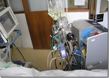
Extracorporeal membrane oxygenation (ECMO) machine in a ICU patient in Santa Cruz Hospital, Lisbon, Portugalj - Wikipedia


# 7270


An update from the Centre Hospitalier Regionale et Universitaire de Lille, in France where two nCoV patients remain in their ICU; the index case who traveled to the UAE during the middle of April, and a second patient who shared a hospital room with him before he was diagnosed.


Checkup of the two patients coronavirus

14 May 2013 - 12

Both patients coronavirus ICU at University Hospital of Lille are still in a poor state of health.


The first patient remains in stable condition. No improvement is recognized by doctors for the moment.


The second patient is still under ventilatory support by ventilator.


His condition is not completely stabilized.


The index case – a 65 y.o. man who was hospitalized on April 23rd -  reportedly remains on an ECMO (Extracorporeal Membrane Oxygenation) machine.  Increasingly, we’ve seen ECMO support used for severe respiratory distress due to pneumonia, avian flu, and the novel coronavirus.


Normally patients with Acute Respiratory Distress Syndrome (ARDS) are placed on a mechanical ventilator and treated with a variety of drugs to reduce infection (antibiotics) and lung inflammation (corticosteroids, Nitric Oxide, etc.).


Despite these measures, ARDS is generally fatal in 50% of patients.


Which is why in 2009 some hospitals tried an expensive and controversial treatment called ECMO on some adult and adolescent patients with H1N1 related pneumonia, and early reports were encouraging.


ECMO is a specialized heart-lung bypass machine used to take over the body’s heart and lung function – for days or weeks if necessary – while the body heals from injury or illness.


ECMO is most commonly used in neonatal intensive care units for newborns in respiratory distress, although it is also used for pediatric and adult patients with severe heart or respiratory deficits.


In the fall of 2009 we saw a report in The Lancet  where UK researchers determined that ARF (Adult Respiratory Failure) patients that received ECMO support as opposed to conventional ventilation had a greater survivability without disability.


While often lifesaving, ECMO resources are extremely limited, and are simply not available in many regions of the world.  


For more on ECMO, and how it has been used for ARDS, you may wish to revisit.


JAMA: H1N1, ECMO, and Survivability
The ECMO Option

Tuesday, November 10, 2009

Cocktails For Flu


# 3985


While the vast majority of H1N1 influenza victims recover quickly and without incident, a very small percentage go on to endure particularly serious and life-threatening symptoms – including ARDS (Acute Respiratory Distress Syndrome).


Last week, at the CHEST 2009 conference (Annual meeting of the American College of Chest Physicians), a study was presented on the results of an antiviral/steroid cocktail administered to ICU patients in Argentina during their recent pandemic wave.


This cocktail consisted of high dose oseltamivir (150mg twice a day), along with one of two steroids.   


For those presenting with ARDS, they were given Methylprednisolone (Medrol) 1 mg/kg/day for 14 days.   All other ICU patients received hydrocortisone 300 mg/day.


The administration of corticosteroids in ARDS has been tried in the past, with varying levels of success.  The experience from SARS in 2003 and H5N1 in the middle of this decade showed short-term improvement, but long-term survival rates were less than encouraging.  


In 2007 the WHO (World Health Organization) advised against the use of steroids in the treatment of Bird flu, stating:


Corticosteroid therapy has failed so far to show effectiveness, and prolonged or high dose corticosteroids can result in serious adverse events in H5N1 patients, including opportunistic infection. Corticosteroids should not be used routinely, except for persistent septic shock with suspected adrenal insufficiency.


But that was then.  This is now.  


I’ve excerpted some passages from a report on Doctor’s Guide that reviews the presentation at the CHEST 2009 Conference. 


It is, admittedly, a small study.  Only 13 patients.  But the results are encouraging.


Follow the link to read it in its entirety.


Oseltamivir Plus Corticosteroids Improves Outcomes in Patients With Acute Respiratory Distress Syndrome, H1N1: Presented at CHEST 2009

By Betty S. Riggs

SAN DIEGO -- November 9, 2009 -- The combination of oseltamivir and prolonged corticosteroid therapy results in clinical improvement in patients with hypoxaemic respiratory failure and influenza A(H1N1), according to a study presented here at CHEST 2009, the annual meeting of the American College of Chest Physicians.


<SNIP>

By day 7 of treatment, patients had significant improvement in lung injury score (LIS) and multiple organ dysfunction syndrome as measured by the Sequential Organ Failure Assessment (SOFA) score. From day 1 to day 7, the LIS decreased from 2.83 +- 0.8 to 2.01 +- 0.5 (P = .003) in H1N1-positive patients and from 3.45 +- 0.3 to 2.15 +- 0.8 (P = .02) in H1N1-negative patients.


From day 1 to day 7, the SOFA score decreased from 5.9 +- 1.6 to 3.3 +- 2.0 (P = .01) in H1N1-positive patients and from 7.4 +- 4.1 to 3.0 +- 3.5 (P = .01) in H1N1-negative patients.


There was 1 death in the H1N1-positive patients (12.5%) thought to be due to pulmonary embolism and 1 death in the H1N1-negative group (20%) due to progression of multiple organ dysfunction syndrome.

(Continue . . . )


In a related story from June of 2008, Hong Kong researchers were investigating zanamivir (Relenza) and two types of NSAIDS (Non-Steroidal Anti-Inflammatory Drugs) used to treat lab mice infected with H5N1.


Survivability increased 4-fold with this cocktail over zanamivir alone.


The NSAIDS used were celecoxib (Celebrex)  and mesalazine, an NSAID used for inflammatory bowel disease. See Research Into Antiviral/NSAID Cocktail.

Wednesday, October 14, 2009

Severely Ill H1N1 Patients At Risk Of PE


# 3833


One of the remarkable findings over the summer regarding the novel H1N1 Swine flu virus has been that, in a very small subset of patients, it can produce absolutely devastating symptoms.  


Not only are some patients experiencing ARDS (Acute respiratory distress syndrome), and requiring ventilator or ECMO support (see The ECMO Option), others are experiencing multi-organ failure and lung damage comparable to what has normally been associated with the H5N1 `bird flu’ virus.


Very early on we began hearing reports of some H1N1 cases involving  Pulmonary Emboli.   Pulmonary embolism (PE) is a blockage of a major artery in the lung by a thrombus (blot clot), or commonly, by an air or fat emboli.


PE can frequently produce a fatal outcome, particularly if untreated.  Diagnosing PE, particularly in patients already experiencing severe respiratory distress from a viral infection, can be difficult.


The July 10, 2009 / 58(Dispatch);1-4  MMWR (Morbidity & Mortality Weekly Report) of the CDC  described 5 such cases observed in Michigan in: Intensive-Care Patients With Severe Novel Influenza A (H1N1) Virus Infection --- Michigan, June 2009


Today, we get a pair of follow up studies in American Journal of Roentgenology, which describe (in detail of most interest to pulmonologists and radiologists) computed tomography (CT) scans of patients with severe H1N1 infection. 


First the press release, where the authors warn clinicians to be watchful for signs of PE in H1N1 patients, and then links to the two studies.


A hat tip to Dutchy on FluTrackers for Posting this link.


American Roentgen Ray Society


CT scans show patients with severe cases of H1N1 are at risk for developing acute pulmonary emboli

Researchers utilizing computed tomography (CT) scans have found that patients with severe cases of the H1N1 virus are at risk for developing severe complications, including pulmonary emboli (PE), according to a study to be published online Oct. 14, 2009, in the American Journal of Roentgenology. The study will be published in the December issue of the AJR.


A pulmonary embolism occurs when one or more arteries in the lungs become blocked. The condition can be life-threatening. However, if treated aggressively, anti-coagulants (blood thinners) can reduce the risk of death.


The study, performed at the University of Michigan Health Service, included 66 patients diagnosed with the H1N1 flu. Two study groups were formed. Group one consisted of 14 patients who were severely ill and required Intensive Care Unit (ICU) admission. Group two consisted of 52 patients who were not severely ill and did not require ICU admission.


All 66 patients underwent chest X-rays for the detection of H1N1 abnormalities. Ten patients from the ICU group and five patients from the largely outpatient group, underwent CT scans. "Pulmonary Emboli were seen on CT in five of 14 ICU patients," said Prachi P. Agarwal, M.D., lead author of the study.


"Our study suggests that patients who are severely ill with H1N1 are also at risk for developing PE, which should be carefully sought for on contrast-enhanced CT scans," she said.


"With the upcoming annual influenza season in the United States, knowledge of the radiologic features of H1N1 is important, as well as the virus's potential complications.The majority of patients undergoing chest X-rays with H1N1 have normal radiographs.

CT scans proved valuable in identifying those patients at risk of developing more serious complications as a possible result of the H1N1 virus, and for identifying a greater extent of disease than is appreciated on chest radiographs," said Dr. Agarwal.


Both articles are available online today, at the links below.


Imaging Findings in a Fatal Case of Pandemic Swine-Origin Influenza A (H1N1)

Chest Radiographic and CT Findings in Novel Swine-Origin Influenza A (H1N1) Virus (S-OIV) Infection