
Credit CDC
# 8944
The open access journal mBio today has published a study today looking at the replication, and pathogenicity of, the avian H7N9 virus in both the upper and lower respiratory tracts of cynomolgus macaques.
Although ferrets and other non-primate animals are often used for influenza transmission research, when you want to study the pathogenicity of a virus, non-human primates offer the closest analog to humans.
The Wisconsin National Primate Research Center explains the rationale behind their use:
Macaques are genetically very similar to humans. They especially share analogous neurological, reproductive and immunological systems with humans. Rhesus and cynomolgous macaques are not endangered in the wild and adapt well to captive housing. Research with rhesus and cynomolgous monkeys, as well as with other nonhuman primates, tells us a great deal about primate biology. Animal studies can be better controlled and can garner more consistent results than human studies, and are often precursors to human studies.
Shortly after the outbreak of H7N9 in Eastern mainland China in the spring of 2013 we learned of some of the research studies that were quickly set in motion to better understand this emerging avian flu virus (see ScienceInsider: Laboratory Plans For H7N9 Virus).
At that time we knew little about the pathogenicity and transmissibility of the virus in humans, and a top stated priority was to test H7N9 on a variety of lab animals, including ferrets and cynomolgus macaques.
Since then, we’ve seen a steady procession of studies, many of which have suggested that the H7N9 virus – perhaps more than any other avian influenza virus we’ve seen to date – has worrying pandemic potential.
- Last year we saw studies suggested H7N9 is unusually well-adapted to mammalian physiology (see mBio: H7N9 Naturally Adapted For Efficient Growth in Human Lung Tissue) – but thus far this virus has failed to demonstrate the ability to transmit efficiently among humans.
- Last May, in EID Journal: H7N9 As A Work In Progress, we looked at a study that found the H7N9 avian virus continues to reassort with local H9N2 viruses, making the H7N9 viruses that circulated in wave 2 genetically distinct from those that were seen during the 1st wave.
- While last June, in Eurosurveillance: Genetic Tuning Of Avian H7N9 During Interspecies Transmission, we saw even more evidence of the genetic diversity, and continual evolution, of the H7N9 virus in Mainland China. Researchers found that at least 26 separate genotypes had emerged, mostly during the first wave, through a process they called `genetic tuning’.
While this virus is apparently on summer hiatus, few expect it to remain so once temperatures begin to decline this fall and winter. Last winter’s outbreak came after an exceptionally quiet summer, and was twice the size of the first wave.
All of which makes learning as much as we can about the pathogenicity, and pandemic potential, of this virus a high priority.
Emmie de Wita, Angela L. Rasmussenb, Friederike Feldmannc, Trenton Bushmakera, Cynthia Martellaroa, Elaine Haddocka, Atsushi Okumurab, Sean C. Prollb, Jean Changb, Don Gardnerc, Michael G. Katzeb,d, Vincent J. Munstera, Heinz Feldmanna,e
ABSTRACT
In March 2013, three fatal human cases of infection with influenza A virus (H7N9) were reported in China. Since then, human cases have been accumulating. Given the public health importance of this virus, we performed a pathogenicity study of the H7N9 virus in the cynomolgus macaque model, focusing on clinical aspects of disease, radiographic, histological, and gene expression profile changes in the upper and lower respiratory tracts, and changes in systemic cytokine and chemokine profiles during infection.
Cynomolgus macaques developed transient, mild to severe disease with radiographic evidence of pulmonary infiltration. Virus replicated in the upper as well as lower respiratory tract, with sustained replication in the upper respiratory tract until the end of the experiment at 6 days after inoculation. Virus shedding occurred mainly via the throat.
Histopathological changes in the lungs were similar to those observed in humans, albeit less severe, with diffuse alveolar damage, infiltration of polymorphonuclear cells, formation of hyaline membranes, pneumocyte hyperplasia, and fibroproliferative changes. Analysis of gene expression profiles in lung lesions identified pathways involved in tissue damage during H7N9 infection as well as leads for development of therapeutics targeting host responses rather than virus replication.
Overall, H7N9 infection was not as severe in cynomolgus macaques as in humans, supporting the possible role of underlying medical complications in disease severity as discussed for human H7N9 infection (H. N. Gao et al., N. Engl. J. Med. 368:2277–2285, 2013, doi:10.1056/NEJMoa1305584).
Curiously, while more pathogenic to macaques than most strains of the 2009 H1N1 pandemic virus, and seasonal H3N2, the H7N9 virus produced less severe symptoms in these non-human primates than has been common observed in humans.
The authors suggest that the high rate of co-morbidities in the – mostly elderly – cohort of cases in China could explain this difference.
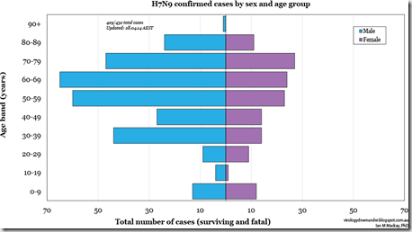
And indeed, the chart below from Dr. Ian Mackay’s VDU Blog shows a pronounced demographic shift towards older, predominantly male, victims in China. Children, and young adults – when they were found to be infected – were more likely to have mild, moderate, or even asymptomatic infections (see Mild Influenza A/H7N9 Infection among Children in Guangdong Province).
Compared to infection with the 1918 H1N1 and H5N1 avian flu viruses (see my three part review of the 2009 Baskin Influenza in Primates Study), H7N9 produced far less lung damage and illness in macaques.
You’ll want to read the entire study for details on methods and materials, and a closer look at the results. In their concluding remarks, the authors summed up their work by saying:
Thus, the emerging H7N9 influenza virus is more pathogenic than seasonal influenza A virus and most isolates of the pandemic H1N1 virus but not as pathogenic as the 1918 Spanish influenza virus and HPAI H5N1 virus in cynomolgus macaques.
However, the pathogenicity of the H7N9 virus may decrease if the virus adapts further to solely using α2,6-linked sialic acids as the receptor for entry, as pandemic influenza viruses to date have done (52–55).
Exclusive attachment to α2,6-linked sialic acids would most likely result in a shift to replication mainly in the upper respiratory tract of humans, likely resulting in less severe disease, as has been described for the 2009 pandemic H1N1 virus (56) and upon adaptation of HPAI H5N1 virus to efficient transmission via respiratory droplets or aerosols (57).
The switch to an `exclusive attachment to α2,6-linked sialic acids’ is one of the evolutionary changes that is believed would make this virus far more easily transmitted between humans, and is probably required if H7N9 were to become a viable pandemic virus.
And while a commensurate decrease in severity would be a welcome result, it is worth noting that a reduction of even a full order of magnitude (30% CFR down to 3%) would still put this virus in the same league as the 1918 Spanish flu.
While the tragedy of Ebola has captured the world’s attention this summer, the most serious pandemic threats are those posed by respiratory viruses, such as influenza, SARS, and MERS.

