Showing posts with label mbio. Show all posts

Tuesday, May 19, 2015

mBio: A Novel Pathogenic Mammalian Orthoreovirus In Diarrheic American Pigs

image

Photo Credit CDC EID Journal


# 10,064


While American poultry farmers are gearing up to battle HPAI, for the past two years North American swine farmers have been dealing with several recently discovered novel coronaviruses causing diarrheal disease, and often death, in young piglets. 


We’ve seen no evidence of human infection due to these swine coronaviruses, so they are not currently considered a zoonotic disease.  Which is not to say that one or both couldn’t someday pose a threat. Coronaviruses – like all RNA viruses – tend to evolve and mutate at a fairly rapid rate (see discussion of new zoonotic coronaviruses).

Porcine epidemic diarrhea has now been reported in 32 states, and unexpectedly has hit some of the same herds more than once, leading researchers to wonder if there might be more pathogens at work than are already identified.  


All of which leads us to a fascinating bit of viral detective work, where researchers have isolated and identified yet another pathogenic diarrhea-inducing virus circulating in American pigs. 


This time, however, the culprit is not a coronavirus – but a novel mammalian orthoreovirus (MRV).


First the abstract from a much larger (open access) mBio research article, after which I’ll return with more:

A Novel Pathogenic Mammalian Orthoreovirus from Diarrheic Pigs and Swine Blood Meal in the United States

Athmaram Thimmasandra Narayanappaa, Harini Sooryanaraina,  Jagadeeswaran Deventhirana, Dianjun Caoa, Backiyalakshmi Ammayappan Venkatachalama,  Devaiah Kambirandab,  Tanya LeRoitha,  Connie Lynn Heffrona,  Nicole Lindstroma,  Karen Halla,  Peter Jobsta,  Cary Sextonc,  Xiang-Jin Menga, Subbiah Elankumarana 

ABSTRACT

Since May 2013, outbreaks of porcine epidemic diarrhea have devastated the U.S. swine industry, causing immense economic losses. Two different swine enteric coronaviruses (porcine epidemic diarrhea virus and Delta coronavirus) have been isolated from the affected swine population. The disease has been reported from at least 32 states of the United States and other countries, including Mexico, Peru, Dominican Republic, Canada, Columbia, Ecuador, and Ukraine, with repeated outbreaks in previously infected herds.

Here we report the isolation and characterization of a novel mammalian orthoreovirus 3 (MRV3) from diarrheic feces of piglets from these outbreaks in three states and ring-dried swine blood meal from multiple sources. MRV3 could not be isolated from healthy or pigs that had recovered from epidemic diarrhea from four states. Several MRV3 isolates were obtained from chloroform-extracted pig feces or blood meal in cell cultures or developing chicken embryos.

Biological characterization of two representative isolates revealed trypsin resistance and thermostability at 90°C. NextGen sequencing of ultrapurified viruses indicated a strong homology of the S1 segment to mammalian and bat MRV3. Neonatal piglets experimentally infected with these viruses or a chloroform extract of swine blood meal developed severe diarrhea and acute gastroenteritis with 100% mortality within 3 days postinfection. Therefore, the novel porcine MRV3 may contribute to enteric disease along with other swine enteric viruses. The role of MRV3 in the current outbreaks of porcine epidemic diarrhea in the United States remains to be determined, but the pathogenic nature of the virus warrants further investigations on its epidemiology and prevalence.


This paper goes on to succinctly describe MRV viruses as:


The family Reoviridae comprises 15 genera of double-stranded RNA (dsRNA) viruses (9). Orthoreoviruses with 10 discrete RNA segments have been isolated from a wide variety of animal species, including bats, civet cats, birds, reptiles, pigs, and humans (10, 11).

Most orthoreoviruses are recognized to cause respiratory infections, gastroenteritis, hepatitis, myocarditis, and central nervous system disease in humans, animals, and birds (11); orthoreovirus genomes are prone to genetic reassortment and intragenic rearrangement (11, 12). The exchange of RNA segments between viruses could lead to molecular diversity and evolution of viruses with increased virulence and host range (13, 14).

MRV serotypes 1 to 3 were associated with enteritis, pneumonia, or encephalitis in swine around the world, including China and South Korea (15– 18). The zoonotic potential of MRV3 has been reported recently (19–21). However, porcine orthoreovirus infection of pigs was unknown previously in the United States.


The entire study is highly detailed, and well worth reading in its entirety.


In recent years we’ve seen increased interest and research into orthoreoviruses, with an particular eye towards their ability to reassort and to perhaps someday pose a greater threat to human health.


In 2007 we saw  the emergence of what is now called the Melaka Virus (see First Bird Flu, Now Bat Flu & Why You Should Catch `Contagion’) – an orthoreovirus carried by bats in Malaysia.


Since then, several additional bat-borne orthoreoviruses have been identified, including the Xi-River, Kampar, Sikamat, HK23629/07 and Broome virus.  As bats are the most abundant and geographically dispersed vertebrates on earth, their ability to carry and vector dangerous diseases without ill-effect (i.e. Rabies, Nipah, Hendra, even influenza) is increasingly a topic of study.


Just last week, PLoS One published a study looking at a potential reassortment of bat, pig, and/or human MRV strains in a bat reservoir in China.


Isolation and Identification of a Natural Reassortant Mammalian Orthoreovirus from Least Horseshoe Bat in China

Lihua Wang,1,2 Shihong Fu,1,2 Lei Cao,1,2 Wenwen Lei,1,2 Yuxi Cao,1,2 Jingdong Song,1,2 Qing Tang,1,2 Hailin Zhang,3 Yun Feng,3 Weihong Yang,3 and Guodong Liang1,2,*

Abstract

Background

Mammalian orthoreoviruses (MRVs) have a wide geographic distribution and can infect virtually all mammals. Infections in humans may be either symptomatic or asymptomatic. This study describes the isolation and identification of a natural reassortant MRV from least horseshoe bats (Rhinolophus pusillu) in China, referred to as RpMRV-YN2012.

<SNIP>

MRV infection in humans has been shown to be fairly common and the infections are often shown to be asymptomatic or associated with mild, self-limiting respiratory or gastrointestinal illness in infants and children [3]. Recent studies have shown that MRV can cause severe illnesses in humans and other mammals, including upper respiratory tract infections, encephalitis, and diarrhea[4, 5, 18].

(Continue . . . )

And a couple of years ago, in the Journal of Virology, we saw:


High Similarity of Novel Orthoreovirus Detected in a Child Hospitalized with Acute Gastroenteritis to Mammalian Orthoreoviruses Found in Bats in Europe

Andrej Steyera, Ion Gutiérrez-Aguireb,e, Marko Kolenca, Simon Korenc, Denis Kutnjakb, Marko Pokornd, Mateja Poljšak-Prijatelja, Nejc Račkib, Maja Ravnikarb,e, Martin Sagadina, Adela Fratnik Steyera and Nataša Toplakc 

ABSTRACT

Mammalian orthoreoviruses (MRVs) are known to cause mild enteric and respiratory infections in humans. They are widespread and infect a broad spectrum of mammals. We report here the first case of an MRV detected in a child with acute gastroenteritis, which showed the highest similarity to an MRV reported recently in European bats.


(Continue . . . )


None of which is to suggest that MRVs are destined to be the next big public health threat, or that the MRV3 virus described in the mBio paper today poses any immediate threat to human health.


But they do serve as a reminder that the constellation of  viruses out there with zoonotic potential is much greater and more widespread than we know, and that these viruses are not static - but are instead moving targets – evolving, reassorting, and spreading geographically at truly impressive rates.


We’ll return to today’s mBio study to close, with the author’s summation of the importance of their findings:


IMPORTANCE Porcine orthoreoviruses causing diarrhea have been reported in China and Korea but not in the United States. We have isolated and characterized two pathogenic reassortant MRV3 isolates from swine fecal samples from porcine epidemic diarrhea outbreaks and ring-dried swine blood meal in the United States. These fecal and blood meal isolates or a chloroform extract of blood meal induced severe diarrhea and mortality in experimentally infected neonatal pigs. Genetic and phylogenetic analyses of two MRV3 isolates revealed that they are identical but differed significantly from nonpathogenic mammalian orthoreoviruses circulating in the United States. The present study provides a platform for immediate development of suitable vaccines and diagnostics to prevent and control porcine orthoreovirus diarrhea.

Tuesday, March 10, 2015

mBio: MRSA – Making A House A Biome

colorized scanning electron micrograph (SEM) of MRSA

MRSA - Photo Credit CDC


# 9804


From the Open Access journal mBio this morning, a report that suggests that MRSA can hang around a household, bounce repeatedly between family members, and evolve into a unique `family’ strain, over a period of several years.


Previously we’ve looked at MRSA colonization, where the CDC quantified colonization by stating:


While 25% to 30% of people are colonized* in the nose with staph, less than 2% are colonized with MRSA (Gorwitz RJ et al. Journal of Infectious Diseases. 2008:197:1226-34.).

*Colonized:
When a person carries the organism/bacteria but shows no clinical signs or symptoms of infection. For Staph aureus the most common body site colonized is the nose.


However, in  Firefighters & Paramedics At Greater Risk Of MRSA and Firefighters & MRSA Revisited we looked at research showing a 10x’s greater incidence of MRSA colonization (20%) among a sampling of firefighters tested in Washington State.


As we learn from an American Society of Microbiology press release today (see MRSA can linger in homes, spreading among its inhabitants), a 2012 study found up to 50% of family contacts in households with one SSTI (Skin or soft Tissue Infection) with MRSA were colonized with the resistant bacteria.


They describe the findings of today’s study as:


Households can serve as a reservoir for transmitting methicillin-resistant Staphylococcus aureus (MRSA), according to a study published this week in mBio®, the online open-access journal of the American Society for Microbiology. Once the bacteria enters a home, it can linger for years, spreading from person to person and evolving genetically to become unique to that household.

<SNIP>

he researchers found that isolates within households clustered into closely related groups, suggesting a single common USA300 ancestral strain was introduced to and transmitted within each household. Researchers also determined from a technique called Bayesian evolutionary reconstruction that USA300 MRSA persisted within households from 2.3 to 8.3 years before their samples were collected, and that in the course of a year, USA300 strains had a 1 in a million chance of having a random genetic change, estimating the speed of evolution in these strains. Researchers also found evidence that USA300 clones, when persisting in households, continued to acquire extraneous DNA.

"We found that USA300 MRSA strains within households were more similar to each other than those from different households," said senior study author Michael Z. David, MD, PhD, an assistant professor of edicine at the University of Chicago. Although MRSA is introduced into households rarely, he said, once it gets in, "it can hang out there for years, ping-ponging around from person to person. Our findings strongly suggest that unique USA300 MRSA isolates are transmitted within households that contain an individual with a skin infection."

(Continue . . .)

A link, and the abstract to the entire mBio report can be accessed below:


Transmission and Microevolution of USA300 MRSA in U.S. Households: Evidence from Whole-Genome Sequencing

Md Tauqeer Alama, Timothy D. Reada,b, Robert A. Petit IIIa, Susan Boyle-Vavrac, Loren G. Millerd, Samantha J. Eellsd, Robert S. Daumc, Michael Z. Davidc,e

ABSTRACT

Methicillin-resistant Staphylococcus aureus (MRSA) USA300 is a successful S. aureus clone in the United States and a common cause of skin and soft tissue infections (SSTIs). We performed whole-genome sequencing (WGS) of 146 USA300 MRSA isolates from SSTIs and colonization cultures obtained from an investigation conducted from 2008 to 2010 in Chicago and Los Angeles households that included an index case with an S. aureus SSTI.

Identifying unique single nucleotide polymorphisms (SNPs) and analyzing whole-genome phylogeny, we characterized isolates to understand transmission dynamics, genetic relatedness, and microevolution of USA300 MRSA within the households. We also compared the 146 USA300 MRSA isolates from our study with the previously published genome sequences of the USA300 MRSA isolates from San Diego (n = 35) and New York City (n = 277). We found little genetic variation within the USA300 MRSA household isolates from Los Angeles (mean number of SNPs ± standard deviation, 17.6 ± 35; π nucleotide diversity, 3.1 × 10⁻⁵) or from Chicago (mean number of SNPs ± standard deviation, 12 ± 19; π nucleotide diversity, 3.1 × 10⁻⁵).

The isolates within a household clustered into closely related monophyletic groups, suggesting the introduction into and transmission within each household of a single common USA300 ancestral strain. From a Bayesian evolutionary reconstruction, we inferred that USA300 persisted within households for 2.33 to 8.35 years prior to sampling. We also noted that fluoroquinolone-resistant USA300 clones emerged around 1995 and were more widespread in Los Angeles and New York City than in Chicago. Our findings strongly suggest that unique USA300 MRSA isolates are transmitted within households that contain an individual with an SSTI. Decolonization of household members may be a critical component of prevention programs to control USA300 MRSA spread in the United States.

IMPORTANCE USA300, a virulent and easily transmissible strain of methicillin-resistant Staphylococcus aureus (MRSA), is the predominant community-associated MRSA clone in the United States. It most commonly causes skin infections but also causes necrotizing pneumonia and endocarditis. Strategies to limit the spread of MRSA in the community can only be effective if we understand the most common sources of transmission and the microevolutionary processes that provide a fitness advantage to MRSA.

We performed a whole-genome sequence comparison of 146 USA300 MRSA isolates from Chicago and Los Angeles. We show that households represent a frequent site of transmission and a long-term reservoir of USA300 strains; individuals within households transmit the same USA300 strain among themselves. Our study also reveals that a large proportion of the USA300 isolates sequenced are resistant to fluoroquinolone antibiotics. The significance of this study is that if households serve as long-term reservoirs of USA300, household MRSA eradication programs may result in a uniquely effective control method.

(Continue . . . )


Thursday, February 19, 2015

mBio: Ebola Virus Transmission (What We Know, What We Don’t)

image

Credit CDC PHIL


# 9729


The open access journal mBio has published an impressively detailed look at what we know about the transmission of the Ebola virus, penned by lead author Michael Osterholm of CIDRAP (along with an impressive international array of co-authors), that also asks a number of questions regarding what is not known about the virus.


Labeled as both an `Opinion’ piece, and a `Hypothesis’, this article also questions some of the assumptions about the behavior of the Ebola virus as it exists today, and as it might exist in the future.


Near the end of this paper you’ll find a clearly marked: RESPIRATORY TRANSMISSION OF EBOLA VIRUSES: A HYPOTHESIS, which asks the question:


Despite the lack of supportive epidemiological data, a key additional question to ask is whether primary pulmonary infections and respiratory transmission of Ebola viruses could be a potential scenario for the future.


Based on some twitter comments this morning, the inclusion of this hypothesis seems to have ruffled some academic feathers. Not being a virologist (or scientist of any stripe), I can’t really speak to the merits of their hypothesis, other than to say:


The path to new discoveries often lies in the willingness to continue to ask those questions that others already consider asked and answered.  


Whether you agree with their hypothesis (which they call `improbable’, but not impossible) or not, this review is well worth reading, and considering, in its entirety.  


Transmission of Ebola Viruses: What We Know and What We Do Not Know

Michael T. Osterholma, Kristine A. Moorea, Nicholas S. Kelleya, Lisa M. Brosseaub, Gary Wongc, Frederick A. Murphyd, Clarence J. Petersd, James W. LeDucd, Phillip K. Russelle, Michel Van Herpf, Jimmy Kapetshig, Jean-Jacques T. Muyembeg, Benoit Kebela Ilungah, James E. Strongc, Allen Grollac, Anja Wolzf, Brima Kargboi, David K. Kargboi, Pierre Formentyj, David Avram Sandersk, Gary P. Kobingerc


  1. ABSTRACT

Available evidence demonstrates that direct patient contact and contact with infectious body fluids are the primary modes for Ebola virus transmission, but this is based on a limited number of studies. Key areas requiring further study include (i) the role of aerosol transmission (either via large droplets or small particles in the vicinity of source patients), (ii) the role of environmental contamination and fomite transmission, (iii) the degree to which minimally or mildly ill persons transmit infection, (iv) how long clinically relevant infectiousness persists, (v) the role that “superspreading events” may play in driving transmission dynamics, (vi) whether strain differences or repeated serial passage in outbreak settings can impact virus transmission, and (vii) what role sylvatic or domestic animals could play in outbreak propagation, particularly during major epidemics such as the 2013–2015 West Africa situation. In this review, we address what we know and what we do not know about Ebola virus transmission. We also hypothesize that Ebola viruses have the potential to be respiratory pathogens with primary respiratory spread.

(Continue . . . )

Wednesday, November 19, 2014

mBio: Not All LPAI Flu Subtypes Are Created Equally

image

Credit NIAID


# 9343


Until avian H7N9 showed up a couple of years ago, LPAI (Low Pathogenic Avian Influenza) in birds was considered to constitute a minor human health threat, and our concerns were mostly they would evolve into HPAI (highly pathogenic) strains – with most of that concern focused on H5 and H7 subtypes.


H7N9 showed us that an avian influenza could be LPAI in birds and yet extremely pathogenic in humans.


Furthermore, in the summer of 2013 Taiwan reported the first known human infection with an avian H6N1 virus, and a few months later mainland China reported the first three cases of H10N8 (two fatal), further widening the range of avian subtypes capable of causing serious illness in humans.


Suddenly, there seemed to be more to keep an eye on than just HPAI H5 and H7.


All of which brings us to a fascinating study, published yesterday in the open-access journal mBio, that attempts to compare the pathogenicity of different HA Subtype LPAI flu strains in mammals, and finds some subtypes are far more virulent than others.


Researchers constructed identical LPAI avian flu viruses – differing each only by their hemagglutinin protein (H1-H16) - and tested them in mice and against human cell cultures.


They found the H1, H6, H7, H10, and H15 HA genes demonstrated enhanced virulence in mice and were destructive to human bronchial epithelial cells (in vitro), while chimeric H2, H3, H5, H9, H11, H13, H14 and H16 subtypes caused no significant disease.


Leading the study is noted NIAID virologist Jeffrey Taubenberger, who was the first to sequence the the genome of the 1918 Spanish Flu virus.  First some excerpts from the mBio article (follow the link to read in its entirety), followed by a link and excerpt from the press release.


Contemporary Avian Influenza A Virus Subtype H1, H6, H7, H10, and H15 Hemagglutinin Genes Encode a Mammalian Virulence Factor Similar to the 1918 Pandemic Virus H1 Hemagglutinin

Li Qia, Lindsey M. Pujanauskia, A. Sally Davisa, Louis M. Schwartzmana, Daniel S. Chertowa,b,  David Baxterc, Kelsey Scherlerc, Kevan L. Hartshornd,  Richard D. Slemonse,  Kathie-Anne Waltersc,  John C. Kasha,  Jeffery K. Taubenbergera

ABSTRACT

Zoonotic avian influenza virus infections may lead to epidemics or pandemics. The 1918 pandemic influenza virus has an avian influenza virus-like genome, and its H1 hemagglutinin was identified as a key mammalian virulence factor. A chimeric 1918 virus expressing a contemporary avian H1 hemagglutinin, however, displayed murine pathogenicity indistinguishable from that of the 1918 virus. Here, isogenic chimeric avian influenza viruses were constructed on an avian influenza virus backbone, differing only by hemagglutinin subtype expressed. Viruses expressing the avian H1, H6, H7, H10, and H15 subtypes were pathogenic in mice and cytopathic in normal human bronchial epithelial cells, in contrast to H2-, H3-, H5-, H9-, H11-, H13-, H14-, and H16-expressing viruses. Mouse pathogenicity was associated with pulmonary macrophage and neutrophil recruitment. These data suggest that avian influenza virus hemagglutinins H1, H6, H7, H10, and H15 contain inherent mammalian virulence factors and likely share a key virulence property of the 1918 virus. Consequently, zoonotic infections with avian influenza viruses bearing one of these hemagglutinins may cause enhanced disease in mammals.

IMPORTANCE Influenza viruses from birds can cause outbreaks in humans and may contribute to the development of pandemics. The 1918 pandemic influenza virus has an avian influenza virus-like genome, and its main surface protein, an H1 subtype hemagglutinin, was identified as a key mammalian virulence factor. In a previous study, a 1918 virus expressing an avian H1 gene was as virulent in mice as the reconstructed 1918 virus. Here, a set of avian influenza viruses was constructed, differing only by hemagglutinin subtype. Viruses with the avian H1, H6, H7, H10, and H15 subtypes caused severe disease in mice and damaged human lung cells. Consequently, infections with avian influenza viruses bearing one of these hemagglutinins may cause enhanced disease in mammals, and therefore surveillance for human infections with these subtypes may be important in controlling future outbreaks.

(Continue . . .)

From the  American Society for Microbiology we get the following press release:


Some flu viruses potentially more dangerous than others

WASHINGTON, DC - November 18, 2014 - Certain subtypes of avian influenza viruses have the potential to cause more severe disease in humans than other avian influenza subtypes and should be monitored carefully to prevent spread of disease, according to a study published this week in mBio®, the online open-access journal of the American Society for Microbiology.

The work, directed by researchers at the National Institute of Allergy and Infectious Diseases in Bethesda, Md., found that flu viruses expressing the low pathogenicity avian H1, H6, H7, H10 or H15 hemagglutinins (genes that encode the major surface protein for the virus) led to fatal infections in mice and caused more cell damage in normal human lung cells grown in culture as compared to avian influenza viruses with other subtypes. The 1918 H1 subtype hemagglutinin has been already identified as a key virulence factor in the pandemic influenza virus of 1918. That virus, which caused the so-called "Spanish flu," spread rapidly around the world, resulting in approximately 50 million deaths.

"Viruses with these avian hemagglutinins have some type of inherent virulence motif to them, in that they induce a marked inflammatory response in mammals including human cells in culture," said senior study author Jeffery K. Taubenberger, MD, PhD, chief of the Viral Pathogenesis and Evolution Section of NIAID's Laboratory of Infectious Diseases. In 2013-2014 there have been close to 400 cases of avian influenza H7N9 infections in people in China, many severe, along with small numbers of severe human infections with H10N8 and H6N1 subtypes. "From a public health and epidemiology standpoint, it's useful to know that avian viruses of these subtypes (for example, H6, H7, or H10) might lead to more severe infections in humans and is something to look out for."

(Continue . . .)

Tuesday, August 12, 2014

mBio: The Pathogenicity Of Avian H7N9 In Cynomolgus Macaques

image

Credit CDC


# 8944


The open access journal mBio today has published a study today looking at the replication, and pathogenicity of, the avian H7N9 virus in both the upper and lower respiratory tracts of cynomolgus macaques. 


Although ferrets and other non-primate animals are often used for influenza transmission  research, when you want to study the pathogenicity of a virus, non-human primates offer the closest analog to humans.


The   Wisconsin National Primate Research Center explains the rationale behind their use:

Why use macaques?

Macaques are genetically very similar to humans. They especially share analogous neurological, reproductive and immunological systems with humans. Rhesus and cynomolgous macaques are not endangered in the wild and adapt well to captive housing. Research with rhesus and cynomolgous monkeys, as well as with other nonhuman primates, tells us a great deal about primate biology. Animal studies can be better controlled and can garner more consistent results than human studies, and are often precursors to human studies.


Shortly after the outbreak of H7N9 in Eastern mainland China in the spring of 2013 we learned of some of the research studies that were quickly set in motion to better understand this emerging avian flu virus (see ScienceInsider: Laboratory Plans For H7N9 Virus).


At that time we knew little about the pathogenicity and transmissibility of the virus in humans, and a top stated priority was to test H7N9 on a variety of lab animals, including ferrets and cynomolgus macaques.


Since then, we’ve seen a steady procession of studies, many of which have suggested that the H7N9 virus – perhaps more than any other avian influenza virus we’ve seen to date – has worrying pandemic potential.


While this virus is apparently on summer hiatus, few expect it to remain so once temperatures begin to decline this fall and winter.  Last winter’s outbreak came after an exceptionally quiet summer, and was twice the size of the first wave.


All of which makes learning as much as we can about the pathogenicity, and pandemic potential, of this virus a high priority. 


Influenza Virus A/Anhui/1/2013 (H7N9) Replicates Efficiently in the Upper and Lower Respiratory Tracts of Cynomolgus Macaques

Emmie de Wita, Angela L. Rasmussenb, Friederike Feldmannc, Trenton Bushmakera, Cynthia Martellaroa, Elaine Haddocka, Atsushi Okumurab, Sean C. Prollb, Jean Changb, Don Gardnerc, Michael G. Katzeb,d, Vincent J. Munstera, Heinz Feldmanna,e

ABSTRACT

In March 2013, three fatal human cases of infection with influenza A virus (H7N9) were reported in China. Since then, human cases have been accumulating. Given the public health importance of this virus, we performed a pathogenicity study of the H7N9 virus in the cynomolgus macaque model, focusing on clinical aspects of disease, radiographic, histological, and gene expression profile changes in the upper and lower respiratory tracts, and changes in systemic cytokine and chemokine profiles during infection.

Cynomolgus macaques developed transient, mild to severe disease with radiographic evidence of pulmonary infiltration. Virus replicated in the upper as well as lower respiratory tract, with sustained replication in the upper respiratory tract until the end of the experiment at 6 days after inoculation. Virus shedding occurred mainly via the throat.

Histopathological changes in the lungs were similar to those observed in humans, albeit less severe, with diffuse alveolar damage, infiltration of polymorphonuclear cells, formation of hyaline membranes, pneumocyte hyperplasia, and fibroproliferative changes. Analysis of gene expression profiles in lung lesions identified pathways involved in tissue damage during H7N9 infection as well as leads for development of therapeutics targeting host responses rather than virus replication.

Overall, H7N9 infection was not as severe in cynomolgus macaques as in humans, supporting the possible role of underlying medical complications in disease severity as discussed for human H7N9 infection (H. N. Gao et al., N. Engl. J. Med. 368:2277–2285, 2013, doi:10.1056/NEJMoa1305584).


Curiously, while more pathogenic to macaques than most strains of the 2009 H1N1 pandemic virus, and seasonal H3N2, the H7N9 virus produced less severe symptoms in these non-human primates than has been common observed in humans. 


The authors suggest that the high rate of co-morbidities in the – mostly elderly – cohort of cases in China could explain this difference.  


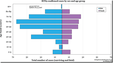
And indeed, the chart below from Dr. Ian Mackay’s VDU Blog shows a pronounced demographic shift towards older, predominantly male, victims in China. Children, and young adults – when they were found to be infected – were more likely to have mild, moderate, or even asymptomatic infections (see Mild Influenza A/H7N9 Infection among Children in Guangdong Province).


image


Compared to infection with the 1918 H1N1 and H5N1 avian flu viruses (see my three part review of the 2009 Baskin Influenza in Primates Study), H7N9 produced far less lung damage and illness in macaques. 


You’ll want to read the entire study for details on methods and materials, and a closer look at the results.   In their concluding remarks, the authors summed up their work by saying:


Thus, the emerging H7N9 influenza virus is more pathogenic than seasonal influenza A virus and most isolates of the pandemic H1N1 virus but not as pathogenic as the 1918 Spanish influenza virus and HPAI H5N1 virus in cynomolgus macaques.

However, the pathogenicity of the H7N9 virus may decrease if the virus adapts further to solely using α2,6-linked sialic acids as the receptor for entry, as pandemic influenza viruses to date have done (5255).

Exclusive attachment to α2,6-linked sialic acids would most likely result in a shift to replication mainly in the upper respiratory tract of humans, likely resulting in less severe disease, as has been described for the 2009 pandemic H1N1 virus (56) and upon adaptation of HPAI H5N1 virus to efficient transmission via respiratory droplets or aerosols (57).


The switch to an `exclusive attachment to α2,6-linked sialic acids’  is one of the evolutionary changes that is believed would make this virus far more easily transmitted between humans, and is probably required if  H7N9 were to become a viable pandemic virus.


And while a commensurate decrease in severity would be a welcome result, it is worth noting that a reduction of even a full order of magnitude (30% CFR down to 3%) would still put this virus in the same league as the 1918 Spanish flu.


While the tragedy of Ebola has captured the world’s attention this summer, the most serious pandemic threats are those posed by respiratory viruses, such as influenza, SARS, and MERS.

Monday, August 04, 2014

mBio: The Risks & Benefits Of `GOF’ Experimentation On Pathogens With Pandemic Potential

image

Credit CDC PHIL


# 8911


Somehow, between the Ebola news, and the fact that it was published on Wednesday (instead of mBio’s normal Tuesday publication date), I missed the following editorial by Arturo Casadevall  and Michael J. Imperiale in the open-access journal mBio.


It is a long, thoughtful look at the complex issues at hand.   Follow the link to read it in its entirety.


1 August 2014

Risks and Benefits of Gain-of-Function Experiments with Pathogens of Pandemic Potential, Such as Influenza Virus: a Call for a Science-Based Discussion

Arturo Casadevall and Michael J. Imperiale

doi:10.1128/mBio.01730-14

Editorial

Influenza virus is one of a handful of infectious disease agents that can cause devastating pandemics with high mortality and morbidity in human populations. The human species is vulnerable to zoonotic infection with new influenza viruses, with the last occurring as recently as 2009. Influenza kills thousands of people each year, and the world is continuously confronting new epidemics. Today the complexity and interconnectivity of our society create vulnerabilities, such that pandemics with even low mortality have the potential to cause widespread suffering and economic disruption. Epidemics can have catastrophic effects on the social order and result in the disruption of benefits that we associate with current society, such as law and order and reliable food distribution (for a vivid and dramatic representation of the effect of epidemics on society, readers are invited to see the movie Contagion, where an outbreak with a new fictional virus leads to the breakdown of the social order). Hence, epidemics pose existential threats to civil society even when morbidity and mortality occur in a fraction of those infected. Given the biological characteristics of the influenza virus that ensure the continuous generation of antigenic variability, this virus poses a continuous extant threat, with the likelihood of new pandemics being determined by variables that remain poorly understood. In this environment, the influenza virus research community is humanity’s best defense against influenza virus. Consequently, anything that impacts influenza virus research is of utmost importance to societal well-being.

(Continue . . . )


For more on this controversial debate, you may wish to revisit:


Scientists For Science: GOF Research `Essential’ & Can be Done `Safely’

Updating The Cambridge Working Group

ECDC Comment On Gain Of Function Research

The Debate Over Gain Of Function Studies Continues

Lipsitch & Galvani: GOF Research Concerns

The Call For Urgent Talks On `GOF’ Research Projects

Tuesday, July 22, 2014

mBio: Airborne Fragments Of MERS-CoV Detected In Saudi Camel Barn

Photo: ©FAO/Ami Vitale

Credit FAO


*** UPDATED with mBIO Link ***


# 8855


A little later today the open access journal mBio will publish a new paper, prepared by researchers from King Abdulaziz University in Saudi Arabia, on  RT-PCR testing of air samples taken from a camel barn during the time of a well studied probable camel-to-human transmission event last November (see CIDRAP: More Evidence for Camel-to-Human MERS-CoV Transmission).


mBio usually posts new articles mid-morning East Coast time every Tuesday, so I’ll update this blog with a link when they do.


Detection of the Middle East Respiratory Syndrome Coronavirus Genome in an Air Sample Originating from a Camel Barn Owned by an Infected Patient

Esam I. Azhar, Anwar M. Hashem, Sherif A. El-Kafrawy, Sayed Sartaj Sohrab, Asad S. Aburizaiza, Suha A. Farraj, Ahmed M. Hassan, Muneera S. Al-Saeed, Ghazi A. Jamjoom and Tariq A. Madani

doi:10.1128/mBio.01450-14


Until that time, we’ve got a press release from the American Society for Microbiology (excerpts below). 


The discovery of fragments of MERS-COV virus in an air sample collected in a camel barn - while an important piece of the the MERS transmission puzzle - is neither totally unexpected nor proof of airborne transmission of the virus.  It only demonstrates a potential route of infection.


First the press release, then I’ll return with more.


Middle East Respiratory Syndrome coronavirus detected in the air of a Saudi Arabian camel barn

Saudi Arabian researchers have detected genetic fragments of Middle East Respiratory Syndrome coronavirus (MERS-CoV) in the air of a barn holding a camel infected with the virus. The work, published this week in mBio®, the online open-access journal of the American Society for Microbiology, indicates that further studies are needed to see if the disease can be transmitted through the air.

<SNIP>

For the study, researchers on three consecutive days last November collected three air samples from a camel barn owned by a 43-year-old male MERS patient who lived south of the town of Jeddah, who later died from the condition. Four of the man's nine camels had shown signs of nasal discharge the week before the patient became ill; he had applied a topical medicine in the nose of one of the ill camels seven days before experiencing symptoms.

Using a laboratory technique called reverse transcription polymerase chain reaction (RT-PCR) to detect gene expression, they found that the first air sample, collected on November 7, contained genetic fragments of MERS-CoV. This was the same day that one of the patient's camels tested positive for the disease. The other samples did not test positive for MERS-CoV, suggesting short or intermittent shedding of the virus into the air surrounding the camels, said lead study author Esam Azhar, PhD, head of the Special Infectious Agents Unit at King Fahd Medical Research Center and associate professor of medical virology at King Abdulaziz University in Jeddah.

Additional experiments confirmed the presence of MERS-CoV-specific genetic sequences in the first air sample and found that these fragments were exactly identical to fragments detected in the camel and its sick owner.

"The clear message here is that detection of airborne MERS-CoV molecules, which were 100% identical with the viral genomic sequence detected from a camel actively shedding the virus in the same barn on the same day, warrants further investigations and measures to prevent possible airborne transmission of this deadly virus," Azhar said.

"This study also underscores the importance of obtaining a detailed clinical history with particular emphasis on any animal exposure for any MERS-CoV case, especially because recent reports suggest higher risk of MERS-CoV infections among people working with camels," he added.

Meanwhile, he said, mounting evidence for camel-to-human transmission of MERS-CoV warrants taking precautionary measures: People who care for camels or who work for slaughterhouses should wear face masks, gloves and protective clothing, and wash their hands frequently. It is also important to avoid contact with animals that are sick or have tested positive for MERS-CoV. Those who visit camel barns, farms or markets should wash hands before and after contact with animals. In addition, pasteurization of camel milk and proper cooking of camel meat are strongly recommended.

(Continue . . . )


Airborne (aerosolized or large droplet) transmission of MERS-CoV in humans is assumed to occur - hence the CDC’s stringent Interim Guidance for Health Professionals   on the use of PPEs – but other routes may be equally important players. 


Last May, in MERS: A Focus On Fomites?  we looked at investigations focusing on the potential  role of inanimate objects and environmental surfaces in the transmission of the virus.


How the virus jumps to man – presumably from camels – and how camels acquire, and spread the virus, is less well mapped out. As Dr. Ian Mackay graphically illustrated last may, there are a lot of options.


Camels at the centre, aerosol all around...

An airborne-centric view of how the camel could be a source of sporadic human infection by MERS-CoV, a virus that is genetically very similar whether found in camels or humans.

The inner ring (orange) is more about droplets and aerosols-if you must differentiate on size. 


These are potential routes by which a human in contact with, or near to, camels might acquire virus from them, when those camels are actively infected.

(Continue . . . )


The detection of fragments of MERS-CoV – using RT-PCR testing – in air samples in a camel barn doesn’t tell us if those fragments were viable, and capable of infecting anyone or anything. 


But this study does show that the opportunity for the virus to spread from camels through the air exists, and thus invites additional research into this plausible route of transmission.


.

Wednesday, July 16, 2014

mBio: The Remarkable Evolution Of Cryptococcus Gatti

image

Credit CDC


# 8837


Although we normally think of viral or bacterial pathogens when we talk about infectious diseases, mycotic (fungal) diseases exert a heavy burden on human health as well.


A little over a year ago, in MMWR: Coccidioidomycosis Rising, we looked at dramatic increase in the number of Valley Fever infections over the past 13 years (1998-2011).


Coccidioidomycosisis – perhaps the best known fungal infection in North America – is caused by the inhalation of spores from one of two soil borne fungi - Coccidioides immitis or C. posadasii - both commonly found in the American Southwest.  Their spores can remain dormant in the desert soil for years, only to become airborne when the earth is disturbed by farming, earthquakes, construction, or windstorms.


Most of the people who live in regions where these fungi are endemic are eventually exposed and either develop brief asymptomatic infections or mild flu-like symptoms. 


But `Cocci’ isn’t alone, as another mycotic disease with a wide range in the United States is called Histoplasmosis, which can be found in the Ohio River Valley and along the lower Mississippi river. The causative agent is Histoplasma capsulatum, a fungus which is found in bird and bat droppings. Its spores can become airborne when these droppings dry out and are picked up by the wind. 

image

Credit Wikipedia

Another fungal threat is Blastomycosis (aka Gilchrist's disease), which is caused by Blastomyces dermatitidis, which is found in decaying leaves and grass.

image


While these mycotic diseases have long been endemic to North America, a new fungus arrived in the Pacific Northwest from the tropics roughly 15 years ago, and has not only adapted to a much colder environment, it has picked up virulence as well. 


It is called Cryptococcus gattii, and it showed up unexpectedly on Vancouver Island in 1999, and has since then has spread into Washington and Oregon.


In 2010 the CDC’s Journal of Emerging Infectious Diseases published a research article on spread spread of C. gattii  in British Columbia (see A Fungus Among Us).  This fungus – a yeast really – is found in a number of species of trees (primarily Douglas fir & Western hemlock) and in the soil.  It can be spread by the wind, particularly during the warmer summer months.


This fungus has a wide host range, having been found to infect humans, cats, dogs, sheep, ferrets, llamas, elk, alpacas, and even porpoises (Cite).


Unlike its better known cousin C. neoformans which generally infects people with weakened immune system – C. gattii often infects those with healthy immune systems.  And unlike its behavior in the South Pacific (primarily Australia & New Guinea) – where it was usually seen causing neurological infections – this new cold-adapted strain tends to cause more pulmonary infections.


This from the open access journal mBio.


Cryptococcus gattii in North American Pacific Northwest: Whole-Population Genome Analysis Provides Insights into Species Evolution and Dispersal

David M. Engelthalera, Nathan D. Hicksa, John D. Gillecea, Chandler C. Roea, James M. Schuppa, Elizabeth M. Driebea, Felix Gilgadob, Fabian Carricondeb,c, Luciana Trillesb,d, Carolina Firacativeb,e, Popchai Ngamskulrungrojb,f, Elizabeth Castañedae, Marcia dos Santos Lazerad, Marcia S. C. Melhemg, Åsa Pérez-Bercoffb,h, Gavin Huttleyh, Tania C. Sorrellb, Kerstin Voelzi,j, Robin C. Mayi,j, Matthew C. Fisherk, George R. Thompson IIIl, Shawn R. Lockhartm, Paul Keima,n, Wieland Meyerb

ABSTRACT

The emergence of distinct populations of Cryptococcus gattii in the temperate North American Pacific Northwest (PNW) was surprising, as this species was previously thought to be confined to tropical and semitropical regions. Beyond a new habitat niche, the dominant emergent population displayed increased virulence and caused primary pulmonary disease, as opposed to the predominantly neurologic disease seen previously elsewhere.

Whole-genome sequencing was performed on 118 C. gattii isolates, including the PNW subtypes and the global diversity of molecular type VGII, to better ascertain the natural source and genomic adaptations leading to the emergence of infection in the PNW. Overall, the VGII population was highly diverse, demonstrating large numbers of mutational and recombinational events; however, the three dominant subtypes from the PNW were of low diversity and were completely clonal. Although strains of VGII were found on at least five continents, all genetic subpopulations were represented or were most closely related to strains from South America.

The phylogenetic data are consistent with multiple dispersal events from South America to North America and elsewhere. Numerous gene content differences were identified between the emergent clones and other VGII lineages, including genes potentially related to habitat adaptation, virulence, and pathology.

Evidence was also found for possible gene introgression from Cryptococcus neoformans var. grubii that is rarely seen in global C. gattii but that was present in all PNW populations. These findings provide greater understanding of C. gattii evolution in North America and support extensive evolution in, and dispersal from, South America.

The good news is that C. gattii infections in the United States remain exceedingly rare, and most people who are exposed (which undoubtedly are millions of people every year) develop no illness or symptoms. 

This from the CDC’s C. gattii cryptococcosis webpage:

C. gattii cryptococcosis Statistics

C. gattii cryptococcosis is considered to be an emerging infection in the United States and is very rare. Approximately 100 infections were documented in the U.S. between 2004 and 2011, almost all of which came from Oregon and Washington.

C. gattii infections are under public health surveillance, and have been reportable in Oregon and Washington since autumn of 2011. This means that healthcare providers and laboratories in these states are required to collect certain information on each case, and report this information to the appropriate local or state public health authority.


The bad news, is there is really not much you can do to prevent infection:

How can I prevent infection with Cryptococcus gattii?

There are no formal recommendations for preventing C. gattii infection. Most people breathe in small amounts of many different types of fungal spores every day but never become sick. However, if you have symptoms that you think may be caused by C. gattii, you should see a doctor.


The authors write:


We provide evidence that the PNW strains originated from South America and identified numerous genes potentially related to habitat adaptation, virulence expression, and clinical presentation. Characterization of these genetic features may lead to improved diagnostics and therapies for such fungal infections. The data indicate that there were multiple recent introductions of C. gattii into the PNW. Public health vigilance is warranted for emergence in regions where C. gattii is not thought to be endemic.

Tuesday, July 08, 2014

mBio: Debating The Dromedary - MERS Coronavirus Connection

Photo: ©FAO/Ami Vitale

Credit FAO


# 8812


For nearly a year we’ve watched the evidence mount up that camels play a major role in the hosting, and probable spread, of the MERS coronavirus which was first discovered two years ago in the Middle East.  Dromedaries aren’t viewed as being the only possible animal reservoir of the virus, nor are they believed responsible for he majority of human cases.


But the virus (or antibodies to the virus) have certainly been found in camels (see Kuwait Tests Camels - Finds 6% Positive For MERS-CoV), and we’ve seen a few instances where camel-to-human transmission is strongly suspected (see CIDRAP: More Evidence for Camel-to-Human MERS-CoV Transmission).


Last month,  in Eurosurveillance: MERS-CoV Antibodies & RNA In Camel’s Milk – Qatar we looked at research supporting the notion that consumption of unpasteurized camel’s milk might be a route to infection.


Some of the most compelling research has been associated with Dr. Ian Lipkin, including a study published last February (see mBio: MERS-CoV In Saudi Arabian Camels) that established MERS-CoV to be a common, likely mild or asymptomatic, infection in young camels in Saudi Arabia and that suggested that they may well be the source of at least some portion of the human infections we’ve seen over the past two years.


Another study, again involving Dr. Lipkin, was published the end of April (see mBio: MERS-CoV Carriage By Dromedaries) that recovered the MERS-CoV from nasal swabs of camels, and demonstrated that whole-genome consensus sequences were indistinguishable from MERS coronaviruses recovered from humans.


An accompanying press release, Columbia University's Mailman School of Public Health, states:

"The finding of infectious virus strengthens the argument that dromedary camels are reservoirs for MERS-CoV," says first author Thomas Briese, PhD, associate director of the Center for Infection and Immunity and associate professor of Epidemiology at the Mailman School. "The narrow range of MERS viruses in humans and a very broad range in camels may explain in part the why human disease is uncommon: because only a few genotypes are capable of cross species transmission," adds Dr. Briese.


Despite the preponderance of evidence, not everyone is ready to accept these findings as `proof’ of the role of camels in the spread of the MERS coronavirus (see Saudi MOA Spokesman: Camel Link Unproven).


Today the open access journal mBio has published a long letter, written by Emad M. Samara Ph.D and Professor Khalid A. Abdoun, both of the Department of Animal Production at King Saud University  that call into question the findings of the two previously mentioned studies involving Dr. Lipkin. 


After listing a number of the findings in these previous studies, the authors argue that they cannot be taken as `conclusive evidence’  that dromedary camels carry the infectious form of MERS-CoV or have ever infected humans. 


They suggest it is equally plausible (albeit, unproven) that humans are the ones who have infected camels.


It’s a long letter – far too long to reproduce here – and so in fairness to their argument,  I would invite my readers to follow the link to read it in its entirety.    When you return, I’ll have a link to a reply from Dr. Lipkin et al.


Concerns about Misinterpretation of Recent Scientific Data Implicating Dromedary Camels in Epidemiology of Middle East Respiratory Syndrome (MERS)

Emad M. Samara, Khalid A. Abdoun 

LETTER

This letter addresses some concerns about two recent articles published by the same authors in mBio (1, 2), specifically many uncertainties regarding the potential applicability of their epidemiological data, which were obtained from dromedary camels (DCs) infected with Middle East respiratory syndrome coronavirus (MERS-CoV), to human public health.

(Continue . . . )


The reply to this critique is fairly brief - where the authors stand by their work - can be read at the following link:


Reply to “Concerns About Misinterpretation of Recent Scientific Data Implicating Dromedary Camels in Epidemiology of Middle East Respiratory Syndrome (MERS)”

Abdulaziz N. Alagailia, Thomas Brieseb, William B. Kareshc, Peter Daszakc, W. Ian Lipkinb


It is absolutely true that one can rarely say `case closed, time to etch our findings in stone’  with any field of scientific research. `Conclusive evidence’  is a very high, sometimes unobtainable, standard of proof. 


And few would argue that more work doesn’t need to be done on the virology, ecology, and epidemiology of the MERS coronavirus. 


But until that can happen, we pretty much have to go by the preponderance of evidence. And for now, despite the misgivings of these authors from King Saud University, that evidence strongly suggests camels play an important role in the hosting, and spread of MERS-CoV.  

Wednesday, June 11, 2014

mBio: Biofilms, Stress Hormones & Heart Attacks

image

Diseased Carotid Arteries - Credit: David Davies, University of Binghamton


# 8729


A couple of months ago, in Post-Disaster Stress Cardiomyopathy: A Broken-Hearted Malady, we looked at a study that found a significant increase in a very rare type of heart problem called Takotsubo cardiomyopathy – also known as broken heart syndrome – following high impact natural disasters.


This stress related syndrome causes acute ballooning of the heart ventricles, and is a well-recognized cause of acute heart failure and dangerous cardiac arrhythmias.  


Johns Hopkins Medicine has a Frequently Asked Questions about Broken Heart Syndrome, that describes the condition:

1. What is “stress cardiomyopathy?”

Stress cardiomyopathy, also referred to as the “broken heart syndrome,” is a condition in which intense emotional or physical stress can cause rapid and severe heart muscle weakness (cardiomyopathy). This condition can occur following a variety of emotional stressors such as grief (e.g. death of a loved one), fear, extreme anger, and surprise. It can also occur following numerous physical stressors to the body such as stroke, seizure, difficulty breathing (such as a flare of asthma or emphysema), or significant bleeding.


We’ve looked at other post-disaster (likely stress related) cardiac problems, including earlier last March in Tulane University: Post-Katrina Heart Attack Rates – Revisited, where an ongoing study finds that heart attack rates remain elevated by 300% in New Orleans six years after that hurricane struck.


While there is plenty of anecdotal evidence showing that stress, fear, grief, or other emotional stressors can cause sudden heart attacks thus far we haven’t had a good model as to how that might happen.


Yesterday the open access journal mBio published a fascinating bit of work that attempts to show how a sudden release of the right stress hormone (norepinephrine) can dissolve bacteria laden biofilm deposits in the carotid artery, potentially initiating a heart attack or stroke.


Bacteria Present in Carotid Arterial Plaques Are Found as Biofilm Deposits Which May Contribute to Enhanced Risk of Plaque Rupture

Bernard B. Lanter, Karin Sauer, David G. Davies

IMPORTANCE The association of bacteria with atherosclerosis has been only superficially studied, with little attention focused on the potential of bacteria to form biofilms within arterial plaques.

In the current work, we show that bacteria form biofilm deposits within carotid arterial plaques, and we demonstrate that one species we have identified in plaques can be stimulated in vitro to undergo a biofilm dispersion response when challenged with physiologically relevant levels of norepinephrine in the presence of transferrin. Biofilm dispersion is characterized by the release of bacterial enzymes into the surroundings of biofilm microcolonies, allowing bacteria to escape the biofilm matrix.

We believe these enzymes may have the potential to damage surrounding tissues and facilitate plaque rupture if norepinephrine is able to stimulate biofilm dispersion in vivo. This research, therefore, suggests a potential mechanistic link between hormonal state and the potential for heart attack and stroke.

(Continue . . . )


The American Society for Microbiology has a press release that explains in layman’s terms the mechanism this study believes it has discovered.


Bacteria help explain why stress, fear trigger heart attacks

WASHINGTON, DC – June 10, 2014 - Scientists believe they have an explanation for the axiom that stress, emotional shock, or overexertion may trigger heart attacks in vulnerable people. Hormones released during these events appear to cause bacterial biofilms on arterial walls to disperse, allowing plaque deposits to rupture into the bloodstream, according to research published in published today in mBio®, the online open-access journal of the American Society for Microbiology.

"Our hypothesis fitted with the observation that heart attack and stroke often occur following an event where elevated levels of catecholamine hormones are released into the blood and tissues, such as occurs during sudden emotional shock or stress, sudden exertion or over-exertion" said David Davies of Binghamton University, Binghamton, New York, an author on the study.

Davies and his colleagues isolated and cultured different species of bacteria from diseased carotid arteries that had been removed from patients with atherosclerosis. Their results showed multiple bacterial species living as biofilms in the walls of every atherosclerotic (plaque-covered) carotid artery tested.

In normal conditions, biofilms are adherent microbial communities that are resistant to antibiotic treatment and clearance by the immune system. However, upon receiving a molecular signal, biofilms undergo dispersion, releasing enzymes to digest the scaffolding that maintains the bacteria within the biofilm. These enzymes have the potential to digest the nearby tissues that prevent the arterial plaque deposit from rupturing into the bloodstream.

According to Davies, this could provide a scientific explanation for the long-held belief that heart attacks can be triggered by a stress, a sudden shock, or overexertion

(Continue . . .)


All of this is a very simplistic summation of a complex, and fascinating paper, one that many will want to read in its entirety. While far from settled science, it proffers a very interesting avenue for further investigation. 

Of note, while some heart attacks and Takotsubo cardiomyopathy are thought to be induced by similar cascades of stress-related hormones, their actual physical effects appears to be quite different.  Johns Hopkins describes the theories behind the cause of stress-cardiomyopathy below:


The precise way in which adrenaline affects the heart is unknown. It may cause narrowing of the arteries that supply the heart with blood, causing a temporary decrease in blood flow to the heart. Alternatively, the adrenaline may bind to the heart cells directly causing large amounts of calcium to enter the cells which renders them temporarily dysfunctional.


The bottom line, I suppose, is that by whatever mechanism, overwhelming stress can take a heavy toll on our physical and mental health.


And in ways that we are only just now beginning to unravel.