Friday, February 28, 2014

Cytokine Storm Chasers

image


# 8338


Readers with good memories will recall that in 2011, in Study: Calming The Cytokine Storm, we looked at research from The Scripps Research Institute  that found a protein located on the surface of endothelial cells, called S1P1, to be largely responsible for flu-associated cytokine storms.


Rather than trying to combat the specific virus – which has a nasty habit of evolving resistance to antivirals – Scripps researchers were looking at ways of reducing the body’s sometimes excessive immune response to viral infection known as a Cytokine Storm.


Cytokines are a category of signaling molecules that are used extensively in cellular communication. They are often released by immune cells that have encountered a pathogen, and are designed to alert and activate other immune cells to join in the fight against the invading pathogen.


This cascade of immune cells rushing to the infection, if it races out of control, can literally kill the patient. Their lungs can fill with fluid (which makes a terrific medium for a bacterial co-infection), and cells in the lungs (Type 1 & Type II Pneumocytes) can sustain severe damage.


Previously, in Swine Flu Sequelae and Cytokine Storm Warnings, we looked at some of the severe lung damage during the 2009 pandemic that was thought to be due to this overreaction of the immune system.


More recently, we looked at a study by Professor Peter Doherty (see PNAS: Genetic Marker & Cytokine Levels Linked To Severity Of Human H7N9 Infection) that linked a specific genetic marker; IFITM3 CC gene variant (aka C/C Genotype)  to hypercytokinemia (aka a `Cytokine Storm’), and a severe outcome, in H7N9 infections.


This genetic marker– while comparatively rare in Caucasians - is far more common in Han Chinese, and may (partially) account for some of the particularly high mortality rates we’ve seen with novel influenza’s in Asia. 


Last month, China’s CDC made specific mention of the role of excess cytokine production in H7N9 infection (see NHFPC: H7N9 Avian Flu Guidance Update) where they warn: H7N9 avian influenza virus after infection the human body, can induce cytokine storm, leading to systemic inflammation, may appear ARDS, shock and multiple organ failure.


Traditionally, ARDS (Acute Respiratory Distress Syndrome) patients end up on mechanical ventilation in ICUs, and are treated with a variety of pharmacological agents to reduce infection (antibiotics) and lung inflammation (corticosteroids, Nitric Oxide, etc.). 


The use of high dose corticosteroids – while fairly common with SARS and and early H5N1 cases – has been discouraged by the WHO and other health agencies due to poor long-term survival rates. 


Hence the need for a better tolerated,  more effective, and targeted drug regimen against the cytokine storm.


All of which serves as prelude to a new report from the The Scripps Research Institute updating their search for a drug to modulate the body’s immune response, and mapping the cytokine signaling and production process.  Their findings appear this week in the early edition of the journal PNAS.

Mapping the innate signaling cascade essential for cytokine storm during influenza virus infection

John R. Teijaroa, Kevin B. Walsha,1, Stephanie Ricea, Hugh Rosenb,c,2, and Michael B. A. Oldstonea,2

Significance

Cytokine storm plays an essential and commanding role in the clinical outcome and pathogenesis of influenza virus infection. We previously documented that a small molecule that activates sphingosine-1-phosphate-1 receptor (S1P1R) signaling is primarily responsible for blunting cytokine storm to protect the infected host from the consequences of influenza infection. In the present study, we map host innate signaling pathways of cytokine storm and chart where along those pathways the drug is effective. We find that the efficacy of S1P1R agonist in blunting cytokine storm is through global inhibition downstream of myeloid differentiation primary response gene 88 and IFN-β promoter stimulator-1 signaling.

(Continue . . .)


Although the bulk of this study is behind a pay wall, we get a pretty detailed overview from the following press release from the Scripps Institute.


News Release

Scripps Research Institute Scientists Describe Deadly Immune ‘Storm’ Caused by Emergent Flu Infections

LA JOLLA, CA—February 27, 2014—Scientists at The Scripps Research Institute (TSRI) have mapped key elements of a severe immune overreaction—a “cytokine storm”—that can both sicken and kill patients who are infected with certain strains of flu virus.

Their findings, published in this week’s online Early Edition of the Proceedings of the National Academy of Sciences, also clarify the workings of a potent new class of anti-inflammatory compounds that prevent this immune overreaction in animal models.

“We show that with this type of drug, we can quiet the storm enough to interfere with the virus-induced disease and lung injury, while still allowing the infected host to mount a sufficient immune response to eliminate the virus,” said John R. Teijaro, an assistant professor in TSRI’s Department of Immunology and Microbial Science and first author of the study.

“This study provides insights into mechanisms that are chemically tractable and can modulate these cytokine storms,” said Hugh Rosen, professor in TSRI’s Department of Chemical Physiology and senior author of the study with Michael B. A. Oldstone, professor in TSRI’s Department of Immunology and Microbial Science.

Calming the Storm

A cytokine storm is an overproduction of immune cells and their activating compounds (cytokines), which, in a flu infection, is often associated with a surge of activated immune cells into the lungs. The resulting lung inflammation and fluid buildup can lead to respiratory distress and can be contaminated by a secondary bacterial pneumonia—often enhancing the mortality in patients.

This little-understood phenomenon is thought to occur in at least several types of infections and autoimmune conditions, but it appears to be particularly relevant in outbreaks of new flu variants. Cytokine storm is now seen as a likely major cause of mortality in the 1918-20 “Spanish flu”—which killed more than 50 million people worldwide—and the H1N1 “swine flu” and H5N1 “bird flu” of recent years. In these epidemics, the patients most likely to die were relatively young adults with apparently strong immune reactions to the infection—whereas ordinary seasonal flu epidemics disproportionately affect the very young and the elderly.

For the past eight years, Rosen’s and Oldstone’s laboratories have collaborated in analyzing the cytokine storm and finding treatments for it. In 2011, led by Teijaro, who was then a research associate in the Oldstone Lab, the TSRI team identified endothelial cells lining blood vessels in the lungs as the central orchestrators of the cytokine storm and immune cell infiltration during H1N1 flu infection.

In a separate study, the TSRI researchers found that they could quiet this harmful reaction in flu-infected mice and ferrets by using a candidate drug compound to activate immune-damping receptors (S1P1 receptors) on the same endothelial cells. This prevented most of the usual mortality from H1N1 infection—and did so much more effectively than the existing antiviral drug oseltamivir, although the combination of both therapies worked even better. “That was really the first demonstration that inhibiting the cytokine storm is protective,” said Teijaro.

(Continue . . . )


This press release goes on to state that the experimental drug – CYM5442 – is now being tested in clinical trials, with uses that extend far beyond just influenza-related ARDS.


An optimized version of CYM5442, initially developed by Rosen and fellow TSRI chemist Ed Roberts, has been licensed to the pharmaceutical company Receptos. It is now in Phase 3 clinical trials for treating relapsing-remitting multiple sclerosis and Phase 2 trials for ulcerative colitis. Other S1P1 receptor agonists are in development for inflammatory conditions. A less-specific S1P receptor agonist—which hits S1P1, but also hits S1P3, S1P4 and S1P5, with potential off-target effects—is already approved for treating multiple sclerosis.


While it is hard to find anything `good’ to say about the emergence of novel viral threats over the past dozen years (H5N1, SARS, H7N9, etc.), it has prompted a remarkable amount of research – not only into the pathogens themselves - but into the complex and far from completely understood inner workings of our own immune system.


Research that has the potential to pay health benefits far beyond simply treating viral infections.

Cambodian MOH Reports Two New H5N1 Cases

image

Kampong Cham province – Credit Wikipedia


# 8337


Today Cambodia’s MOH is reporting their 4th & 5th confirmed H5N1 cases of 2014, that of two young girls (ages 10 & 11) from Kampong Cham Province (albeit villages in different districts). Unlike most of the H5N1 cases we’ve seen, these girls appear to have suffered relatively mild to moderate illness, and have both recovered.


While this brings the official case count to five for the year, we also had a strongly suspected case – that of a sibling of a confirmed case who died, but was never tested – from Kratie Province earlier this month (see Cambodia: 2 Deaths - 1 Confirmed H5N1, 1 Probable).


First some excerpts from the MOH statement, after which I’ll have a bit more.


4th and 5th New Human Case of Avian Influenza H5N1 in Cambodia in 2014

27 February 2014

The Ministry of Health (MoH) of the Kingdom of Cambodia wishes to advise members of the public that two (2) new human cases of avian influenza have been confirmed for the H5Nl virus. These are the 4th and 5th cases this year and the 51st and 52nd persons to become infected with the H5Nl virus in Cambodia. The cases are from Kampong Cham (newly named Tboung Khmun) province. Of the 52 confirmed cases, 40 were children under 14, and 29 of the 52 were female. In addition, since the first case happened in Cambodia in 2005 there were 18 cases survived.


The 4th case, a 10-year-old girl from Rorveang village, Knor Damborng commune, Cheung Prey district, Kampong Cham (newly named Tboung Khmun) province, was detected by the Naval Medical Research Unit 2 (NAMRU-2) through fever surveillance on 20th February and confirmed positive by Institut Pasteur du Cambodge on 20th February 2014. The girl had onset symptom of fever on 26th January 2014. Her mother bought medicine in the village. From the 27th January, she continued to develop symptoms and had fever, running nose, cough, and abdominal pain. On the 29th January, the health staff of the NAMRU-2 project took samples from the girl. Upon confirmation of H5Nl, she was referred toKampong Cham Provincial Hospital on 20th February and Tamiflu was administered on the same day. Currently, she recovered and had normal activities.The case had direct exposure with dead and sick poultry. Farm ducks in the village started to suddenly die around 15th January 2014.  The mother of the case brought sick/dead ducks on the 25th January and the family prepared them, with the help of the case, for food the same day.


The 5th case, an 11-year-old girl from La Ork Village, Krek Commune, Ponhea Krek district, Kampong Cham (newly named Thoung Khmum) province, was detected by the Naval Medical Research Unit 2 (NAMRU-2) through fever surveillance on 20th February and confirmed positive by Insitut Pasteur du Cambodge on 29th February 2014.  The girl had onset of symptoms of fever and cough on 9th February 2014. The health staff at the NAMRU-2 project took samples from the girl on the 10th February.  Upon confirmation of H5N1, she was referred to Kampong Cham Provincial Hospital on 20th February and Tamiflu was administered on the same day.  Currently, she recovered and had normal activities.

From the 7th to 10th February, all 30 chickens owned by family died around the house.  The relatives reported that the girl had no direct contact, but the chickens died in close proximity to the case.

(Continue. . . .)


NAMRU-2 mentioned above is the Naval Medical Research Unit (#2) which previously had been based in Jakarta for nearly 40 years, but became ensnared in former Indonesian Health Minister Supari’s increasingly absurd political theatre, where she eventually accused the lab of espionage and ordered it closed (see NAMRU-2 Debate Turns Ugly)..


Diplomatic efforts to reopen the lab ultimately failed, and so the lab was temporarily relocated to Pearl Harbor in 2010, and then moved on to Cambodia last year.


Given the sudden uptick of human H5N1 cases in Cambodia over the past 3 years, the presence of NAMRU – which has dealt extensively with avian H5N1 previously in both Indonesia and Egypt (NAMRU-3) – is a decided plus.  You can visit NAMRU’s website at the link below:

image


Despite the dramatic uptick in H5N1 cases reported out of Cambodia over the past couple of years, their sporadic nature and broad geographic distribution show no evidence of sustained or efficient community-level transmission, and the assumption is that the vast majority of these infections came from exposure to infected birds.


The World Health Organization’s most recent public health assessment on the H5N1 virus reads:

Overall public health risk assessment for avian influenza A(H5N1) viruses: Whenever influenza viruses are circulating in poultry, sporadic infections or small clusters of human cases are possible, especially in people exposed to infected household poultry or contaminated environments. This influenza A(H5N1) virus does not currently appear to transmit easily among people. As such, the risk of community-level spread of this virus remains low. 


But viruses can change over time, and so could this assessment.

Thursday, February 27, 2014

Study: Probable Longer Incubation Period For H7N9

image


# 8336


Although individual host responses can vary considerably, public health officials are constantly looking to define the `outside’  duration of viral incubation, as to know when to sound the `all clear’  when someone who has been exposed is out of danger.


The incubation period of novel Influenza – which can be carried sub-clinically, yet potentially still passed on to others – is of particular concern, as those exposed may be quarantined to protect the public. 


And someone with travel history to an area where they might have been exposed to a specific pathogen will more likely be tested if they fell ill within its known incubation period.


The incubation period of viruses varies widely, with seasonal influenza running 1 to 4 days (avg. 2 days), measles running 7 to 21 days (avg. 14 days), MERS-CoV up to 15 days. In late April 2013, just over a month after the first H7N9 cases were announced from China, Hong Kong’s CHP sent a letter to doctors warning that the incubation period could be up to 10 days.


In view of the latest best available evidence, the longest incubation period of human infection with avian influenza A(H7N9) virus has been revised from 7 days to 10 days and the epidemiological criteria of the reporting criteria has been revised accordingly (see attached). Please also note that Taiwan has not been included as an affected area as the case recorded was classified as an imported infection.


Hong Kong has continued to use 10 days as an `outside’ incubation period, for testing and quarantine purposes.  


Just last month, however, in a major release from China’s NHFPC: H7N9 Avian Flu Guidance Update, the incubation period for H7N9 was described as:


According to the incubation period of influenza and human infection with H7N9 avian influenza existing case findings, the incubation period is generally 3 to 4 days.


It is axiomatic that the more cases that are examined, the more studies that are published, the better becomes our knowledge. Which brings me to a new study, appearing this week in the journal Epidemiology & Infection (h/t Crof) , that seems to bear out Hong Kong’s early - more conservative stance - finding the H7N9’s incubation period can range up to 10 days.


Epidemiol Infect. 2014 Feb 24:1-7. [Epub ahead of print]

Probable longer incubation period for human infection with avian influenza A(H7N9) virus in Jiangsu Province, China, 2013.


Huang Y1, Xu K2, Ren DF3, Ai J2, Ji H2, Ge AH2, Bao CJ2, Shi GQ4, Shen T4, Tang FY2, Zhu YF2, Zhou MH2, Wang H2.

Abstract

SUMMARY Human infection with the emerging avian influenza A(H7N9) virus in China in 2013 has raised global concerns. We conducted a retrospective descriptive study of 27 confirmed human influenza A(H7N9) cases in Jiangsu Province, to elaborate poultry-related exposures and to provide a more precise estimate of the incubation periods of the illness.

The median incubation period was 6 days (range 2-10 days) in cases with single known exposure and was 7·5 days (range 6·5-12·5 days) in cases with exposures on multiple days, difference between the two groups was not significant (Z = -1·895, P = 0·058). The overall median incubation period for all patients was estimated to be 7·5 days (range 2-12·5 days).

Our findings further highlight the necessity for public health authorities to extend the period of medical surveillance from 7 days to 10 days.

ECDC: GIS Timeline Of H7N9 Cases In China

image

ECDC GIS For Disease Prevention


# 8335


GIS or Geographic Information Systems, are a powerful way to look at emerging disease information, and the ECDC has certainly embraced the technology.  Below you’ll find some snapshots from a terrific interactive timeline map showing the spread of the H7N9 virus over the past year. 


Follow THIS LINK to view/manipulate this handy frequently updated resource.


This first view shows the opening week of the outbreak, with cases (eventually) reported from four provinces.

image


By mid-summer the first wave had ended, with roughly 130 cases reported.

image


New cases began to appear in October of 2013.  Here is the geographic spread as of the end of 2013.

image


And lastly, a view of the major uptick of reported cases in the first 7 weeks of 2014, including the exportation of a case to Malaysia.


image

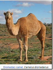
EID Journal: MERS Coronaviruses in Dromedary Camels, Egypt

image

Photo Credit Wikipedia


# 8334


Adding to the stampede of MERS-CoV in Camel studies coming out in recent months (see mBio: MERS-CoV In Saudi Arabian Camels & The Lancet: Identification Of MERS Virus In Camels), today we have a new Dispatch from the CDC’s EID Journal that relates the finding of a 99% match to the MERS coronavirus in Egyptian camels.


While just 4 (3.6%) of nasal swabs (out of 110 tested) were positive for the MERS-CoV virus (via RT-PCR testing) – suggesting current or active infection - 48 (92.3%) of 52 dromedary serum samples revealed titers from between 20 to >640, indicating prior infection. 


Serological testing of 179 camel abattoir workers, however, revealed no signs of previous MERS infection. Leading the researchers to conclude that while MERS infection in camels is common in Egypt, thus far it is rarely transmitted on to humans. 


I’ve only excerpted the Abstract & Conclusions below. Follow the link to read the entire study.


MERS Coronaviruses in Dromedary Camels, Egypt

Daniel K.W. Chu1, Leo L.M. Poon1, Mokhtar M. Gomaa, Mahmoud M. Shehata, Ranawaka A.P.M. Perera, Dina Abu Zeid, Amira S. El Rifay, Lewis Y. Siu, Yi Guan, Richard J. Webby, Mohamed A. Ali, Malik PeirisComments to Author , and Ghazi KayaliComments to Author
Abstract

We identified the near-full-genome sequence (29,908 nt, >99%) of Middle East respiratory syndrome coronavirus (MERS-CoV) from a nasal swab specimen from a dromedary camel in Egypt. We found that viruses genetically very similar to human MERS-CoV are infecting dromedaries beyond the Arabian Peninsula, where human MERS-CoV infections have not yet been detected.

<Big SNIP>

Conclusions

Our findings confirm that MERS-CoV infects dromedary camels and that this virus is genetically very similar to a MERS-CoV that is infecting humans. The detection of MERS-CoV in nasal swab specimens of camels in 2 of 12 sampling occasions in abattoirs, taken together with the high seropositivity to MERS-CoV in dromedaries previously reported, supports the contention that MERS-CoV infection is common in dromedaries.

Studies of dromedaries within camel herds and through the animal marketing system supplying abattoirs are needed to define the epidemiology of the infection. Our findings strengthen the plausibility that dromedaries may be a potential source of human infection and emphasize the need for detailed epidemiologic investigation of the exposure histories of humans with MERS.

However, the lack of serologic evidence of infection of humans working in these abattoirs suggests that transmission of this virus to humans is uncommon. The detection of MERS-CoV in dromedaries in Egypt, in animals imported from Sudan and Ethiopia, suggests that cases may occur in humans beyond the Arabian Peninsula. MERS CoV diagnostic tests should be considered for all patients with unexplained severe pneumonia in Egypt, northeastern Africa, and beyond.

Referral: A Couple Of Graphic MERS Posts From Ian Mackay

image

Credit Dr. Ian Mackay VDU Blog


# 8333


Since I can’t draw a straight line with a ruler, I’m always happy to see the latest, and greatest graphics coming from Dr. Ian Mackay’s VDU blog.   Over the past 24 hours Ian released five new MERS-CoV related graphs, which you can view on the following two blog entries.


Snapdate: Middle East respiratory syndrome coronavirus (MERS-CoV)
Coming back to MERSerable data...


I’m also pleased to note that I’m not the only fan of Ian’s artistic talent.  For the second time, one of his graphics has been cited in a scientific paper (see  Editor's Note #14: VDU in the scientific literature).


Congrats!