Showing posts with label EID Journal. Show all posts

Thursday, May 07, 2015

EID Journal: Influenza A(H5N6) Virus Reassortant, Southern China, 2014

image


# 10,020


Although we’ve yet to see a definitive move by any of the recently emerged novel flu viruses towards becoming a global public health threat, the rapid increase in the number of these viral contenders over the past three years has been remarkable. 


Not so very long ago we really only had one HPAI virus of genuine concern; H5N1.


But as both LPAI and HPAI viruses like H9N2,  H7s, H6’s, H5’s, and others have comingled in poultry operations and wild birds, and have reassorted and spread, they’ve evolved into new – sometimes highly pathogenic versions. 


Recently emerged viruses include  H7N9, H5N8, H5N2, H5N3, H5N5, H5N6, H6N1, H10N8 . . . .


Prime among these new viral upstarts has been the H5N6 virus, which first came to prominence just one year ago, after it emerged in a Sichuan China poultry flock, and infected (fatally) one man in Nanchong City.  Since then we are aware of at least 2 additional human infections (1 survived) in China, along with dozens of outbreaks in China and Vietnam.


Six months after it appeared, we saw an  FAO-EMPRES Report On The Emergence And Threat Of H5N6, with our first detailed look at H5N6, along with a short list of other newly emerged HPAI H5 viruses (including H5N8 & H5N3), that presciently warned

image


Today we have a letter appearing in the CDC’s EID Journal, which describes the more-or-less simultaneous appearance of novel H5N6 in both China and Laos in March of 2014, and describes its evolution as:  `reassortants of wild duck H5N1 and H6N6 viruses, both of which have pathogenic and potential pandemic capacity in southern China’.


I’ve snipped some of the more technical sections and have re-paragraphed some of it to improve readability, so many of you will want to follow the link below to read this letter in its entirety.


Volume 21, Number 7—July 2015
Letter

Influenza A(H5N6) Virus Reassortant, Southern China, 2014

Hanqin Shen1, Boliang Wu1, Yimin Chen, Yingzuo Bi, and Qingmei XieComments to Author

To the Editor: Avian influenza A viruses generally do not cause disease in aquatic birds, the natural reservoir of these viruses (1). Influenza A(H5N6) was first isolated from mallards by García et al. in 1975 (2). Influenza viruses continue to evolve and reassort to generate novel, highly pathogenic viruses. Novel H5 highly pathogenic avian influenza virus subtypes, such as H5N2, H5N5, and H5N8, have been reported (3,4).

Highly pathogenic influenza A viruses are endemic to many countries (http://www.oie.int/en/animal-health-in-the-world/update-on-avian-influenza/2015/), cause tremendous economic losses to the poultry industry, and represent a serious threat to public health.

In March 2014, an influenza A(H5N6) outbreak caused the death of 457 birds in Laos (http://www.oie.int/wahis_2/public%5C..%5Ctemp%5Creports/en_imm_0000015052_20140507_182757.pdf).

During the same month, a flock of ducks in Guangdong Province in southern China exhibited typical respiratory signs of influenza A virus infection. This flock also had 70% decreased egg production and a slightly increased mortality rate. Throat swab specimens were taken from the symptomatic and dead ducks, and the samples were used to inoculate chicken embryos for virus isolation.

Hemagglutination (HA) and neuraminidase (NA) inhibition assays were performed to identify the subtype of the isolated virus, which was designated A/duck/Guangdong/GD01/2014 (H5N6) (GD01/2014). The complete RNA genome was amplified by reverse transcription PCR and cloned into the pMD-19T vector for sequencing (5). The complete genome sequence of the GD01/2014 virus was submitted to GenBank (accession nos. KJ754142–KJ754149).

<SNIP>

In summary, in 2014, outbreaks of H5N6 virus occurred in China, Laos, and Vietnam and caused the deaths of infected humans in Sichuan province, China (http://www.oie.int/en/animal-health-in-the-world/update-on-avian-influenza/2014/; http://www.wpro.who.int/china/mediacentre/releases/2014/20140507/en/).

We characterized the novel reassortant H5N6 virus in China and found that it was the same genome type as and was highly homologous with the H5N6 virus in Laos. However, the adaptation, host range, and virulence of this reassortant H5N6 virus are still unclear and should be further investigated. Furthermore, the potential for infection, outbreaks, and pandemic in other poultry and mammals should be carefully monitored.

(Continue . . . )


Tuesday, May 05, 2015

EID Journal: Seropositivity For H6 Influenza Viruses In China

image

Flu Virus binding to Receptor Cells – Credit CDC


# 10,011


In the summer of 2013 Taiwan reported the first known human infection with an avian H6N1 virus, in a a 20-year-old female who was hospitalized with mild pneumonia on the May 8th, treated with oseltamivir (Tamiflu ®), and released from the hospital on the 11th.


Were it not for the enhanced surveillance for H7N9, which had only recently broken out in Mainland China, there is a pretty good chance this novel flu infection would have gone unnoticed.


And that’s why we really don’t have a good handle on just how often these novel flu viruses jump to humans. Often, these infections present just like any other flu or respiratory infection, and only rarely are the right tests done to determine the cause.


Over the years we’ve seen heated debates and scientific `guesstimates’ that have attempted to quantify the number of people who have been infected with novel H5N1 (see The Great CFR Divide), H3N2v (see CID Journal: Estimates Of Human Infection From H3N2v (Jul 2011-Apr 2012), and H7N9 (see Lancet: Clinical Severity Of Human H7N9 Infection) with varying results.


Although difficult to mount, expensive, and subject to some limitations – serological testing of a large cohort of individuals is undoubtedly the best way to determine what the level of exposure to a particular virus a community has experienced.  Infections – even mild or asymptomatic ones – generally leave behind strain-specific antibodies which may be detectable months or even years later.


Assuming your test is sensitive enough to detect the right antibodies, and specific enough not to produce false positives, you can get a pretty good idea how many people in a community have had a previous exposure.  


Categorize your test subjects by age, gender, location, and occupation – and you can not only get an idea how common infection with this novel virus is across a population, you can begin to tease out some interesting information about relative exposure risks.


Which is exactly what researchers in China have done, looking for evidence of previous H6 influenza exposure among a cross section of people from both Northern and Southern China.  Yesterday, the EID Journal published the following letter, which describes a small, but significant number of people in their serological study who tested positive for H6 influenza antibodies (indicating previous exposure).


While the overall number of positives was low (298 by HI, 63 by MN) out of 15,689 samples, not unexpectedly, people with frequent exposure to poultry and/or live birds were more likely to test positive. 


Seroprevalence was noticeably higher in the southern provinces than in the north.  Interestingly, this same study found a far lower seropositivity for H5N1 (only 2 positive results), but a much greater seropositivity for H9N2 (3.4% positive).


Follow the link below to read this letter, and view its data, in its entirety:


Volume 21, Number 7—July 2015
Letter

Seropositivity for Avian Influenza H6 Virus among Humans, China

Li Xin, Tian Bai, Jian Fang Zhou, Yong Kun Chen, Xiao Dan Li, Wen Fei Zhu, Yan Li, Jing Tang, Tao Chen, Kun Qin, Jing Hong Shi, Rong Bao Gao, Da Yan Wang, Ji Ming Chen, and Yue Long Shu

To the Editor: Influenza virus subtype H6 was first isolated from a turkey in 1965 in the United States (1) and was subsequently found in other parts of the world (2). Over the past several decades, the prevalence of H6 virus has dramatically increased in wild and domestic birds (24). In China, highly pathogenic influenza A(H5N1), low pathogenicity influenza (H9N2), and H6 are the most prevalent avian influenza viruses among poultry (5). Although only 1 case of H6 virus infection in a human has been reported worldwide (6), several biological characteristics of H6 viruses indicate that they are highly infectious to mammals. Approximately 34% of H6 viruses circulating in China have enhanced affinity to human-like receptors (ɑ-2,6 NeuAcGal) (2). H6 viruses can also infect mice without prior adaptation (2,7), and some H6 viruses can be transmitted efficiently among guinea pigs (2). To evaluate the potential threat of H6 viruses to human health, we conducted a systematic serologic study in populations occupationally exposed to H6 viruses.

During 2009–2011, a total of 15,689 serum samples were collected from live poultry market workers, backyard poultry farmers, large-scale poultry farmers, poultry-slaughter factory workers, and wild bird habitat workers in 22 provinces in mainland China. A/chicken/Y94/Guangdong/2011 (H6N2), a representative isolate of predominant H6 viruses in mainland China, was used for the serologic testing (Technical Appendix[PDF - 155 KB - 4 pages] Table 1). Hemagglutination inhibition (HI) assay was performed for all serum samples, and samples with an HI titer ≥20 were verified by a microneutralization (MN) assay, as indicated by World Health Organization guidelines (8). An MN result of ≥20 was considered positive.

The HI result was ≥20 for H6N2 virus in 298 of the 15,689 specimens, and the MN result was positive in 63 of the 298 specimens (overall seropositivity range 20–320, mean 32.7, 0.4%) (Technical Appendix[PDF - 155 KB - 4 pages] Table 2). The proportion of group members who were seropositive differed significantly according to occupational exposure (p = 0.0125). Seropositivity was highest among workers in live poultry markets, backyard poultry farmers, and workers in wild bird habitats (s0.66%, 0.42%, and 0.51%, respectively) (Table). According to χ2 test results, seropositivity among workers in live poultry markets was significantly higher than that among large-scale poultry farmers (p = 0.0015, adjusted ɑ = 0.005. Analysis by unconditional logistic regression model showed that exposure to live poultry markets was a risk factor for human infection with avian influenza H6 virus (odds ratio 2.1, 95% CI 1.27–3.47).

Seropositivity did not differ significantly among male and female persons tested (p = 0.08) (Table). No children were positive for the H6N2 virus. For other age groups, seropositivity ranged from 0.25% to 0.45%, but differences were not significant (p>0.05) (Table).

Of the 22 provinces from which serum specimens were collected, 11 were northern provinces and 11 were southern provinces. Positive specimens were detected in all southern provinces. In northern China, no seropositive results were detected in Henan, Liaoning, or Jilin Provinces. According to χ2 test results, seropositivity in southern China was significantly higher than seropositivity in northern China (p = 0.0375) (Table).

Human infection with influenza H6 virus in mainland China has not been reported, but 63 serum specimens tested in our study were positive for the H6 virus. This level of seropositivity is much higher than that for highly pathogenic avian influenza A(H5N1) virus, for which only 2 of the serum specimens we tested were positive (data not shown), but much lower than the seropositivity level for low pathogenicity avian influenza A(H9N2) virus; 3.4% of the samples tested were positive for A/Chicken/Hong Kong/G9/1997(H9N2)–like virus (data not shown). A previous US study has reported H6N2-positive antibodies in veterinarians (9). Our results and the veterinarian study indicate that the H6N2 virus could infect humans.

In our study, positive samples were detected in 19 of 22 provinces and in all tested worker populations, suggesting that the H6 virus has been broadly circulating in birds in China. Live poultry market exposure is the major risk factor for human infection with avian influenza H6 virus. The limitation of this study is that antigen selection may not accurately detect neutralization antibodies for different subtypes of H6 viruses. Surveillance of the H6 virus in birds and occupationally exposed populations should be strengthened for pandemic preparedness.

(Continue . . . )


Friday, May 01, 2015

EID Journal: The Stability Of The Ebola Virus On Surfaces & In Fluids

image

October 15th  Outside Nina Pham’s Apartment


# 9998


One of the critical issues that emerged during the Ebola epidemic in Western Africa is how little we actually knew about the stability and persistence of the virus in the environment, and how that might vary across different climates and settings. 


Variations in temperature, humidity, duration and strength of UV exposure, the types of fluids (including their pH), and the types of surfaces were all potentially mitigating factors. 


Last August, a CDC Interim Guidance and FAQ (see Interim Guidance for Environmental Infection Control in Hospitals for Ebola Virus) had this to say about the research to date:


6. How long does the Ebola virus persist in indoor environments?

Only one laboratory study has been reported, which was done under environmental conditions that favor virus persistence. This study found that under these ideal conditions, Ebola virus could remain active for up to six days.1 In a follow-up study, Ebola virus was found, relative to other enveloped viruses, to be quite sensitive to inactivation by ultraviolet light and drying; yet subpopulations did persist in organic debris.2

In the only study to assess contamination of the patient care environment during an outbreak, conducted in an African hospital under "real-world conditions," Ebola virus was not detected by either nucleic acid amplification or culture in any of 33 samples collected from sites that were not visibly bloody. Virus was detected on a blood-stained glove and bloody intravenous insertion site by nucleic acid amplification, which may detect nonviable virus, but not by culture for live, infectious virus.3 Based upon these data and what is known regarding the environmental infection control of other enveloped RNA viruses, the expectation is that with consistent daily cleaning and disinfection practices in U.S. hospitals, the persistence of Ebola virus in the patient care environment would be short, with 24 hours3 considered a cautious upper limit.


That said, the CDC adopted some very strict guidance on dealing with potential environmental contamination from the Ebola virus, outlined in Interim Guidance for the U.S. Residence Decontamination for Ebola Virus Disease (Ebola) and Removal of Contaminated Waste  and CDC Interim Ebola Guidance: Mortuary Removal and Handling.


Last February, in EID Journal: Post Mortem Stability Of The Ebola Virus, we saw a study that found that viable Ebola virus could be isolated 7 days post-mortem in cynomolgus macaques, and that viral RNA continued to be detectable reliably for 3 weeks and sporadically for up to 10 weeks


The authors wrote, `. . .  viable virus can persist for >7 days on surfaces of bodies, confirming that transmission from deceased persons is possible for an extended period after death.’


Today the same team of NIH researchers are back with another EID Dispatch, this time looking at the persistence of the Ebola virus in the environment.


Two of their most striking findings were;

  1. That the Ebola virus lived longer on surfaces (stainless steel, plastic, or Tyvek) roughly twice as long in a climate controlled environment (temp 21°C, 40% RH) than it did in a  tropical environment (27°C, 80% relative humidity (RH)).
  2. The Ebola virus remains viable in water for as long as 3 days at 27°C  or 6 days at 21°C


I’ve only excerpted part of the study, follow the link below to read it in its entirety.


Volume 21, Number 7—July 2015
Dispatch

Ebola Virus Stability on Surfaces and in Fluids in Simulated Outbreak Environments

Robert Fischer1, Seth Judson1, Kerri Miazgowicz, Trenton Bushmaker, Joseph Prescott, and Vincent J. MunsterComments to Author

Author affiliations: National Institutes of Health, Hamilton, Montana, USA


Abstract

We evaluated the stability of Ebola virus on surfaces and in fluids under simulated environmental conditions for the climate of West Africa and for climate-controlled hospitals. This virus remains viable for a longer duration on surfaces in hospital conditions than in African conditions and in liquid than in dried blood.

<SNIP>

We report stability of EBOV with a current outbreak strain from Guinea (Makona-WPGC07) (9) on 3 clinically relevant surfaces: stainless steel, plastic, and Tyvek (Dupont, Wilmington, DE, USA). We also determined the stability of EBOV in water, spiked human blood, and blood from infected nonhuman primates (NHPs). These experiments were conducted in 2 environmental conditions, 21°C, 40% RH, and 27°C, 80% RH, to simulate a climate-controlled hospital and the environment in West Africa, respectively.

Conclusions

We found that EBOV can persist on surfaces common in an ETU, highlighting the need for adherence to thorough disinfection and doffing protocols when exiting the ETUs and careful handling of medical waste. In addition, EBOV maintains viability for a longer duration in liquid than in dried blood. EBOV in blood of experimentally infected NHPs persists for a similar duration as EBOV in spiked human blood. A recent study showed that blood in the body cavity of an NHP contained viable EBOV for up to 7 days after death (13). We detected viable EBOV in drying blood for up to 5 days at both environmental conditions in human and NHP blood. Therefore, dried and liquid blood from an infected person in their home or ETU should be treated as potentially infectious. The finding that EBOV remains viable in water for as long as 3 (27°C) or 6 (21°C) days at the experimental concentration warrants further investigation into the persistence of the virus in aqueous environments, such as in wastewater or sewage canals. Viable EBOV has been isolated from urine (14) but not from human stool (8). Therefore, the potential for dissemination of EBOV through wastewater remains unknown.

This study is subject to several limitations. First, because standard volumes for samples were used, different volumes or matrices could influence the stability of EBOV under the tested conditions. Second, blood samples from the NHPs might have different immunologic or biochemical conditions, which can potentially influence virus stability. Third, the experimental conditions in the laboratory are sterile, but in disease-endemic areas and ETUs, bacteria or chemicals could influence EBOV viability.

Overall, we found that different environmental conditions, fluids, and surfaces influence the persistence of EBOV. These findings demonstrate that such factors are crucial in understanding transmission and improving safety practices.

Thursday, April 30, 2015

EID Journal: Extensively Drug Resistant NDM Bacteria In The Environment – Dhaka, 2012

image

E. coli – Photo Credit CDC


# 9994


It’s been nearly 5 years since The Lancet published a study (see NDM-1: A New Acronym To Memorize)  by Walsh, Toleman, Livermore, et al. that awakened the world to the emergence and growing prevalence of the NDM-1 (New Delhi metallo-β-lactamase) enzyme that can make many types of bacteria resistant to a wide spectrum of antibiotics - including Carbapenems.


Carbapenems are newer generation beta-lactam antibiotics (a class that includes penicillins, cephalosporins, cephamycins, and carbapenems) that are usually reserved as an antibiotic of last resort.


Complicating matters, this enzyme is carried by a plasmid – a snippet of portable DNA  - that can be transferred to other types of bacteria (see Study: Adaptation Of Plasmids To New Bacterial Species) in a shared environment. 


In 2011 (see Lancet Study: NDM-1 In New Delhi Water Supply), Timothy Walsh, Janis Weeks, David M Livermore, and Mark A Toleman  published a study that looked for – and found – bacteria carrying the NDM-1 enzyme in New Delhi's drinking water supply.


A snippet from the press release stated ominously (emphasis mine):


Resistant bacteria were found in 4 per cent of the water supplies and 30 per cent of the seepage sites. The researchers identified 11 new species of bacteria carrying the NDM-1 gene, including strains which cause cholera and dysentery.


In an interview for Reuters, co-author Mark Toleman of Britain’s Cardiff University School of Medicine stated that as many as 500,000 residents of New Delhi may be carrying the NDM-1 resistance gene in their gut flora. Like with MRSA, carriage of NDM  bacteria does not necessarily mean infection, and even asymptomatic carriers can still spread it to contacts and to the environment.


Since then, scattered variants of NDM-1 have begun to emerge (see First Imported Case Of NDM-4 Reported In Hong Kong) around the globe, often in travelers recently returned from the Indian Subcontinent (see VOA News  report  Concerns Mount Over India's Role In Incubating Drug-Resistant Bacteria).


India’s response to all of this negative publicity was initially angry denial, but in 2014 – bowing to immense international pressure – India introduced new regulations designed to halt the unregulated sale of more than 3 dozen cheap and powerful (Schedule H1) antibiotics.


Recent media reports (see 515 chemists lose licences in Pune div) suggest less than full compliance, so it is difficult to know how much of an impact these laws are having.


Last year, in EID Journal: Acquisition of Drug Resistant Genes Through International Travel, we looked at a study from the Netherlands tested that 122 healthy travelers both before and after making an international trip for evidence that they carried one of (several) antimicrobial resistance-inducing genes.


They found a high rate of resistance genes in the commensal gut bacteria of returning travelers – particularly those visiting Southeast Asia and the Indian subcontinent .


All of which serves a prelude to a new Dispatch in the EID Journal  – again from Dr. Toleman et al.  - that looked for, and found, NDM encoding bacteria prevalent in the Dhaka’s environment.  They also found evidence - that while rife today - this carbapenemase is a relatively recent arrival to Bangladesh.


I’ve only excerpted the abstract and conclusions, so follow the link to read this report in it its entirety.


Dispatch

Extensively Drug-Resistant New Delhi Metallo-β-Lactamase–Encoding Bacteria in the Environment, Dhaka, Bangladesh, 2012

Mark A. TolemanComments to Author , Joachim J. Bugert, and Syed A. Nizam

Abstract

Carriage of the New Delhi metallo-β-lactamase variant 1 (NDM-1) enables drug resistance to move between communities and hospitals. In Bangladesh, we found the blaNDM-1 gene in 62% of environmental waters and in fermentative and nonfermentative gram-negative bacteria. Escherichia coli sequence type (ST) 101 was most commonly found, reflecting a common global relationship between ST101 and NDM-1.

<SNIP>

Conclusions

Our findings indicate that NDM-1 is widespread in the Dhaka environment. We detected 241 NDM-1–encoding bacterial isolates; they were found in all 7 sampled regions and at 36 (62%) of the 58 sampling sites. This high level of environmental blaNDM-1 contamination is of concern, especially because drinking water in Bangladesh usually carries high levels of sewage-derived bacteria (11). It is therefore likely that blaNDM-1 carriage rates will rise rapidly. Future environmental studies could provide indicators of epidemics of emerging resistant bacteria before they are realized in hospitals.

Despite the widespread presence of NDM-1 in Dhaka, it appears that this carbapenemase has recently emerged in the Bangladesh environment. Studies in northern Bangladesh did not find NDM-1 in wild ducks and poultry in 2009 (9) or in crow and gull feces in 2010 (10). Similarly, NDM-1 was not detected in drinking water in Dhaka during 2008–2009 (11) even though all samples had high levels of fecal and blaCTX-M-15 contamination. Furthermore, a study of 1,879 clinical E. coli and Shigella spp. isolates collected during 2009–2010 in Bangladesh did not detect blaNDM-1 (12). The first known clinical isolates date from 2008 (12), and the first evidence of human gut carriage of blaNDM-1 was found in samples collected in Dhaka (13) a month before our study.

Because E. coli is the leading cause of human urinary tract infections, bloodstream infections, and neonatal meningitis, the ability of NDM-1 to give this bacterium clinical resistance to carbapenems is of concern (14). E. coli is also universally carried in the human gut. Therefore, we focused on this species because it is likely to be the greatest threat to human health. E. coli encoding NDM-1 were found in 3 of the 7 sampled regions, and genotyping showed they belonged to only 3 STs: ST648, ST101, and ST405. These same 3 E. coli genotypes are responsible for 80% of clinical NDM-1–encoding E. coli isolates in the United Kingdom (15). Furthermore, ST101 is the most common E. coli genotype in the Bangladesh environment (10.3% prevalence) and in clinical isolates from the United Kingdom (50%). Results of a literature search for NDM-1–encoding E. coli belonging to ST101 showed that this genotype has been detected in 15 nations (Figure 2). Thus, E. coli ST101 appears to be a successful global genotype that is often associated with NDM-1. This association with a single global genotype is analogous to the association between E. coli ST131 and the cephalosporinase CTX-M-15. Because of the critical nature of extensively drug-resistant bacteria, we are investigating the underlying factors responsible for the success of these particular antimicrobial drug–resistant strains


While still relatively rare – at least in the United States and Europe – this ever expanding rogues gallery of new, multi-drug resistant organisms continues to gain traction around the world, threatening an early demise for much of our current antibiotic arsenal. 


In early 2012 World Health Director-General Margaret Chan expressed a dire warning about our dwindling antibiotic arsenal (see Chan: World Faces A `Post-Antibiotic Era’). A year later CDC Director Thomas Frieden called it a `nightmare bacteria’ during the release of a major US report on the threat (see MMWR Vital Signs: Carbapenem-Resistant Enterobacteriaceae (CRE)).


For more on the growing threat of antibiotic resistant bacteria, you may wish to revisit:


AAP/CDC: New Guidance On For Antibiotics For Children

The Lancet: Antibiotic Resistance - The Need For Global Solutions

UK CMO: Antimicrobial Resistance Poses `Catastrophic Threat’

Thursday, April 23, 2015

EID Journal: The Rapidly Expanding Range Of HPAI Viruses

image

# 9971


What a difference a year makes.


In January of 2014 a new, HPAI H5N8 virus (which had only rarely been sighted in China before) turned up in a big way in South Korea’s poultry and wild bird population, and proceeded to infect dozens of farms, resulting in the culling of millions of birds.  


The virus briefly appeared in Southern Japan (see Japan: Detection Of H5 Avian Flu At Poultry Farm) in April of last year, but was pretty much considered an `Asian’ problem – and one that was far less well distributed across the landscape than was H5N1.


But early last November the world awoke to find HPAI H5N8 had made its way to western Europe, when a farm in Germany reported the virus (see Germany Reports H5N8 Outbreak in Turkeys), followed 10 days later  by reports from the Netherlands (see
Netherlands: `Severe’ HPAI Outbreak In Poultry), and again from Japan (see Japan: H5N8 In Migratory Bird Droppings).

image

H5N8 Branching Out To Europe & Japan


Suddenly H5N8 was on the move, in a manner which we hadn’t seen since the great H5N1 diaspora of 2006 – when that virus spread out of Asia and into Europe, Africa, and the Middle East.


Soon the UK, Italy, China and Russia would be added to the list of nations where H5N8 was showing up, as would Taiwan towards the end of the year. 

But the biggest surprise came when HPAI H5 virus literally crossed oceans and turned up – first in Canada’s Pacific Northwest (see Fraser Valley B.C. Culling Poultry After Detecting H5 Avian Flu) in early December – and then began spreading across the western United States (see EID Journal: Novel Eurasian HPAI A H5 Viruses in Wild Birds – Washington, USA).

.

And somewhat ominously, as H5N8 has arrived in Taiwan, Canada, and the United States, it  reassorted with local avian flu viruses and produced unique reassortant viruses (H5N2 and H5N1 in North America, H5N2, H5N3 in Taiwan).

image

How viruses shuffle their genes (reassort)


Of these, H5N2 appears to be spreading the fastest, and causing the most damage to the poultry industry.  But the possibility of seeing additional reassortments emerge is real, and their behavior – and their pathogenicity in birds and humans – is quite frankly, impossible to predict.


Yesterday the EID Journal published a dispatch on the recent arrival of these HPAI H5 viruses, their evolution to date, and how their propensity for viral reassortment may lead to the creation of additional subtypes in the future.  I’ve only posted some excerpts, follow the link to read it in its entirety.


Dispatch

Rapidly Expanding Range of Highly Pathogenic Avian Influenza Viruses

Jeffrey S. HallComments to Author , Robert J. Dusek, and Erica Spackman

Author affiliations: US Geological Survey National Wildlife Health Center, Madison, Wisconsin, USA (J.S. Hall, R.J. Dusek); US Department of Agriculture, Athens, Georgia, USA (E. Spackman)

Abstract

The movement of highly pathogenic avian influenza (H5N8) virus across Eurasia and into North America and the virus’ propensity to reassort with co-circulating low pathogenicity viruses raise concerns among poultry producers, wildlife biologists, aviculturists, and public health personnel worldwide. Surveillance, modeling, and experimental research will provide the knowledge required for intelligent policy and management decisions.

The recent introduction of highly pathogenic avian influenza (HPAI) subtype H5N8 virus into Europe and North America poses major risks to poultry industries, zoologic collections, and wildlife populations; thus, this introduction warrants continued and heightened vigilance.

First discovered in early 2014 in poultry and wild birds in South Korea, HPAI H5N8 virus apparently arose in China from reassortment events between HPAI subtype H5N1 virus (clade 2.3.4.4) and several low pathogenicity viruses (LPAIVs) (13). The H5N8 virus was subsequently detected in waterfowl in Russia in September 2014, and since then, H5N8 virus and reassortants have been detected in poultry and wild birds in Europe (Netherlands, Germany, Italy, the United Kingdom, Hungary, and Sweden), Taiwan, Japan, Canada (British Columbia), and the western and central United States (Washington, Oregon, California, Idaho, Utah, Minnesota, Missouri, Arkansas, Kansas, Wyoming, and Montana).

Wild waterfowl are a primary natural host for LPAIVs, and infection rates in these populations peak at autumn migratory staging locations, where large numbers of immunologically naive juvenile birds congregate (4). The HPAI H5N8 virus has apparently adapted to wild waterfowl hosts: few or no clinical signs or adverse effects are apparent in these hosts when infected with the virus. Thus, it seems probable that the virus was disseminated out of Russia into Europe, East Asia, and North America by migrating waterfowl during autumn 2014 (5).

The HPAI H5N8 virus has encountered, interacted with, and reassorted with co-circulating LPAIVs in migratory and overwintering waterfowl populations, creating new HPAI viruses (HPAIVs). In Taiwan, new Eurasian lineage reassortant HPAIVs (i.e., H5N2 and H5N3 subtypes) and the parental H5N8 subtype virus have been detected in poultry and wild birds (6). In North America, HPAI H5N8 virus continues to circulate among waterfowl and commercial and backyard poultry flocks. In addition, new HPAIV reassortants (i.e., H5N2 and H5N1 subtypes) that are combinations of HPAI H5N8 virus and genetic elements from Eurasian and North American viruses are also circulating in these populations (7,8) (Figure).

<SNIP>

As HPAIVs continue spreading and evolving, the questions posed here, along with many more questions, will need to be answered to understand the risks to agriculture, zoologic collections, wildlife, and, potentially, human populations. As other researchers have recently pointed out, robust, targeted surveillance programs among wild birds (11) and poultry, modeling of the movements of HPAIV-infected wild birds, and experimental research studies will provide the knowledge required for intelligent policy and management decisions regarding agriculture, wildlife, and public health.

(Continue . . .)


While we can’t know what new reassortments may appear next fall or winter in North America, the idea that somehow we in North America are somehow insulated from the Asian and Eurasian avian flu strains by oceans and distance seems pretty well demolished.


For more on how these viruses may be able to cross oceans and continents, you may wish to revisit:

USGS: Alaska - A Hotspot For Eurasian Avian Flu Introductions

Erasmus Study On Role Of Migratory Birds In Spread Of Avian Flu

PNAS: H5N1 Propagation Via Migratory Birds

EID Journal: A Proposed Strategy For Wild Bird Avian Influenza Surveillance

PLoS One: North Atlantic Flyways Provide Opportunities For Spread Of Avian Influenza Viruses

Wednesday, April 08, 2015

EID Journal: Rapid Emergence Of Novel HPAI H5 Subtypes

image

How viruses shuffle their genes (reassort)


# 9912


After more than 17 years of relative stability (1996-2013) - during which time we only had one major HPAI H5 avian virus (H5N1) to concern ourselves with - we’ve seen a sudden and unprecedented expansion of highly pathogenic H5 avian subtypes around the globe.


The first minor crack in H5’s veneer appeared in 2008, when two mallard ducks in Eastern China tested positive for a new subtype H5N5 (see Novel H5N5 Avian Influenza Detected In China).


The account of its discovery appeared in the 2011 EID Journal Dispatch called Novel Reassortant Highly Pathogenic Avian Influenza (H5N5) Viruses in Domestic Ducks, China, where they identified the likely parental viruses (H5N1 and H6N5) both circulating in local domestic ducks.


While never a huge `player’ in the avian flu world, H5N5 demonstrated that H5N1 could reassort into a novel subtype, and suggested that domestic ducks could serve as `mixing’ vessels for creating new subtypes of influenza viruses. 

In an instance of particularly good timing – in the middle of March of 2013, just two weeks before we learned of the emergence of H7N9 in China – the EID Journal published a research Article on Predicting Hotspots for Influenza Virus Reassortment


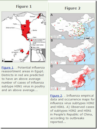
While the northern plains of India, the western Korean Peninsula and southwestern Japan were mentioned, their two biggest hotspots were Eastern China, and the Nile Valley of Egypt – both regions that have produced either new subtypes, or new clades (see Emergence Of A Novel Cluster of H5N1 Clade 2.2.1.2), of HPAI H5 viruses over the past two years.


image


In January of 2014, an emerging HPAI H5N8 appeared in South Korea, and rapidly spread through that nation’s poultry and wild bird population.  It showed up in Japan in April, and was subsequently reported in China.  


During the winter of 2014-15, H5N8 migrated to Russia, Western Europe, and even North America – and along the way spawned additional reassortants (H5N2, H5N3, H5N1) when it mixed with local avian flu subtypes.  Thus far, none of these H5N8 derived viruses have proven pathogenic in humans.


During the Spring of 2014 another HPAI H5 appeared in Southeast Asia; H5N6.   Unlike the H5N8 virus (and its descendents), H5N6 has caused serious (even fatal) human illness (see China Reports 3rd H5N6 Case (Fatal) – Yunnan Province).


Suddenly we’ve gone from one HPAI H5 virus of concern, to a half dozen.  And as these viruses spread, and mingle with other viruses, more novel subtypes may yet emerge.   And as the following dispatch from the EID Journal points out, their future behavior may be unpredictable.


Dispatch

Rapid Emergence of Highly Pathogenic Avian Influenza Subtypes from a Subtype H5N1 Hemagglutinin Variant

Erik de VriesComments to Author , Hongbo Guo1, Meiling Dai1, Peter J.M. Rottier, Frank J.M. van Kuppeveld, and Cornelis A.M. de Haan  Abstract

In 2014, novel highly pathogenic avian influenza A H5N2, H5N5, H5N6, and H5N8 viruses caused outbreaks in Asia, Europe, and North America. The H5 genes of these viruses form a monophyletic group that evolved from a clade 2.3.4 H5N1 variant. This rapid emergence of new H5Nx combinations is unprecedented in the H5N1 evolutionary history.

A highly pathogenic avian influenza (HPAI) A(H5N1) virus (A/goose/Guangdong/1/1996) was first detected in China in 1996. Multiple clades, defined by phylogenetic characterization of the H5 hemagglutinin (HA) (1), have evolved and spread across Asia, Africa, and Europe, causing enormous losses to the poultry industry. A total of 694 human infections (death rate 58%) were recorded during 2003–2014 (2).

During the evolution of HPAI H5N1 viruses, reassortment events involving the 6 internal gene segments have often been detected (reviewed in [3]), but novel subtypes (i.e., combinations of HPAI H5 with other N subtypes) have rarely been isolated. In 2014, a novel highly virulent reassortant HPAI H5N6 virus (4) caused multiple outbreaks in Southeast Asia and 1 lethal human infection, which led the Food and Agricultural Organization of the United Nations to issue a warning (5). Outbreaks of novel HPAI H5N8 virus in South Korea (6,7), China (8), and Japan raised further concern, and in November 2014, this subtype emerged outside Eastern Asia, causing outbreaks in poultry farms in Germany, the Netherlands, the United Kingdom, Canada, and the United States.

<SNIP>

Conclusion

(Excerpt)

In this study, we exclusively focused on the unique occurrence of new HA–NA combinations. Recent publications have already described the reassortment events of the internal gene segments of several of the viruses mentioned above (68,1114). In contrast to novel HA–NA combinations, novel constellations of internal gene segments are far from unique and have frequently been observed for HPAI H5N1 viruses (3). Our analysis indicates that new HPAI viruses have emerged that carry H5 proteins capable of matching with multiple NA subtypes. Whether the formation of new HA–NA combinations confers a selective advantage that contributed to the emergence of these novel subtypes is not known and requires elaborate research. However, the balance between HA (receptor binding) and NA (receptor cleavage) protein activities is known to be critical to cell entry and host tropism and may be an important factor that lead to the emergence of new HA–NA combinations. In contrast to HPAI H5N1, the novel clade 2.3.4.4 viruses, excluding H5N6 viruses, have not caused human infections. However, it is unknown to what extent the repeated acquisition of a new NA proteins could enhance the rate of evolution of the HA protein. Obviously such changes could further affect host and tissue specificity, potentially having serious consequences. Therefore, surveillance is required to monitor further spread, evolution, and potential changes in host range.


Given the recent emergence of H5N8, H5N6, H5N3 in Asia and novel reassortants of H5N2 and H5N1 in North America, and it comes as little surprise that the World Health Organization recently released a pointed warning that H5 Is Currently The Most Obvious Avian Flu Threat.


Saturday, April 04, 2015

EID Journal: Emerging Norovirus GII.17 - Guangdong, China

image


# 9900


Noroviruses - which are often mistakenly called `stomach flu’ - are single-stranded RNA viruses that (like influenza) are able to evolve rapidly. So we typically see a new dominant norovirus strain emerge every two or three years. In 2009, we saw the emergence of the New Orleans strain of GII.4, while in 2012, the Sydney strain appeared.


Victims usually experience nausea, frequent vomiting & diarrhea, and stomach pain – and may also suffer from headache, fever, and body aches.


Outbreaks from these viruses are the bane of cruise ships, schools, and hospitals – anywhere large numbers of people congregate.  While the illness usually runs its course in 1 to 3 (very long) days - among those who are aged or infirmed -the virus can take a heavy toll.

According to the CDC, in the United Sates each year the norovirus:

  • causes about 21 million cases of acute gastroenteritis (inflammation of the stomach or intestines or both)
  • contributes to about 70,000 hospitalizations and 800 deaths, mostly among young children and the elderly

With new strains appearing every few years, and most requiring a ridiculously low infectious dose, the ability for norovirus to spread is legendary.  In Vomiting Larry And His Aerosolized Norovirus, we looked at work done by the UK’s Health and Safety Laboratory, where they created a robot that  . . . you guessed it . . . vomits.

image

Credit UK’s Health & Safety Laboratory

By adding a florescent dye marker to robot’s `vomitus’, researchers have determined that droplets – too small to be readily seen – can end up as far as 3 meters away from the source.


The `standard’ mode of norovirus transmission is considered to be the fecal-oral route, but limited airborne transmission is increasingly being considered a factor.


The CDC – in a an MMWR report from 2011 called Updated Norovirus Outbreak Management and Disease Prevention Guidelines describes transmission thusly:

Transmission

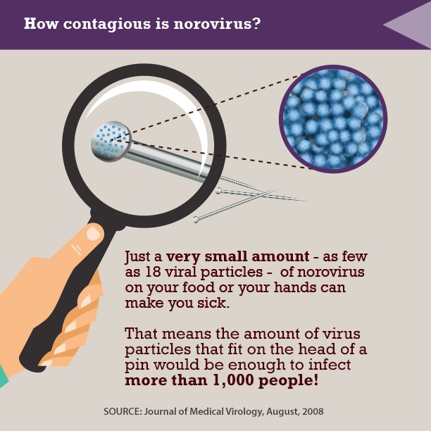
Norovirus is extremely contagious, with an estimated infectious dose as low as 18 viral particles (41), suggesting that approximately 5 billion infectious doses might be contained in each gram of feces during peak shedding. Humans are the only known reservoir for human norovirus infections, and transmission occurs by three general routes: person-to-person, foodborne, and waterborne.

Person-to-person transmission might occur directly through the fecal-oral route, by ingestion of aerosolized vomitus, or by indirect exposure via fomites or contaminated environmental surfaces.


While the numbers change almost years, there are currently 6 identified genogroups of norovirus (GI-GVI).  GI and GII genogroups are known to infect humans, and GI has 9 known genotypes, while GII has 22. 


The most common source of human infection for more than a decade has come from Genogroup II, genotype 4 (abbreviated as GII.4).


Yesterday, however, the CDC’s EID Journal carried a dispatch describing a recently emerged GII.17 strain.  Although sporadic GII.17 cases have been previously reported in Africa, Korea, Taiwan, and Japan, the first major outbreak was documented in Guangzhou last November, and it is now reportedly spreading rapidly across China. 


Gastroenteritis Outbreaks Caused by Norovirus GII.17, Guangdong Province, China, 2014–2015

Jing Lu1, Limei Sun1, Lin Fang, Feng Yang, Yanling Mo, Jiaqian Lao, Huanying Zheng, Xiaohua Tan, Hualiang Lin, Shannon Rutherford, Lili Guo, Changwen Ke, and Li HuiComments to Author

Abstract

In the past decade, the most prevalent norovirus genotype causing viral gastroenteritis outbreaks worldwide, including China, has been GII.4. In winter 2014–15, norovirus outbreaks in Guangdong, China, increased. Sequence analysis indicated that 82% of the outbreaks were caused by a norovirus GII.17 variant.

Norovirus infection is a leading cause of nonbacterial gastroenteritis outbreaks in industrialized and developing countries (1,2). On the basis of amino acid identity in viral protein 1, noroviruses can be divided into at least 6 genogroups (GI–GVI). GI and GII infect humans and can be further classified into genotypes; at least 9 genotypes belong to GI and 22 belong to GII (3). During the past decade, most reported norovirus outbreaks were caused by GII.4 norovirus (4,5). New variants of GII.4 have emerged approximately every 2–3 years and have caused norovirus gastroenteritis pandemics globally (6). Since 1999, the major circulating genotype in mainland China has been GII.4, accounting for 64% of all genotypes detected (7). In winter 2014–15, norovirus outbreaks in Guangdong Province, China, increased. Sequence analyses showed that the major cause of continuous gastroenteritis outbreaks in the region was a rarely reported norovirus genotype: GII.17.

<SNIP>

Sequence comparison with archived GII.17 strains from GenBank suggests that the GII.17 genotype identified in Guangdong is a newly emerged variant, differing from GII.17 strains detected before 2011. The recent detection of this new variant in samples from patients with sporadic cases in several regions of Asia (e.g., Korea, Japan, and Taiwan) and from groundwater in Kenya (11) suggests that this variant of GII.17 has circulated in a wide range of areas in recent years. For GII.17, most (66 [83%] of 80) sequences from the GenBank database are restricted to region C, the short conserved sequences of the N terminus of the capsid gene. This conserved region has been widely used for genotyping strains (12) and phylogenetic studies (13). To include more reference strains and to illustrate the relationship between GII.17 from Guangdong and other regions, we mainly used region C for phylogenetic analyses in this study. Similarly, phylogenetic analysis based on the nearly full length of capsid sequences also showed that the newly emerged GII.17 variant in Guangdong clustered with the strains from Japan and Taiwan in 2013 and 2014 and differed from GII.17 strains detected before 2011 (Technical Appendix[PDF - 159 KB - 2 pages] Figure 2).

In conclusion, a norovirus genotype GII.17 variant emerged in winter 2014–15 and caused outbreaks in multiple cities in Guangdong Province, China. The distribution of GII.17 genotype among patients with sporadic cases of gastroenteritis remains unknown. In future studies, epidemiologic and virologic surveillance should be broadened to better clarify virologic, clinical, and epidemiologic patterns of this newly emerged norovirus.


Of note, GII.17 made news last summer in Norovirus GII.17 Predominates in Selected Surface Water Sources in Kenya and was suspected as the cause of an outbreak recently in Taiwan. 


Whether GII.17 spreads as rapidly, and as thoroughly, as the Sydney GII.4 strain that emerged in 2012  remains to be seen, but the timing is right for seeing a new strain take hold. 


One of the keys to prevention is good hand hygiene, unfortunately, unlike with many other bacteria and viruses, alcohol gel doesn’t do a particularly good job of killing the virus (see CMAJ: Hand Sanitizers May Be `Suboptimal’ For Preventing Norovirus). 


The CDC offers this advice to help prevent the spread of this virus.

image

Friday, April 03, 2015

EID Journal: H5N1 In Nigerian Poultry – 2015

image

H5 Clade Diversity Around the Globe – Credit WHO


# 9899


We’ve discussed the immense, and growing, diversity of avian H5N1 often over the years, including just last week in WER: Development Of Candidate Vaccine Viruses For Pandemic Preparedness, where we saw two new H5 virus candidates (both H5N1 & H5N8) proposed by the World Health Organization.


The continual evolution of HPAI H5 viruses over the past dozen years has resulted in more than 2 dozen different candidate vaccines to either be developed or proposed in order to keep up with the newly emerging clades and subclades of the virus.


Earlier today, in Eurosurveillance: Emergence Of A Novel Cluster of H5N1 Clade 2.2.1.2, we looked a recently formed branch in H5N1’s family tree that is spreading rapidly across Egypt.


Simply put, there are a number of subclades of H5N1 that circulate around the globe, and there can be considerable variability in each strain’s ability to infect, and kill (see Differences In Virulence Between Closely Related H5N1 Strains).  


You can get a sense of how H5N1 has expanded from one clade in 1996, to more than 20 clades and subclades today, by the chart below (note: not all continue to circulate).

    image

(click to load larger image)  (Note: Chart only goes through 2011)


By identifying and charting these variations on an avian H5 theme, you can often track down where a specific clade originated, where it has traveled, and get some sense of its `biological fitness’ - its ability to compete with other strains of the virus. 


All of which serves as prelude to a new report, appearing in the EID Journal, that examines the H5N1 virus which has turned up again in Nigeria after an absence of 7 years, and finds that the recently arrived clade appears to have originated in China.


Specifically clade 2.3.2.1c, which was also isolated from a Nurse who returned to Alberta, Canada from a trip to China (see Alberta Canada Reports Fatal (Imported) H5N1 Infection), and has also been reported in Vietnam, India, Bulgaria, and Indonesia over the past several years.


How clade 2.3.2.1c managed to hopscotch thousands of kilometers and show up in Nigerian poultry last December remains an epidemiological mystery. 


Letter

Highly Pathogenic Avian Influenza A(H5N1) Virus in Poultry, Nigeria, 2015

Isabella Monne1Comments to Author , Clement Meseko1, Tony Joannis, Ismaila Shittu, Mohammed Ahmed, Luca Tassoni, Alice Fusaro, and Giovanni Cattoli

To the Editor: In Nigeria, from February 2006 through July 2008, outbreaks of highly pathogenic avian influenza (HPAI) subtype H5N1 virus infection in poultry negatively affected animal and public health as well as the agricultural sector and trade. These outbreaks were caused by viruses belonging to genetic clades 2.2 and 2.2.1 (1). In January 2015, seven years after disappearance of the virus, clinical signs of HPAI (swollen head and wattles, hemorrhagic shank and feet) and increased mortality rates were observed among backyard poultry in Kano and in a live bird market in Lagos State, Nigeria. The virus was isolated from 2 samples independently collected from the poultry farm (parenchymatous tissues) and the market (tracheal swab), and H5 subtype virus was identified by reverse transcription PCR. The samples were adsorbed onto 2 Flinders Technology Associates cards (GE Healthcare Life Sciences, Little Chalfont, UK), which were sent to the World Organisation for Animal Health/Food and Agriculture Organization of the United Nations Reference Laboratory for Avian Influenza in Italy for subtype confirmation and genetic characterization. Influenza A(H5N1) virus was detected in both samples, and sequencing of the hemagglutinin (HA) gene showed that the viruses possessed the molecular markers for HPAI viruses with a multibasic amino acid cleavage site motif (PQRERRRKR*G).

<SNIP>

The results obtained from whole-genome analysis provide evidence that a novel clade of the A(H5N1) virus, specifically clade 2.3.2.1c, has reached Nigeria. Although ascertaining how and exactly when this has happened is difficult, it seems most likely that the virus entered the country in December 2014, as evidenced by unverified accounts of increased poultry deaths in some live bird markets in Lagos, after the birds had been moved from the north (Kano) to the south during the festive season. The identification of genetic clustering between the strains from Nigeria analyzed here and the HPAI A(H5N1) viruses originally identified in Asia suggests an unknown epidemiologic link between these regions, probably associated with human activities, migratory bird movements, or both.

Considering that this virus is an intersubtype reassortant and has already caused infection in humans, we believe that complete characterization of the strain in terms of virulence and host range is of high priority. Furthermore, because the reemergence of subtype H5N1 virus was followed by epidemiologic amplification (≈265 outbreaks in 18 states as of February 2015; T. Joannis, pers. comm., 2015) for which virus genetic characterization is not yet available, local veterinary and public health services and international organizations should take necessary measures to identify critical control points and stop circulation of this virus.

Wednesday, April 01, 2015

EID Journal: The Transmission Potential Of A(H7N9) In China

image


# 9892


Whether you cast your gaze towards Egypt (H5N1), or China (H7N9), recent outbreaks of avian flu among humans continue to raise concerns over their pandemic potential.  Neither virus has demonstrated an ability to spread efficiently from human-to-human, but both continue to get fresh opportunities to try to figure us out.


Last month, in WHO: H5 Currently The Most Obvious Avian Flu Threat, the World Health Organization – while not ignoring H7N9’s potential - weighed in on the risks posed by the recent and dramatic surge in both the variety, and spread, of HPAI H5 viruses around the globe.


A couple of weeks later, in Nature: Dissemination, Divergence & Establishment of H7N9 In China, we saw renewed warnings over the evolutionary path that the H7N9 virus has been taking over the past two years.  A topic we revisited the next day, in H7N9: Primus Inter Pares?


To this short list, we must also add a growing variety of reassortant avian and swine viruses (H5N8, H5N6, H5N2, H10N8, H3N8, H3N2v, etc.), many of which have at least some potential to adapt to humans. 


Today the EID Journal has published a new review of the transmissibility of the avian H7N9 virus in China, and while no evidence of sustained transmission was detected, they found:

  • `evidence of a small but significant amount of transmission between humans in the first and second waves’
  • `evidence of increased transmission potential in the second wave’

First a few excerpts from the study (follow the link to read it in its entirety), after which I’ll be back with a bit more.


Volume 21, Number 5—May 2015
Dispatch

Transmission Potential of Influenza A(H7N9) Virus, China, 2013–2014

Adam J. Kucharski1Comments to Author , Harriet L. Mills1, Christl A. Donnelly, and Steven Riley
Abstract

To determine transmission potential of influenza A(H7N9) virus, we used symptom onset data to compare 2 waves of infection in China during 2013–2014. We found evidence of increased transmission potential in the second wave and showed that live bird market closure was significantly less effective in Guangdong than in other regions.

From February 19, 2013, through April 22, 2014, a total of 429 cases of influenza A(H7N9) virus infection in humans in China were reported and occurred in 2 outbreak waves. During the first wave in spring 2013, live bird markets were closed in several parts of China (1,2); these market closures substantially reduced the risk for infection in affected regions (3). During a second wave in autumn 2013 (4), markets were again closed in some provinces (57). Analysis of the largest clusters of subtype H7N9 virus infection in 2013 suggested that the basic reproduction number (R0, the average number of secondary cases generated by a typical infectious host in a fully susceptible population) was higher in some clusters than in others (8,9), although the absence of sustained transmission implied that R0 was less than the critical value of 1. To determine the transmission potential of influenza A(H7N9) virus in the first and second waves in 2013, we compared symptom onset data. We also measured the extent to which market closures in 2014 reduced spillover hazard (i.e., risk for animal-to-human infection).

<SNIP Study Details>

Conclusions

We found no evidence of reduced human-to-human transmission between the 2 waves. For a serial interval of 7 days, we estimated that R0 increased in Zhejiang. Furthermore, the effectiveness of live bird market closures varied between regions; short-term closures were substantially less effective than interventions in other regions. These results emphasize the value of prompt and sustainable control measures during outbreaks of influenza A(H7N9) virus infection.


Last summer, in Eurosurveillance: Genetic Tuning Of Avian H7N9 During Interspecies Transmission, we saw evidence of the genetic diversity, and continual evolution, of the H7N9 virus in Mainland China.  Researchers found that at least 26 separate genotypes had emerged, mostly during the first wave, through a process they called `genetic tuning’.


The Nature report, mentioned above, expands that H7N9 universe to 48 genotypes, spread across three major clades.


Not only are the incarnations of H7N9 continuing to grow, the H7N9 virus has also reassorted into at least two new subtypes on the Chinese mainland; H7N7 (see  Nature: Genesis Of The H7N9 Virus) and a new H7N6 virus described in the recent Nature report.


This malleable H7N9 virus, which is spreading asymptomatically and stealthily in China’s poultry, could also potentially reassort with a human influenza virus like H3N2 or H1N1.  Something that Hong Kong’s CHP Director has discussed repeatedly this winter (see  HK’s Dr. Ko Wing-man On Flu Reassortment Concerns).


While there are seemingly a lot of ways for H7N9 to become a pandemic threat, it is also possible that there is some – as yet unidentified - `species barrier’  that prevents avian flu viruses from adapting well enough to humans to pose a serious threat.


The progression of human influenza pandemics over the past 130 years has been H2, H3, H1, H2, H3, H1, H1 . . . .  and while that doesn’t prove that  an H5 or an H7 virus couldn’t adapt to humans (or hasn’t in the past), it has led some researchers to wonder whether a non H1, H2, or H3 virus has the `right stuff’ to spark a pandemic.


While some scientists believe that may be a possibility (see Are Influenza Pandemic Viruses Members Of An Exclusive Club?), few are willing to bet the farm on our being that lucky.