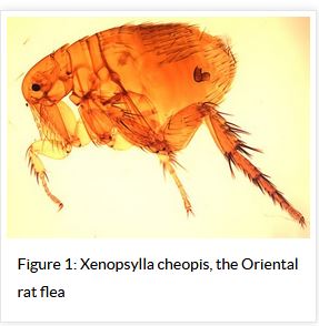
Thursday, November 30, 2017

Texas: DSHS Issues Murine Typhus Alert

Credit CDC


#12,933


Murine typhus, aka flea-borne typhus, is a bacterial disease caused Rickettsia typhi - and while not terribly common in most of the United States - it is most often reported in California, Texas and Hawaii. Although some cases  will recover without treatment, others can become sick enough to require hospitalization, and deaths are not unheard of.
Not to be confused with Typhoid Fever - which is caused by a Salmonella Typhi infection that can (rarely) be transmitted from one person to another - Typhus is acquired from contact with infected fleas that live on rodents, possums, raccoons and cats.
While I've mentioned the occasional outbreak (see Disease Transmission At The Human-Animal Interface), Typhus - for many decades - had been pretty rare in the United States.  But like so many other emerging and reemerging zoonotic diseases, we are increasingly  seeing it crop up in new, or unusual locations.

Texas reports anywhere from a couple of dozen to a couple of hundred typhus cases each year, but until recently most cases have been reported in the south and central part of the state, along the coastline.
Over the past few years, that geographic distribution has begun to change.
After going 8 decades without a case - in 2012 Galveston, Texas reported two cases prompting an epidemiological investigation that led to the identification of 12 more cases in 2013 (see MMWR Reemergence of Murine Typhus in Galveston, Texas, USA, 2013).

The authors wrote:
Dynamic shifts in the epidemiology and transmission of murine typhus are not unprecedented. Although the rat-to-rat cycle of transmission by fleas is often referred to as an urban cycle, the rural South experienced high rates of murine typhus in the 1940s as a result of a proliferation of rats after a change in crop production from cotton to peanuts, because rats were attracted to the peanuts as a source of food (13).
In southern California, opossums infested with R. typhi– and R. felis–infected cat fleas (C. felis) have been associated with a shift of fleaborne rickettsioses from the urban center of Los Angles to suburban areas (14). This suburban cycle of transmission involving C. felis plays a recognized role in Corpus Christi, Texas, a coastal city ≈220 miles southwest of Galveston (1). Additionally, this cycle has been suspected in a recent outbreak of murine typhus in the central Texas city of Austin (15).
This year, the number of Typhus cases in Texas is expected to well exceed last year's (21st century) record of 364 cases, and the DSHS notes increased activity in the urban areas of both Dallas-Fort Worth and Houston.

Since the signs of symptoms of Typhus are similar to many other diseases, the DSHS has today issued the following alert to health care providers to increase their clinical suspicion, and to urge prompt treatment with antibiotics (even before lab results are returned).

HEALTH ALERT
Increased Flea-borne (Murine) Typhus Activity in Texas
November 30, 2017

Due to increased reports of flea-borne (murine) typhus during 2017 from multiple areas of Texas, DSHS is requesting that healthcare providers increase their clinical suspicion for patients presenting with fever and one or more of the following: headache, myalgia, anorexia, rash, nausea/vomiting, thrombocytopenia, or any hepatic transaminase elevation. The diagnosis of flea-borne typhus relies on a high index of clinical suspicion and on results of specific laboratory tests.

Flea-borne typhus is caused by infection with the bacterium Rickettsia typhi (or R. felis). Rat and cat fleas are the primary vectors. Transmission to humans can occur when infected flea feces are scratched into a bite site or another abrasion in the skin, or rubbed into the conjunctiva. Rats, opossums, and cats are thought to be the primary reservoirs for the disease in Texas.

People with typhus report non-specific symptoms including fever, headache, chills, malaise, anorexia, myalgia, rash, nausea, and vomiting. Laboratory findings may include thrombocytopenia, hyponatremia, elevated hepatic transaminases, and CSF abnormalities consistent with aseptic meningitis. Although flea-borne typhus is often a mild, self-limited illness, more than 60% of reported cases are hospitalized. Since 2003, eight deaths have been attributed to flea-borne typhus infection in Texas. When left untreated, severe illness can cause damage to one or more organs, including the liver, kidneys, heart, lungs, and brain. As with other rickettsial infections, prompt antibiotic treatment is recommended; treatment should not be delayed pending diagnostic tests. Additional clinician guidance on typhus can be accessed at the CDC website: https://www.cdc.gov/typhus/murine/index.html

Over 2,800 cases of flea-borne typhus were reported in Texas between 2000 and 2016 [median = 157 cases/year; max = 364 cases/year (2016); min = 22 cases/year (2001)], and over 400 reported cases are expected for 2017. In previous years, typhus was primarily reported from South Texas, along the Gulf Coast (Nueces County), and Central Texas (Travis and Bexar counties). In 2017, increased typhus activity has been noted in the Dallas-Fort Worth and Houston areas. Cases are reported year-round, but the majority of typhus cases occur between May and July, with another peak in December and January. Typhus can occur in any age group, but over 25% of cases are reported among those between 6-15 years of age.
Laboratory Diagnosis of Flea-borne Typhus Infection

Laboratory testing is required to confirm a diagnosis of flea-borne typhus. The most efficient and readily available diagnostic method to confirm infection with R. typhi is detection of IgG antibodies to R. typhi using an indirect fluorescent antibody (IFA) test in acute and convalescent serum specimens collected at least 3 weeks apart. However, because antibodies for rickettsial diseases can be cross-reactive, specimens should be tested against a panel of Rickettsia antigens, including, at a minimum, R. rickettsii and R. typhi, to differentiate between spotted fever group and non-spotted fever group Rickettsia spp. Many commercial laboratories offer rickettsial serology testing, but it should be noted that ELISA or EIA tests are not reliable for rickettsial disease diagnosis. Molecular testing is a more definitive testing option. Whole blood collected within a few days of illness onset may be tested by PCR in an attempt to detect Rickettsia spp.

Rickettsial panel IFA testing is available at the DSHS Laboratory. It is desirable to submit a volume of 2 mLs of serum per specimen. Serum samples may be refrigerated for transport if the specimens will be tested within 48 hours. If not, ship frozen at 2°-8°C. If molecular testing is preferred, whole blood samples may be routed through the DSHS Laboratory to CDC. Guidance for the submission of specimens to the DSHS Laboratory for typhus testing can be found at: http://www.dshs.texas.gov/lab/mrs_mic_test_t2.htm#Typhus. Please contact the DSHS Laboratory at (512) 776-7514 during regular business hours with any questions pertaining to typhus diagnostic testing.
Disease Reporting

Flea-borne typhus cases are required to be reported to the local health department (LHD) within one week. If there is no LHD, reports can be made to the Regional DSHS Zoonosis Control Office. Contact information for Regional Zoonosis Control staff is available at: http://www.dshs.texas.gov/idcu/health/zoonosis/contact/
 Last updated November 30, 2017  
You can find more information on the CDC's Typhus Home page. 


Genome Announce.: Novel Avian H3N2 Virus Isolated From Domestic Ducks - China, 2016


#12,932


The progression of human influenza pandemics over the past 130 years is believed to have been H2, H3, H1, H2, H3, H1, H1 . . .
  • H2N2 sparked two pandemics (1898 and 1957) roughly 69 years apart.   
  • H3 viruses unleashed two pandemics (1900 and 1968) roughly 68 years apart
  • And H1N1 caused the great 1918 pandemic only to return in the pseudo-pandemic of 1977, 59 years later (and then reinvented itself in the 2009 Pandemic).
While we keep a wary eye on avian H5 and H7, this repeating pattern of the H1, H2, H3 pandemics is hard to ignore, even if the sample size is small (see Are Influenza Pandemic Viruses Members Of An Exclusive Club?). Granted, we've no clue what influenza subtypes might have circulated in humans before 1890, and there is some guesswork surrounding those prior to 1918.
There are already two H3N2 viruses (H3N2 swine variant [A/Indiana/08/2011]) and (Canine H3N2 [A/canine/Illinois/12191/2015]) on the CDC's IRAT list of novel flu subtypes/strains that currently circulate in non-human hosts that pose a potential threat to human health.
H1, H2, and H3 influenza viruses are generally thought to have `less far to jump' to humans than do H5 or H7 viruses, so we take particular notice whenever we see a new one emerging in the wild. A few recent blogs on this special subset of flu viruses include:
H2N2: Everything Old Is Flu Again

MMWR: Investigation Into H3N2v Outbreak In Ohio & Michigan - Summer 2016 
J. Virol: Novel Reassortant Human-like H3N2 & H3N1 Influenza A Viruses In Pigs

PNAS: The Pandemic Potential Of Eurasian Avian-like H1N1 (EAH1N1) Swine Influenza
 

All of which brings us to a new announcement today of the isolation of a novel avian H3N2 virus from domestic ducks in China, with contributions from H1N1, H3, and H7 AIVs.

Detected at a live bird market in Jiangsu last year, this is another reminder just how quickly new reassortant viruses can emerge, particularly when many birds are housed in close quarters.

Novel Reassortant H3N2 Avian Influenza Virus Isolated from Domestic Ducks in Eastern China in 2016

Wenqiang Sun, Jiaxin Li, Jiao Hu, Daxiu Jiang, Zhichuang Ge, Chaonan Xing, Xiaoquan Wang, Min Gu, Xiaowen Liu, Shunlin Hu, Xiufan Liu

ABSTRACT

H3 subtype avian influenza virus (AIV) poses a great threat to public health, and so investigating its epidemiology is of great importance. A novel reassortant H3N2 AIV strain was isolated from a live poultry market in eastern China. The strain’s genes originated from H1N1, H3, and H7 AIVs. Thus, the genome information of the H3N2 isolate will help to investigate further the epidemiology of H3 subtype AIVs in China.

GENOME ANNOUNCEMENT

Avian influenza viruses (AIVs) have a wide range of hosts, from birds to mammals (17). Notably, multiple subtypes of influenza viruses (H3, H4, H6, H9, H10, etc.) have also circulated and evolved in live poultry markets (LPMs), which are considered to be recombinant sources of AIVs in China (810). Meanwhile, highly pathogenic AIVs (HPAIVs) and low pathogenic AIVs (LPAIVs) frequently undergo reassortment with each other (79, 1113). In particular, H3 subtype AIVs can be detected in LPMs in China (9). As we know, H3 subtype AIVs have a wide host range, including birds, humans, pigs, dogs, and horses (3, 4, 1417). H3 subtype AIVs can serve their gene segments to other HPAIVs and also reassort with other AIV subtypes (10, 1820). So, the monitoring of H3 subtype AIVs is of great significance to public health.

An H3N2 strain of AIV was isolated from a duck in September 2016 in Jiangsu, China, and named A/duck/Jiangshu/YZ916/2016(H3N2). RT-PCR was performed by universal primers (21), followed by sequencing (Nanjing Genscript). The DNA sequences were edited with Lasergene version 7.1 software (DNASTAR), and genetic evolution analysis of the isolate was performed with MEGA7 software.

The complete genome of the isolate included eight genes, polymerase basic 2 (PB2), polymerase basic 1 (PB1), polymerase acidic (PA), hemagglutinin (HA), nucleoprotein (NP), neuraminidase (NA), matrix (M), and nonstructural (NS) proteins. The open reading frame lengths of these segments are 2,280, 2,277, 2,151, 1,701, 1,497, 1,401, 982, and 838 nucleotides, respectively. PEKQTR↓G at the cleavage site of the isolate indicated that the virus was an LPAIV. The potential glycosylation sites of the isolate revealed that there were five potential N-linked glycosylation sites in HA (positions 22, 38, 165, 285, and 483) and six in NA (positions 61, 69, 86, 146, 200, and 402). Moreover, there were amino acid residues, Q226 and G228, at the receptor binding site in the HA protein, which indicated that the isolate preferentially binds to a receptor of avian origin. In PB2, there were no mutations for E627K and D701N, which are associated with mammalian host adaptation (22).

Genetic evolution analysis of the eight genes of the isolate indicated that the HA gene showed a high similarity (97%) with isolate A/duck/Jiangsu/J2186/2014(H3). The NA gene showed high homology (99%) with isolate A/chicken/Ganzhou/G243/2016(H3N2). The PB2 gene showed the highest homology (98%) with isolate A/duck/Jiangxi/21224/2013(H7N3). The PB1 gene showed the highest homology (99%) with isolate A/chicken/Huzhou/3916/2013(H7N3). The PA gene showed the highest homology (99%) with isolate A/duck/Mongolia/520/2015(H1N1). The NP gene showed the highest homology (99%) with isolate A/duck/Jiangxi/21710/2013(H7N3). The M gene showed the highest homology (99%) with isolate A/muscovy duck/Vietnam/LBM348/2013(H3N8). The NS gene was most closely related to isolate A/environment/Korea/W478/2014(H7N7) with 99% homology. Overall, the isolate was a novel reassortant H3N2 AIV originating from H1N1, H3, and H7.

H3 subtype AIVs can pose a great threat to public health, and thus the genome information reported here can help further epidemiological investigations of H3 subtype AIVs in China.

Accession number(s).This genome sequence has been deposited in GenBank under the accession numbers MG021165 to MG021172.

Hong Kong Holds Novel Flu Pandemic Exercise `Garnet'

Credit HK CHP


#12,931


A decade ago, when the concept of an avian flu pandemic was relatively fresh in the minds of the public,  health officials, and all levels of government  . . .  a week scarcely went by without our seeing one or more high profile `pandemic exercise' somewhere around the world.

These drills were held by both the public and private sector, and ranged from multi-national  exercises all the way down to exercises held by local hospitals, and city governments. A few (out of scores of) blogs from that `golden age' of pandemic preparedness include:
Singapore To Begin 2-Week Financial Sector Pandemic Drill
Merced County California Flu Drill
Scottsdale Arizona: Pandemic Drill

Idaho BlogEx : July 28th


UK: Lessons Learned From Winter Willow 


UK Exercise: PPE Usage In A Pandemic

While not all of these drills were terribly realistic (i.e. practicing for rapid delivery of a vaccine that was unlikely to exist for months), they did serve to increase awareness - both for those taking part, for the public who heard about them in the media.

Although pandemic threats are arguably greater today than ever, pandemic exercises like the ones listed above have become far less common, particularly among the private sector or at the local level.
Drills - when they are held - rarely get a mention in the media.  This despite repeated warnings (see World Bank: World Ill-Prepared For A Pandemic and Pandemic Unpreparedness Revisited) that we remain woefully unprepared for a severe pandemic.
There are exceptions, of course. Mainland China, being at ground zero for both H7N9 and H5N6, drills at both the national and province level several times each year. Hong Kong is another of those places that not only holds drills every year, they publicize it heavily (see Hong Kong Conducts Avian Flu Drill).

Today Hong Kong's CHP reports on their latest `novel flu' drill, called exercise `Garnet'.   First their announcement, then I'll return with a post script.
     The Government tested its preparedness for possible detection of a novel influenza case today (November 30) during an exercise code-named "Garnet", organised by the Centre for Health Protection of the Department of Health (DH) in collaboration with other government departments and organisations at Wo Che Plaza, Sha Tin.

     Exercise "Garnet" was aimed at assessing the interoperability of government departments and organisations in response to the detection of a case of novel influenza, testing their execution of the Preparedness Plan for Influenza Pandemic, as well as enhancing the alertness and readiness of relevant stakeholders in guarding Hong Kong against novel influenza and the threat of the spread of communicable disease.

     About 50 participants from relevant government departments and organisations took part in the exercise, with 11 experts from the Mainland and Macau health authorities as observers. The exercise consisted of two parts. The first part was a table-top exercise conducted on November 15, in which relevant departments and organisations discussed and co-ordinated the communicable disease response measures required in the simulated scenario of detection of a confirmed case of novel influenza in Hong Kong.

     The second part, conducted today, was a ground movement exercise. Under the exercise simulation, a female staff member of a telecommunications company working at Wo Che Plaza tested positive for novel influenza virus. Initial epidemiological investigations revealed that while she had no poultry contact locally, she had travelled to a country with a novel influenza outbreak. Among other staff members who had close contact with her, some had also developed symptoms of novel influenza. 

     The DH responded immediately and co-ordinated with relevant departments and organisations to formulate and implement corresponding measures. In addition to conducting on-site assessment and epidemiological investigations by its Public Health Team, the DH under this exercise also held a briefing for the staff members concerned, advised them on infection control measures, prescribed antiviral prophylaxis for the close contacts, and instructed the shopping mall operator and its cleaning services company to disinfect the contaminated areas.

     "Apart from the seasonal influenza viruses which circulate among humans, many other influenza A viruses are found in birds and other animal species. These viruses are distinct from human seasonal influenza viruses, and new types of virus may appear from time to time. Some of them can infect humans, causing disease ranging from mild conjunctivitis to severe pneumonia and even death. They are collectively termed novel influenza viruses, against which the human population generally lacks immunity. If a novel influenza virus acquires the capacity to spread easily from person to person through genetic changes, an influenza pandemic can occur. As such, we should stay vigilant to better prepare ourselves for these emerging infections," a DH spokesman said.

     "The DH conducts public health exercises regularly to test and assess the effectiveness of the Government's preparedness and response plans as well as procedures for communicable diseases, and carry out preventive measures with dedication. We will continue to conduct interdepartmental exercises with government departments, organisations and stakeholders concerned in close partnership, with the goal of enhancing Hong Kong's overall preparedness in the control and prevention of communicable diseases," the spokesman added.

Ends/Thursday, November 30, 2017

Issued at HKT 17:10

Encouragingly, 2017 has seen a bit of a renaissance in pandemic planning at the highest levels of government (see ECDC: Guide To Revising The Influenza Pandemic Preparedness Plan  & HHS 2017 Influenza Pandemic Update: Planning Assumptions) - along with recent events like the Smithsonian `Next Pandemic' Webinar.
Hopefully some of that will start to filter down to the state and local level, and into the private sector as well. 
Given the speed at which a pandemic could spread in our highly mobile 21st century society, we might find ourselves with only days or perhaps a few weeks to prepare once the threat becomes obvious. 
Going flatfooted into a mild pandemic would be regrettable, doing so with a severe pandemic virus could be disastrous.  Simply put, if we hope to be prepared, we need to start now.
 For more on pandemic preparedness, you may wish to revisit these older blogs.
WHO: Candidate Vaccines For Pandemic Preparedness - Sept 2017
#NatlPrep : Pandemic Planning Considerations

Upcoming Webinar: The Strategic National Stockpile
Are We Prepared to Help Low-Resource Populations Mitigate a Severe Pandemic?


Wednesday, November 29, 2017

Nigeria: NCDC Monkeypox Sitrep #9

http://www.ncdc.gov.ng/themes/common/files/sitreps/aab2a3eac6b6b0009323043123f767f6.pdf


#12,930


Although we just saw a report a yesterday (see Nigeria: CDC Monkeypox Update - Week 48) - which was dated November 19th - the Nigerian CDC has just released another update - current through November 23rd - which shows a sharp jump in confirmed cases. 
The previous report (Sitrep #8) cited 148 total (suspected & confirmed) cases, with 42 lab confirmed cases, while today's Sitrep #9 bumps that number to 56 (out of 155).
Both reports are listed on the Nigerian Disease Situation Report page as week 48, although today's internal date says epi-week 47.

Aside from the jump in confirmed cases, we also learn that 3 more states have confirmed cases (Imo,  Katsina  and  Nasarawa), and genetic testing suggests multiple introductions of the virus into the country.  

Some highlights from today's report includes:
  • The  Nigeria  Centre  for  Disease  Control  (NCDC)  continued  to  coordinate  the response  through its activated Emergency Operations Centre (EOC)
  • Fifty-six (56) confirmed cases recorded since the onset of the outbreak
  • Three  (3)  new  States  (Imo,  Katsina  and  Nasarawa)  recorded  confirmed  cases  in  the  reporting week
  • Thirteen (13) new suspected cases reported in the reporting week 
  • NCDC National Reference Laboratory, Abuja continued laboratory diagnosis of monkeypox with improved turn-around time 
  • There  has  been  a  decline  in  the  number  of  new  suspected  cases  reported  over  the  last  four weeks
  • Since  the  onset  of  the  outbreak,  a  total  of  155  cases  (suspected  and  confirmed)  have  been recorded from 21 States and the Federal Capital Territory (FCT)
  • No death attributable to monkeypox has been recorded so far
  • Epidemiologist deployed to five (5) more States

EPI SUMMARY

  • Fourteen new confirmed cases were recorded in the reporting week from Benue (1), Cross River (3), Edo (1), FCT (2), Imo (2), Katsina (1), Nasarawa (1) and Rivers (3) state
  • Out of a total of 155 suspected cases reported from 21 States and the FCT 56 cases have been confirmed in 13 States  
  • Clustering of cases was demonstrated in some instances, however no known evidence yet of epidemiological linkages across States
  • Preliminary genetic sequencing suggests multiple sources of introduction of monkeypox virus into human population
  • The male to female ratio for confirmed cases is 3:1
  • The most affected age group is 21-40 years (Median Age = 30)
  • No death recorded among cases so far

 For more background on Monkeypox, you may wish to revisit Yesterday's blog.

Saudi MOH: Burying The Lede

https://www.moh.gov.sa/en/CCC/PressReleases/Pages/default.aspx

#12,929


When you hit, as I do, a hundred or more websites every day searching for infectious disease stories . . . it is sometimes easy to miss things.
For reasons I can't explain, the Saudi MOH - after not posting updates for several days (a common occurrence), began putting new daily MERS updates half way down the list. Had Lisa Schnirring at CIDRAP not clued me in, I might still be oblivious. 
In any event, over the past 6 days the Saudis have reported 4 new MERS cases, the details of which follow:

From the 24th:

https://www.moh.gov.sa/en/CCC/PressReleases/Pages/statistics-2017-11-24-001.aspx


 From the 28th:

https://www.moh.gov.sa/en/CCC/PressReleases/Pages/statistics-2017-11-28-001.aspx


From Today:

https://www.moh.gov.sa/en/CCC/PressReleases/Pages/statistics-2017-11-29-001.aspx

Tottori University: Shimane HPAI H5N6 A New Reassortment

Credit Tottori University


#12,928


On Monday, in South Korea: Gochang HPAI H5N6 A New Reassortant Virus, we learned (somewhat cryptically) that South Korea's recent outbreak of H5N6 was not simply a return of last year's Asian-origin virus, but an entirely new reassortment comprised of last year's European H5N8 virus and the NA from a European H3N6.
Today we learn from testing at Tottori University that the virus samples collected two weeks ago from Shimane Prefecture on the east coast of Japan (see Japan: MOE Raises Surveillance Alert Level As More Dead Birds Are Discovered) are a similar reassortant virus. 
Not only does this announcement provide us with additional details on these genetic changes - by turning up at locations more than 500km apart within days of each other - it suggests this new reassortant virus may be more than just a flash in the pan.


First some (translated) excerpts from the University's press release (bolding mine), then I'll return with a bit more.

New information
 
This season's highly pathogenic avian influenza virus domestic first separation New!
 

Nov 28, 2017
List of new information

This season's highly pathogenic avian influenza virus gene properties of first isolates in Japan

~ Viral origin and virulence ~
Overview

In November 2017, the H5N6-type highly pathogenic avian influenza virus was first isolated in this season from the corpse of the Cobb Swan recovered in Shimane Prefecture. As a result of the analysis of the full-length gene of the virus isolated in the university, the virus has been found to be a different virus from last season's domestic strains.
The virus was also a gene re-assembly of the waterfowl-derived N6-type virus, widely distributed in Europe and other popular H5-type viruses in the last season, and was presumed to have invaded the country in the new season. Furthermore, the virus's genes are not likely to infect people directly because mutations that are considered to be involved in the infection to the person reported so far have not been recognized.
Background

In Korea last season, the epidemic of h5n6-type highly pathogenic avian influenza virus spread across the country, totaling 383 cases, and the largest outbreak in the past over 37 million feathers of culled. On the other hand, in Japan, at about the same time, it was confirmed that the outbreak occurred at 12 farms total of 9 road prefectures, and the occurrence of 218 cases, which is the largest in the past, was reported in birds and other countries. This h5n6-type virus It was discovered for the first time in Jiangsu, China in 2013, and it is believed that the next day in 2014, the epidemic spread to Southeast Asian countries such as Viet Nam and Laos, which have already spread widely in China.
On the other hand, in European countries, H5N8-type viruses have been prevalent in countries since 2014, and last season, 23 countries have already been confirmed as Italy and Russia in this season.
History

On November 5, 2017, in the trachea and total excretion swab specimens taken from the corpse of the Cobb Swan found in Matsue City, Shimane Prefecture, the positive response was observed in the type a influenza simple diagnostic kit, the Ministry of the Environment " It is one of the confirmed diagnostic agencies of avian influenza virus possession survey in birds. At the Center for Epidemiology of Common Infectious Diseases in the University of Tsukuba, the analysis of full-length genes was performed along with the implementation of virus separation, sub-type identification and virulence testing from the same specimen.
Content and significance

The influenza virus gene consists of eight RNAs, called segments, which are called PB1, PB2, PA, HA, NP, NA, M, and NS genes, depending on the major proteins coded by each gene. The eight genetic segments of this separation virus (hereafter Shimane 2017) it was found that the homology of the h5n6-type domestic epidemic strains of last season was 94% or less, and that this virus was a different virus from last season's domestic epidemic strain.
In addition, seven gene segments other than the NA gene of the virus last year showed the highest homology of over 99% with those of H5N8-type viruses prevalent in European countries The NA gene also showed more than 97% homology with those of the N6 virus derived from wild waterfowl widely distributed in Eurasia. From the above, the virus has been found to be a gene re-aggregation virus with the H5-type virus and wild waterfowl-derived n6 virus prevalent in poultry in Europe, etc. Last season.
According to the method prescribed by the OIE (International World Secretariat), as a result of the pathogenic test of the chicken in the intravenous inoculation, since the death was confirmed within 48 hours of all feathers, it has been confirmed that the virus is highly pathogenic avian influenza virus that indicates a high virulence against chickens. In addition, in the experiment of the infection by the nasal inoculation to chickens considered to be close to the natural infection path, with 106 virus shots, all of them died within three days and all of them were left in the 104 inoculation group, and the virus was compared to last season's domestic stock It was considered to be a virus with the same degree of virulence.
When the influenza virus infects cells, the HA protein is adsorbed on the receptor of the cell surface, but it is known that the receptor is different in birds and mammals. The amino acid residues involved in the adsorption of the HA protein in the Shimane 2017 strain on host cells were an array of specificity for bird-type receptors. In the amino acid sequence of other viral proteins, amino acid substitution was not allowed to acquire the infection to mammals and people. In addition, the amino acid substitution involved in the neuraminidase inhibitor resistance was not observed in the NA protein. From the above results, it was estimated that the virus would be less likely to infect people directly.

Future plans

The gene sequence information of this virus will be released to the international nucleotide sequence database. We will continue to analyze the genetic properties of other domestic isolates and Korea isolates, as well as to analyze the antigenic nature of the virus in detail. We will also clarify the sensitivity, virulence, and propagation power of chickens, ducks, and wild baby animals, etc. The results of these researches are expected to lead to an increase in the future of epidemic forecasting and prevention measures.
(Continue . . . )

One of the questions we've been pondering over the summer is exactly what virus would return to Europe, Asia, and possibly North America with this fall's migration.  
During the first three HPAI H5 transcontinental waves (2005-2006, 2009-2010, and 2014-2015) HPAI H5N1 was the culprit, and while the clades changed over time, the basic subtype remained stable (see Sept 2016 FAO Empres  Watch).
The FAO has pegged the emergence and global spread of HPAI H5N8 as the `fourth wave of intercontinental movement of H5 HPAI virus since 2005 - but unlike venerable H5N1 - this new clade (2.3.4.4.) of H5 has proved itself to be highly promiscuous, spinning off scores of genotypes and multiple subtypes as it has spread across Asia and Europe.
Given that the original H5N8 subtype still circulates and interacts with LPAI viruses in Europe, Africa, and Russia - as does Asian H5N6 in China - the genetic diversity of clade 2.3.4.4 H5 is only likely to increase over time. 

The good news is that viruses spun off from H5N8 are currently viewed as less likely to infect humans than those with an H5N6 origin.  That said, we've seen a number of lab studies suggesting that H5N8 could potentially pick up mammalian adaptations that could increase its threat to humans.
Vet. Research: Synergistic AA Changes That Enhance Virulence Of H5N8 In Mice
J. Virulence : Altered Virulence Of (HPAI) H5N8 Reassortant Viruses In Mammalian Models

Study: Virulence Of HPAI H5N8 Enhanced By 2 Amino Acid Substitutions

The bottom line is that avian flu viruses continue to spread and reinvent themselves, and that kind of diversity means we'll have to remain alert for changes to both their genetics, and to their behavior.

Stay tuned. 


Tuesday, November 28, 2017

Sci. Ttl. Enviro.: Cold-Dry Days Favor H7N9 Transmission

Credit FAO


#12,927


As the epi chart at the top of this blog shows, while H7N9 declines sharply over the summer months in China, it surges again during the late fall and early winter. Some years, that surge comes as early as October, while other years it doesn't appear until January.

While it is no secret that influenza (and many other respiratory) viruses are more common during the winter months, influenza spreads year round in the tropics, and as we saw during the opening months of the 2009 pandemic, a novel flu can do pretty well during the summer as well.
There is still obviously a lot we don't yet understand about how influenza spreads, either via droplets, aerosols, or fomites. 
Over the years we've looked at a number of studies (on a variety of viruses) attempting to find what environmental conditions favor transmission, and what indoor environmental changes might reduce transmission, including:
Study: Survival Of Aerosolized Coronavirus In The Ambient Air
B&E: Assessing The Airborne Spread Of Avian Influenza From LPMs

NIH Study: Climate & Influenza Transmission

A 2012 study (see Influenza Virus Survival At Opposite Ends Of The Humidity Spectrum) found both extremely low and extremely high humidity were conducive to flu transmission – at least when it resides in mucus and respiratory fluids like those found in your nose, throat, or lungs.

Essentially, these researchers inoculated droplets of simulated respiratory fluids (containing salts & proteins) with influenza viruses, and tested their survivability at different humidity levels.
  • At low humidity (< 50%) the droplets evaporated quickly, and the virus survived well in a dry environment.
  • At high humidity (near 100%), the droplets were stable, and the virus survived as well.
But at humidity levels in-between, the droplets slowly dried out, increasing the concentration of salts and proteins to which the viruses were exposed, decreasing their survival rate.

The following year, in PLoS One: High Humidity Reduces Flu’s Infectivity, we saw a study that suggested that maintaining a higher indoor humidity level during flu season may curb influenza transmission.
As a side note, the Chinese have long boiled vinegar in their homes to ward off respiratory ailments, such as influenza. The noxious odor was supposed to `purify' the air inside the home.
While the use of vinegar was unlikely to have any beneficial effect, vinegar is 95% water, and boiling it undoubtedly raised the humidity inside their homes.  A spike in the sale of vinegar in China in 2002-2003 was credited as an early sign of their growing (SARS) epidemic.

Since different viruses may favor one environment over another, it is important to determine how a potential pandemic threat like H7N9 reacts to ambient temperature and humidity.

Over the past 18 months we've seen some concerns that H7N9 has become more `heat tolerant' (see June 2016  HK CHP Statement On Recent Mainland H7N9 Cases), and indeed in 2017 we saw human cases extend far later into the summer than in previous years.
But we are now 10 weeks since the last case (Sept 15th) reported by the Chinese government. As that lull isn't expected to last much longer, based on the following study recently published by the Journal Science of The Total Environment, we would do well to be keeping a close eye on the weather reports coming out of China.

Although the full study is behind a payway, we get a pretty good synopsis from the abstract. 

Independent and interactive effects of ambient temperature and absolute humidity on the risks of avian influenza A(H7N9) infection in China
Tao Liua, 1, Min Kangb, 1, Bing Zhanga, Jianpeng Xiaoa, Hualiang Lina, Yongqian Zhaoa, Zhao Huanga, Xiaojie Wanga, Yonghui Zhangb, Jianfeng Heb, Wenjun Maa, , 

https://doi.org/10.1016/j.scitotenv.2017.11.226


Highlights•

  • The emergence of H7N9 virus poses a pandemic threat to human beings.•
  • Temperature and absolute humidity were negatively associated with H7N9 infection.•
  • The risks of H7N9 infection were significantly higher in cold-dry days.•
  • The risky windows of H7N9 were different in the northern and southern areas.•
  • Meteorological factors should be integrated into the establishment of preventive actions and precautionary measures.
Abstract

The emergence of avian influenza A(H7N9) virus poses a pandemic threat to human beings. It was proposed that meteorological factors might be important environmental factors favoring the occurrence of H7N9 infection, but evidence is still inadequate.

In this study, we aimed to investigate the independent and interactive effects of ambient temperature (TM) and absolute humidity (AH) on H7N9 infection risks in China. The individual information of all reported H7N9 cases and daily meteorological data in five provinces/municipality (Zhejiang, Jiangsu, Shanghai, Fujian, and Guangdong) in China during 2013–2016 were collected. 


We employed a case-crossover study design, in which the 7–10 days before the onset date of each H7N9 case was defined as the hazard period, and 4 weeks before the hazard period was taken as the control period. The average levels of meteorological factors were calculated during the hazard and control periods. A Cox regression model was used to estimate the independent and interactive effects of TM and vapor pressure (VP), an indicator of AH, on H7N9 infection risks. 

A total of 738 H7N9 cases were included in the present study. Significantly nonlinear negative associations of TM and VP with H7N9 infection risks were observed in all cases, and in cases from northern and southern regions. There were significant interactive effects between TM and VP on H7N9 infection risks, and the risks of H7N9 infection were higher in cold-dry days than other days. We further observed different risky windows of H7N9 infection in the northern (TM: 0–18 °C, VP: 3 13 mb) and southern areas (TM: 7–21 °C, VP: 3–17 mb). 


We concluded that ambient temperature and absolute humidity had significant independent and interactive effects on H7N9 infection risks in China, and the risks of H7N9 infection were higher in cold-dry days. The risky windows of H7N9 infection were different in the northern and southern areas.

And, for reference, the weather this week in Beijing is forecast to be:

https://weather.com/weather/today/l/39.90,116.41

Nigeria: CDC Monkeypox Update - Week 48

http://www.ncdc.gov.ng/themes/common/files/sitreps/72cb569d609098f0998f92d253c85e58.pdf


#12,926


In early October, in Nigerian CDC: Suspected Monkeypox Outbreak in Baylesa State, we saw the first report of suspected Monkeypox in the West African nation of Nigeria in nearly 40 years.  Five days later the Nigerian CDC Activated Their Emergency Operation Centre as the number of reported cases climbed. 
The first suspect case was an 11 year old male patient in Baylesa State, but  within days 11 other suspect cases were identified.
Unable to test for the virus locally, samples were sent to the WHO Regional Laboratory in Dakar, Senegal - where - after 11 days (Oct 16th), we learned that 3 cases had tested positive for the virus, while 12 others were negative.

At that time the CDC also reported `a total of 43 other SUSPECTED cases have been reported from eight other States (Akwa Ibom, Cross River, Ekiti, Lagos, Enugu, Nasarawa, Rivers, and FCT)'. Four cases from Lagos State had already tested negative, and the CDC predicted that many of those cases would test negative as well. 
Despite this reassurance - and the fact that no deaths had been reported - the level of panic and the number of conspiracy theories in the local press and on social media began to swell.
As Nigeria had not reported a case of Monkeypox since 1978, the finding of any lab-confirmed cases was unexpected, and some states were slow to respond. By the beginning of November (week 45), however, the Nigerian CDC's epi summary read:
  • A total of 116 suspected cases have been reported from 20 States (Abia, Akwa-Ibom, Bayelsa, Benue, Cross River, Delta, Ekiti, Edo, Enugu, Imo, Kano, Katsina, Kwara, Kogi, Lagos, Ondo, Nasarawa, Niger, Oyo, Rivers) and the FCT
  • Of these, 38 laboratory confirmed cases from Akwa Ibom, Bayelsa, Delta, Edo, Ekiti, Enugu, Lagos, Rivers and FCT
  •  A total of 103 samples have been collected and sent to National Reference Laboratory (NRL), Lagos for testing
  • Male to female ratio is 2:1
  • The most affected age group is 21-40 years (Median Age = 31)
The name `monkeypox’ is a bit of a misnomer. It was first detected (in 1958) in laboratory monkeys, but further research has revealed its host to be rodents or possibly squirrels. It produces a remarkably `smallpox looking'  illness in humans, albeit not as deadly.  The CDC's Monkeypox website states: 
The illness typically lasts for 2−4 weeks. In Africa, monkeypox has been shown to cause death in as many as 1 in 10 persons who contract the disease.
Humans can contract it in the wild from an animal bite or direct contact with the infected animal’s blood, body fluids, or lesions, but consumption of under cooked bushmeat is also suspected as an infection risk. Human-to-human transmission is also possible.

This from the CDC’s Factsheet on Monkeypox:
The disease also can be spread from person to person, but it is much less infectious than smallpox. The virus is thought to be transmitted by large respiratory droplets during direct and prolonged face-to-face contact. In addition, monkeypox can be spread by direct contact with body fluids of an infected person or with virus-contaminated objects, such as bedding or clothing.
Monkeypox is most commonly found in the Central Africa countries of the DRC and the CAR - where outbreaks have been on the rise for years (see here and here) - presumably because smallpox vaccinations (which provided up to 85% protection) were halted in the late 1970s.
   
As the percentage of vaccinated members of the community dwindles, the risks of outbreaks is only expected to increase (see 2010 PNAS study Major increase in human monkeypox incidence 30 years after smallpox vaccination campaigns cease in the Democratic Republic of Congo).

Nigeria's CDC has published a new Sitrep (#8), which provides updates through November 19th.  While suspected cases continue to be investigated in multiple states, the number of new suspect cases had declined over the past few weeks, and the number confirmed cases now stands at 42.
Monkeypox Outbreak in Nigeria Situation Report (No. 008) (November 19, 2017)
Highlights

  • The Nigeria Centre for Disease Control (NCDC) continued to coordinate the response through its activated Emergency Operations Centre (EOC)
  • Four new positive results were recorded from previously reported cases in Cross River (2), Imo (1) and Delta (1) states
  • Fourteen new suspected cases reported in the last one week
  • NCDC National Reference Laboratory, Abuja continued laboratory diagnosis of monkeypox.
  • There has been a decline in the number of new suspected cases reported over the last three weeks
  • Since the onset of the outbreak, a total of 146 cases have been recorded from 21 States and the Federal Capital Territory (FCT)
  • No death attributable to monkeypox has been recorded so far
  • Laboratory investigation has so far confirmed 42 cases of monkeypox in ten states (Akwa Ibom, Bayel sa, Cross River, Delta, Edo, Ekiti, Enugu, Imo, Lagos, Rivers) and the FCT
  • Suspected cases reported from other States are being investigated
  • In furtherance of the one health approach to the outbreak response, a multi-sectoral team of human and animal epidemiologists have been deployed to some affected states to conduct detailed outbreak investigations among human and animal populations
http://www.ncdc.gov.ng/themes/common/imgs/flat/pdf.png

http://www.ncdc.gov.ng/themes/common/imgs/flat/pdf.png


While the lack of deaths (which are rare with the West African clade of Monkeypox) has been reassuring, Monkeypox - like all viruses - continues to evolve and diversify, as discussed in a 2014 EID Journal article Genomic Variability of Monkeypox Virus among Humans, Democratic Republic of the Congo where the authors caution:
The global effects of the emergence of MPXV strains that are highly adapted to humans could be devastating. Importation of MPXV by infected vertebrates is of concern because of the potential for establishment of new reservoirs outside Africa. In fact, American ground squirrels have been found to be susceptible to infection (39), suggesting that other rodent species worldwide might also be susceptible.
Small genetic changes could favor adaptation to a human host, and this potential is greatest for pathogens with moderate transmission rates (such as MPXV) (40). The ability to spread rapidly and efficiently from human to human could enhance spread by travelers to new regions.
Therefore, active disease surveillance should continue to be used monitor MPXV for changes that are consistent with increasing adaptation to humans. Continued active surveillance of the Sankuru District, and expansion to all other regions where the virus is known or predicted to circulate, would help determine the true geographic range of this virus.
Given the apparent rapid evolution of this virus, when suspected or confirmed cases in humans are observed, health authorities in presently unaffected areas should become vigilant and actively prepare to take immediate action.

Given the unusual location and size of this Nigerian outbreak, and the limits of surveillance in this part of the world, this outbreak certainly bears watching.