Tuesday, April 30, 2019

EID Journal: Equine Influenza - A Neglected, Reemergent Disease Threat

Credit NIAID


#14,038

While equine influenza (H3N8 for the past 50+ years), isn't considered to be a zoonotic disease, it has jumped species (to dogs in 2004), and has been shown experimentally capable of infecting both pigs (see J.Virol.: Experimental Infectivity Of H3N8 In Swine) and cats (see Equine influenza A(H3N8) virus infection in cats).
In 2016, in Epizootics, Host Ranges, and Conventional Wisdom, we looked at the (limited) scientific and historical evidence that suggests that equine influenza may have infected humans in the past, and could possibly do so again someday.
This is a scenario we looked at again in 2018 in Equine H3N8: Looking At A long-shot In The Pandemic Sweepstakes. Canine and equine flu are of particular interest because the H3N8 and H3N2 subtypes they carry are similar to pandemic strains of the past (see chart below).


There is a lot of debate over these pre-1900 influenza pandemics, with conflicting views over whether the 1889-93 `Russian flu’ was due to the H2N2, H3N2, or H3N8 virus, but with most attributing the 1900 outbreak to H3N8 (see Transmissibility and geographic spread of the 1889 influenza pandemic).
Already in 2019 we've looked at two high profile equine influenza epidemics (see UK BHA: Update On Equine Influenza Outbreak and OIE: Senegal Equine Influenza - 275 Outbreaks, 2700 Horses Lost), which makes the following EID Journal Historical Review particularly timely.
I've only posted a few excerpts from a much longer review, so follow the link to read it in its entirety.

Volume 25, Number 6—June 2019
Historical Review
Equine Influenza Virus—A Neglected, Reemergent Disease Threat Metric Details

Alexandra Sack, Ann Cullinane, Ulziimaa Daramragchaa, Maitsetseg Chuluunbaatar, Battsetseg Gonchigoo, and Gregory C. Gray
.C. Gray)
Abstract

Equine influenza virus (EIV) is a common, highly contagious equid respiratory disease. Historically, EIV outbreaks have caused high levels of equine illness and economic damage. Outbreaks have occurred worldwide in the past decade. The risk for EIV infection is not limited to equids; dogs, cats, and humans are susceptible.
In addition, equids are at risk from infection with avian influenza viruses, which can increase mortality rates. EIV is spread by direct and indirect contact, and recent epizootics also suggest wind-aided aerosol transmission. Increased international transport and commerce in horses, along with difficulties in controlling EIV with vaccination, could lead to emergent EIV strains and potential global spread. We review the history and epidemiology of EIV infections, describe neglected aspects of EIV surveillance, and discuss the potential for novel EIV strains to cause substantial disease burden and subsequent economic distress.

(SNIP)
Humans are also a potential host for EIV. Experimental infection of antibody-negative human volunteers in the 1960s saw >60% of them seroconvert and have positive virus cultures from throat swabs collected 2–6 days after nasal inoculation. Most of the human volunteers also shed virus from day 2 through day 5 but rarely shed past day 6 (34,35).
In the same study, horses became infected by strains of EIV passed through humans (34). During 1958–1963, human serum samples were tested in the Netherlands for EIV antibodies. Less than 0.5% of people < 60 years of age had elevated antibody titers, but 11.5% of people > 60 years of age had elevated EIV antibodies, with > 40% EIV antibody elevation among people >70 years of age.  .
The authors surmised that a virus resembling the 1963 EIV strain infected humans during 1896–1900 (36). The study was performed before the human H3 influenza virus was recorded and determined to have crossed from ducks to humans in 1965, although the equine H3 strain is older (37). The evidence suggests past equine-to-human interspecies transmission.
(SNIP)
Future Challenges

Equine influenza is a highly contagious virus with the potential to cause global harm. The 2007 EIV outbreak in Australia demonstrated the economic impact the virus can have when introduced into a previously unexposed equine population (18). Furthermore, potential novel and virulent avian influenza virus strains could cross into horses and rapidly spread despite previous equid vaccinations (31,33). Risk from avian strains is compounded by EIV’s potential for infecting humans (13).
Although the role of humans in EIV evolution is unknown, historical and serologic evidence suggest EIV has zoonotic potential and is known to infect other nonhuman species (26,28,30). Historical review suggests the 1889 human influenza pandemic might have been of equine origin, with equids playing the role that swine play in modern outbreaks (12). With all this in mind, we posit that EIV should be recognized as a potential epidemic, if not pandemic, threat.
At the time of this research, Dr. Sack was a postdoctoral associate with the School of Medicine and the Global Health Institute at Duke University. She is now a postdoctoral fellow at Tufts Clinical and Translational Science Institute, Boston, Massachusetts, USA. Her primary research interests include zoonoses, specifically involving, human, livestock, and wildlife interactions.

For a particularly fascinating account of the 1972 equine epizootic, and its possible spillover into poultry, and humans, you may wish to revisit 2010's Morens and Taubenberger: A New Look At The Panzootic Of 1872.

Highly recommended.  


WHO Global Influenza Update #340


#14,037


Late April is generally a quiet time for influenza activity anywhere in the world. While flu circulates - albeit at low levels - year-round in the tropics, the Northern Hemisphere is usually done with their winter flu season by late March, and the Southern Hemisphere doesn't generally crank up before June, or sometimes July.
Always the contrarian, influenza A continues its streak of deviating from the expected.
Two summers ago we saw Hong Kong - which generally sees its biggest flu season between January and April (often with a smaller late summer `second peak') - saw an unusually severe summer flu season - which began in early May - and claimed hundreds of lives over the following 90 days (Hong Kong's Summer Flu Appears To Have Peaked).
A similar `out-of-season' flu epidemic was hitting Taiwan, Macau, and Southern China at the same time (see Taiwan's Summer H3N2 Epidemic Continues Near Peak and Macao, Hong Kong & Guangdong Province All Reporting Heavy Flu Activity).
The strangeness continued during the 2017-2018 Northern Hemisphere, which saw Asia and parts of Europe dominated by Influenza B, while the United States experienced the worst H3N2 flu season - and death toll - since the 1968 pandemic (see CDC: More Than 900,000 Hospitalizations & 80,000 Deaths In Last Winter's Flu Season).

While milder, the current (2018-19) U.S. flu season (which started off as a milder H1N1, and switched to a harsher H3N2) was the longest lasting flu season in a decade.  At the same time, Australia - which normally reports very little flu during their summer (Dec - May) - has seen a surprising number of flu cases in early 2019 (see Australia: An Early Surge In Flu Cases).

https://www.sahealth.sa.gov.au/wps/wcm/connect/0572038042ec8c1f8e9abe9d0fd82883/Item+2_Influenza.pdf?MOD=AJPERES&CACHEID=ROOTWORKSPACE-0572038042ec8c1f8e9abe9d0fd82883-mDwFUW1

Yesterday the World Health Organization published their latest global influenza report - current through April 14th - which found most of the Northern Hemisphere's waning flu activity centered in the United States and Saudi Arabia, and notes the early start to Australia's flu season.

Influenza update - 340
29 April 2019 - Update number 340, based on data up to 14 April 2019

Information in this report is categorized by influenza transmission zones, which are geographical groups of countries, areas or territories with similar influenza transmission patterns. For more information on influenza transmission zones, see the link below:
Summary
  • In the temperate zone of the northern hemisphere influenza activity decreased overall.
  • In North America, influenza activity continued to decrease with influenza A(H3N2) the dominant virus, followed by influenza B.
  • In Europe, influenza activity decreased across the continent. Both influenza A viruses co-circulated; influenza A(H3N2) was the most frequently identified subtype.
  • In North Africa, influenza detections were low across reporting countries.
  • In Western Asia, influenza activity appeared to decrease overall, with exception of Saudi Arabia where activity remained elevated.
  • In East Asia, influenza activity was reported in some countries, with influenza B viruses most frequently detected, followed by influenza A(H3N2). A second wave of influenza activity was reported in the Republic of Korea.
  • In Southern Asia, influenza activity was low overall.
  • In the Caribbean, Central American countries, and the tropical countries of South America, influenza and RSV activity were low in general.
  • In West and Middle Africa, influenza activity was low across reporting countries. Influenza activity continued to be reported from Eastern Africa although in decreasing trend with predominantly influenza A(H3N2) followed by B detections.
  • In the temperate zones of the southern hemisphere, influenza detections increased in southern Australia and South Africa. The influenza activity in South America remained at inter-seasonal levels.
  • Worldwide, seasonal influenza A viruses accounted for the majority of detections.

National Influenza Centres (NICs) and other national influenza laboratories from 124 countries, areas or territories reported data to FluNet for the time period from 01 April 2019 to 14 April 2019 (data as of 2019-04-26 03:51:00 UTC). The WHO GISRS laboratories tested more than 137187 specimens during that time period. A total of 20772 were positive for influenza viruses, of which 17422 (83.9%) were typed as influenza A and 3350 (16.1%) as influenza B.
Of the sub-typed influenza A viruses, 1917 (32.8%) were influenza A(H1N1)pdm09 and 3922 (67.2%) were influenza A(H3N2). Of the characterized B viruses, 108 (8.3%) belonged to the B-Yamagata lineage and 1196 (91.7%) to the B-Victoria lineage.

Of particular note, the percentages of Influenza A viruses characterized as either H1N1pdm vs H3N2 very nearly swapped places since the March 5th report.  Today, 2/3rds are now H3N2.

How all of this plays out over the next few months in Australia, or what dominates next fall in the Northern Hemisphere, is far from clear.  But it does remind us to expect flu to do the unexpected.

Often with very little warning.


Friday, April 26, 2019

FluView Week 16: ILI Visits Drop Below National Baseline 1st Time Since November


#14,036

While there is undoubtedly still a bit of flu around, the CDC is reporting the first week (epi week 16) where ILI (Influenza-like Illness) doctors visits fell below the national baseline since mid-November of last year.
Our extended, double-humped flu season began as a relatively mild H1N1 season, and appeared on its way to an early demise right after the New Year, but an emerging H3N2 clade 3C.3a began to spread rapidly starting in January.
This H3N2 virus not only produced an extended flu season, it was more severe - particularly among the elderly - than the strain it replaced. While only estimates, the CDC graphic below suggests the winter 2018-19 flu season took a substantial toll. 
 
https://www.cdc.gov/flu/about/burden/preliminary-in-season-estimates.htm

We should get a much better accounting and flu-season wrap-up and assessment from the CDC in the next couple of months.

I've posted some excerpts from the week 16 FluView Summary below. Follow the link to read the report in its entirety.

2018-2019 Influenza Season Week 16 ending April 20, 2019

All data are preliminary and may change as more reports are received.

An overview of the CDC influenza surveillance system, including methodology and detailed descriptions of each data component, is available at http://www.cdc.gov/flu/weekly/overview.htm.
 
Synopsis:

Influenza activity continues to decrease in the United States. Influenza A(H1N1)pdm09 viruses predominated from October to mid-February, and influenza A(H3N2) viruses have been more commonly identified since late February. Small numbers of influenza B viruses also have been reported. 

Below is a summary of the key influenza indicators for the week ending April 20, 2019:
  • Viral Surveillance:The percentage of respiratory specimens testing positive for influenza viruses in clinical laboratories decreased. During the most recent three weeks, influenza A(H3) viruses were reported more frequently than influenza A(H1N1)pdm09 viruses nationally, and in all 10 HHS Regions.
  • Virus Characterization:The majority of influenza A(H1N1)pdm09 and influenza B viruses characterized antigenically are similar to the cell-grown reference viruses representing the 2018–2019 Northern Hemisphere influenza vaccine viruses. However, the majority of influenza A(H3N2) viruses are antigenically distinguishable from A/Singapore/INFIMH-16-0019/2016 (3C.2a1), a cell-propagated reference virus representing the A(H3N2) component of 2018-19 Northern Hemisphere influenza vaccines.
  • Antiviral Resistance:The vast majority of influenza viruses tested (>99%) show susceptibility to oseltamivir and peramivir. All influenza viruses tested showed susceptibility to zanamivir.
  • Influenza-like Illness Surveillance:The proportion of outpatient visits for influenza-like illness (ILI) decreased to 2.1%, which is below the national baseline of 2.2%. This is the first week ILI activity was below the national baseline since mid-November 2018. Four of 10 regions reported ILI at or above their region-specific baseline level.
  • ILI State Activity Indictor Map: Puerto Rico experienced high ILI activity; one state experienced moderate ILI activity; nine states experienced low ILI activity; New York City, the District of Columbia and 40 states experienced minimal ILI activity; and the U.S. Virgin Islands had insufficient data.
  • Geographic Spread of Influenza: The geographic spread of influenza in five states was reported as widespread; Puerto Rico and 17 states reported regional activity; 19 states reported local activity; the District of Columbia, the U.S. Virgin Islands and nine states reported sporadic activity; and Guam did not report.
  • Influenza-associated Hospitalizations A cumulative rate of 64.2 laboratory-confirmed influenza-associated hospitalizations per 100,000 population was reported. The highest hospitalization rate is among adults 65 years and older (214.1 hospitalizations per 100,000 population).
  • Pneumonia and Influenza Mortality: The proportion of deaths attributed to pneumonia and influenza (P&I) was below the system-specific epidemic threshold in the National Center for Health Statistics (NCHS) Mortality Surveillance System.
  • Influenza-associated Pediatric Deaths: Five influenza-associated pediatric deaths were reported to CDC during week 16.
             (Continue . . . . )


Editor's Note: I'm going to be out of town, and on the road, for the next three days and while I'll have my laptop with me, my opportunities to blog will be limited.  I expect to be back, and blogging my regular schedule by Monday night or early Tuesday. 

In the meantime, to get your infectious diseases fix, I recommend checking in with Crof's Blog,  FluTrackersand Dr. Ian Mackay's Virology Down Under Blog.  

I know I will. 


OFID: Avian H5, H7 & H9 Contamination Before & After China's Massive Poultry Vaccination Campaign

image
Zhejiang Province – Credit Wikipedia


#14,035


In the summer of 2017, following a disastrous spring surge in H7N9 infections and the emergence of a new HPAI strain, China's MOA announced plans to test a new experimental H5+H7 poultry vaccine in two provinces (Guangdong & Guangxi).
With fears that H7N9 was creeping ever closer to becoming a pandemic strain, less than a month later the MOA Ordered HPAI H7N9 Vaccine Deployed Nationwide that fall. 
While previous poultry vaccination programs had yielded varying levels of success, China's dramatic drop in human infections, reported outbreaks in poultry, and virus detection from routine surveillance has exceeded all expectations.

Illustrating the effectiveness, last October we looked at an EID Journal Dispatch that found a remarkable reduction in H7 virus detection in Guangdong Province:
Volume 25, Number 1—January 2019
Dispatch
Influenza H5/H7 Virus Vaccination in Poultry and Reduction of Zoonotic Infections, Guangdong Province, China, 2017–18
Jie Wu1, Changwen Ke1, Eric H.Y. Lau, Yingchao Song, Kit Ling Cheng, Lirong Zou, Min Kang, Tie Song2 , Malik Peiris, and Hui-Ling Yen2

Abstract

We compared the detection frequency of avian influenza H7 subtypes at live poultry markets in Guangdong Province, China, before and after the introduction of a bivalent H5/H7 vaccine in poultry. The vaccine was associated with a 92% reduction in H7 positivity rates among poultry and a 98% reduction in human H7N9 cases.
Given that the immediate goal was preventing an H7N9 pandemic, this was an extraordinary turnaround.  But nothing happens in a vacuum, and the impact of China's H5+H7 vaccination program on other avian flu subtypes had yet to be determined.

And while avian flu activity in China has been greatly suppressed for more than a year, we've seen a few cracks in the veneer.  
All of which brings us to a new study, published two days ago, that looks at the impact of China's H5+H7 vaccination program on prevalence of H5, H7, and H9 viruses in Zhejiang Province.
As with the above study, this first introduction of an H7 poultry vaccine in China reduced H7N9 detection by well over 90% in Zhejiang Province. 
Today's report also adds that detection of H5 viruses remains pretty much unchanged, while the prevalence of H9N2 viruses has increased significantly. First the abstract (follow the link for the full Provisional PDF), then I'll return with a bit more.

Comparison of avian influenza virus contamination in the environment before and after massive poultry H5/H7 vaccination in Zhejiang province, China

Wei Cheng Ka Chun Chong Steven Yuk-Fai Lau Xiaoxiao Wang Zhao Yu Shelan Liu Maggie Wang Jinren Pan Enfu Chen
Open Forum Infectious Diseases, ofz197, https://doi.org/10.1093/ofid/ofz197

Published: 23 April 2019

PDF

Abstract
Background

Information regarding comparison of the environmental prevalence of avian influenza virus (AIVs), before and after massive poultry vaccinations, is limited. Our study aimed to detect differences in the prevalence of AIVs type A and subtypes H5, H7, and H9 before and after the September 2017 massive poultry vaccination, across different sampling places and types.
Methods

We collected 55,130 environmental samples from 11 cities in Zhejiang Province (China) between March 2013 and December 2018. Multivariate logistic regression analyses were conducted to determine the prevalence of AIV type A and subtypes H5, H7, and H9 across different sampling places and types, before and after massive poultry vaccination.
Results
After the vaccination, contamination risk of AIV type A (aOR=1.08, 95%CI: 1.03-1.14) and subtype H9 (aOR=1.58, 95%CI: 1.48-1.68) increased, and that of subtype H7 (aOR=0.12, 95%CI: 0.10-0.14) decreased.
Statistically significant decreased risk for H7 subtype contamination and increased risk for H9 subtype contamination were observed in backyard poultry flocks, live poultry markets, and slaughtering/processing plants.
Swabs from poultry cages and slaughtering tables showed a statistically significant increased risk for H5 subtype contamination. The prevalence of H7 subtype decreased statistically significantly, while that of H9 subtype increased across the five sample types (poultry cages swabs, slaughtering table swabs, poultry feces, poultry drinking water, and poultry sewage).
Conclusions

Despite the sharp decrease in H7 subtype prevalence, reduction measures for avian influenza virus circulation are still imperative, given the high type A prevalence and the increase in H9 subtype contamination across different sampling places and types.
        (Continue . . . )


These results mirror pretty much what we've observed over the past year.  
H7N9 cases and outbreaks are down dramatically, H5N6 cases and outbreaks have remained roughly unchanged, and we've seen an increasing number of H9N2 human cases since December of 2017.
The authors offer several possible explanations for the H5 and H9 results, and take note of H9N2's modest pandemic potential and reputation for being a gene donor to many HPAI reassorted viruses.

A few past blogs on this include:
Virology: Receptor Binding Specificity Of H9N2 Avian Influenza Viruses
J. Infect & Public Health: High Seroprevalence Of Avian Influenza H9 Among Poultry Professionals In Pakistan
Vet. Sci.: The Multifaceted Zoonotic Risk of H9N2 Avian Influenza
All of which means that while H7N9 has been - at least temporarily - suppressed, there are other viral gears in motion in China, and the battle is far from won.  New reassorted viruses are likely to arise over time, H5N6 could gain additional human adaptations, and H7N9 could stage a comeback.

Because with novel influenza A viruses, the story is always evolving.

Thursday, April 25, 2019

Saudi MOH: (Week 17) 2nd Primary MERS Case In Medina


#14,034

For the second time in 4 days, the Saudi MOH has announced a primary MERS case from Medina (aka Madinah), which is a important pilgrimage destination for Umrah and Hajj visitors.

Both patients are male (ages 56 & 76), with the latest (76 y.o.) reportedly having recent camel contact. 

https://www.moh.gov.sa/en/CCC/events/national/Documents/Epiwk17-19.pdf
The start of the Holy month of Ramadan is now just 10 days away, when roughly 1 million religious pilgrims with make the journal to Mecca and other Holy sites (including Medina) to perform Umrah.

Despite the slowdown in MERS reports the past few weeks, KSA is now only 1 case short of matching their entire 2018 total (n=137).
 

OIE Notification: HPAI H5N8 In Turkeys - Israel


#14,033


While news of HPAI H5N8 has been scarce of late, the virus continues to circulate in parts of the Middle East and Africa, and has recently been reported in Bulgaria and Russia.

With the spring northbound migration now well underway, and millions of birds abandoning the tropics for their high latitude summer roosting grounds, we occasionally see outbreaks along the flyways. 


Today the OIE has been notified of an HPAI H5N8 outbreak on a turkey farm in Israel, which lies beneath two major migratory bird routes (see map above); The Black Sea-Mediterranean and the East Asian-East Africa flyways.

Under Epidemiological Comments, it notes:
Israel is on the migration route of wild birds coming from Africa to Europe. The farm is located in an aquaculture area with multiple fish ponds, attracting migrating birds. 
A study, published in 2016 (see Sci Repts.: Southward Autumn Migration Of Waterfowl Facilitates Transmission Of HPAI H5N1), suggests that waterfowl can pick up new HPAI viruses in the spring (likely from poultry or terrestrial birds) on their way to their summer breeding spots - where they can potentially spread and evolve - and then redistribute them on their southbound journey the following fall.
 
This reshuffling of the avian flu deck every six months is one of the big reasons why we can see so much variance in activity - and avian flu subtypes - from one season to the next.


http://www.oie.int/wahis_2/public/wahid.php/Reviewreport/Review?page_refer=MapFullEventReport&reportid=30291&newlang=en


ECDC: Influenza Virus Characterisation, March 2019


#14,032


With the 2018-19 Northern Hemisphere just now winding down, we are already looking ahead to what could be a rocky 2019 Southern Hemisphere flu season, and the return of seasonal flu north of the equator next fall.  
All flu seasons bring a certain degree uncertainty, but over the past few years the growing diversity of H3N2 viruses has added additional complexity to the twice annual selection of flu vaccine strains.
So much so, that last February - when the WHO normally decides on what strains to put in next fall's vaccine - they opted to delay their decision on the H3N2 component for 30 days (see WHO: (Partial) Recommended Composition Of 2019-2020 Northern Hemisphere Flu Vaccine).
At issue was the sudden rise of H3N2 clade 3C.3a reported in the United States (and other places), which had started last fall's season as a minor component of what appeared on track to being a relatively mild H1N1 season.
By early 2019 we'd switched into a moderately severe H3N2 season with clade 3C.3a leading the pack (see CDC HAN #0418: Influenza Season Continues with an Increase in Influenza A(H3N2) Activity).


Making matters worse, in many other regions of the world, other antigenically distinct H3N2 clades continue strong, making the selection of next fall's H3N2 vaccine component even more difficult.

The most recent ECDC Influenza Characterisation report lists the current H3N2 players on the field:
Viruses in clades 3C.2a and 3C.3a have circulated since the 2013–14 northern hemisphere influenza season, with clade 3C.2a viruses having dominated since the 2014–15 influenza season, notably subclade 3C.2a2 viruses, though subgroup 3C.2a1b viruses have predominated in recent months (Figure 2).
The HA gene sequences of viruses in both clades continue to diverge. Notably, clade 3C.3a viruses have evolved to carry HA1 amino acid substitutions of L3I, S91N, N144K (loss of a N-linked glycosylation motif at residues 144-146), F193S and K326R compared to A/Stockholm/6/2014 and the number of detections in January 2019 has increased in certain WHO European region countries (Belgium, France, Germany, Israel, Netherlands and Spain; Figure 2) and North America. New genetic groups have also emerged among the clade 3C.2a viruses, designated as subclades/subgroups. Amino acid substitutions that define these subclades/subgroups are:
  • Clade 3C.2a – L3I, N144S (resulting in the loss of a potential glycosylation site), F159Y, K160T (in the majority of viruses, resulting in the gain of a potential glycosylation site) and Q311H in HA1 and D160N in HA2, e.g. A/Hong Kong/7295/2014 a cell culture-propagated surrogate for A/Hong Kong/4801/2014 (a former vaccine virus).
  • Subclade 3C.2a1 – those in clade 3C.2a plus N171K in HA1 and I77V and G155E in HA2; most also carry N121K in HA1, e.g. A/Singapore/INFIMH-16-0019/2016 (2018–19 northern hemisphere vaccine virus).
  • Subgroup 3C.2a1a – those in subclade 3C.2a1 plus T135K in HA1, resulting in the loss of a potential glycosylation site, and also G150E in HA2, e.g. A/Greece/4/2017.
  • Subgroup 3C.2a1b – those in subclade 3C.2a1 plus K92R and H311Q in HA1, e.g. A/La Rioja/2202/2018, with many viruses in this subgroup carrying additional HA1 amino acid substitutions.
  • Subclade 3C.2a2 – those in clade 3C.2a plus T131K, R142K and R261Q in  HA1, e.g.• A/Switzerland/8060/2017 (2019 southern hemisphere vaccine virus).
  • Subclade 3C.2a3 – those in clade 3C.2a plus N121K and S144K in HA1, e.g. A/Cote d’Ivoire/544/2016
  • Subclade 3C.2a4 – those in clade 3C.2a plus N31S, D53N, R142G, S144R, N171K, I192T, Q197H and A304T in HA1 and S113A in HA2, e.g. A/Valladolid/182/2017.
  • Clade 3C.3a – T128A (resulting in the loss of a potential glycosylation site), R142G and N145S in HA1 which defined clade 3C.3 plus A138S, F159S and N225D in HA1, many with K326R, e.g. A/England/538/2018.
In late March the WHO decided to switch to the surging Clade 3C.3a H3N2  virus, betting that it will become the dominant H3 strain worldwide by next fall.  Meanwhile, Australia and New Zealand will face their winter flu season using last year's selected H3N2 vaccine candidate (Subclade 3C.2a2).

As if things we're complicated enough, for the past 5 years H3N2 viruses have been very difficult to analyze, as explained by the ECDC below:
As described in many previous reports, influenza A(H3N2) viruses have continued to be difficult to characterise antigenically by HI assay due to variable agglutination of red blood cells (RBCs) from guinea pigs, turkeys and humans, often with the loss of ability to agglutinate any of these RBCs. As was first highlighted in the November 2014 report 3 , this is a particular problem for most viruses that fall in genetic clade 3C.2a.
All of which brings us to the latest highly detailed ECDC characterization report (PDF File).  I've reproduced the summary below, but this is only a small fraction of a much larger report. 
As you will see, H1N1 viruses in circulation continue to appear to be a good match to the vaccine strain, but most of the H3N2 viruses that they tested were poorly recognized by the existing flu vaccine. 
I'll have a bit more after the break.
Summary
 
This is the fifth report for the 2018–19 influenza season. As of week 14 in 2019, 197 027 influenza detections across the WHO European Region had been reported. Detections were 99.1% type A viruses, with A(H1N1)pdm09 prevailing over A(H3N2), and 0.9% type B viruses, with 72 (67%) of 108 ascribed to a B/Yamagata-lineage.


Since the February 2019 characterisation report 1 , a further five shipments of influenza-positive specimens from EU/EEA countries have been received at the London WHO CC, the Francis Crick Worldwide Influenza Centre (WIC). A total of 1 037 virus specimens, with collection dates after 31 August 2018, have been received.


A total of 103/105 (98.1%) A(H1N1)pdm09 test viruses characterised antigenically since the February 2019 characterisation report showed good reactivity with antiserum raised against the 2018–19 vaccine virus, A/Michigan/45/2015 (clade 6B.1). The 304 test viruses with collection dates from week 40 of 2018 genetically characterised at the WIC, including an H1N2 reassortant, have all fallen in a 6B.1 subclade, designated 6B.1A, defined by HA1 amino acid substitutions of S74R, S164T and I295V. Of these recently circulating viruses, 273 also have HA1 S183P substitution, often with additional substitutions in HA1 and/or HA2.


Since the last report, only 46 A(H3N2) viruses successfully recovered had sufficient HA titre to allow antigenic characterisation by HI assay in the presence of oseltamivir. These viruses were poorly recognised by antisera raised against the currently used vaccine virus, egg-propagated A/Singapore/INFIMH-16- 0019/2016, in HI assays.
Of the 247 viruses with collection dates from week 40 of 2018 genetically characterised at the WIC, 224 were clade 3C.2a (with 29 3C.2a2, nine 3C.2a3, five 3C.2a4 and 181 3C.2a1b) and 23 were clade 3C.3a.

Recent clade 1A B/Victoria-lineage viruses carry HA genes that encode HA1 amino acid substitutions of I117V, N129D and V146I were compared to a previous vaccine virus, B/Brisbane/60/2008. Groups of viruses defined by deletions of two [Δ162–163, 1A(Δ2)] or three [Δ162–164, 1A(Δ3)] amino acids in HA1 have emerged, with the triple deletion group having subgroups of Asian and African origin. HI analyses with panels of post-infection ferret antisera have shown these virus groups to be antigenically distinguishable. 


One virus that was characterised since the last report is of the Asian [Δ162–164, 1A(Δ3)] subgroup. Of the five viruses characterised from EU/EEA countries, one was Δ162–163 and four Δ162–164 (three African and one Asian subgroup).

Including the three B/Yamagata-lineage viruses reported here, nine from the 2018–19 season have been characterised. All have HA genes that fall in clade 3 and encode HA1 amino acid substitutions of L172Q and M251V compared to the vaccine virus B/Phuket/3073/2013, but remain antigenically similar to the vaccine virus that is recommended for use in quadrivalent vaccines for current and subsequent northern hemisphere influenza seasons.

In Epi Week 15 Clade 3C.3a comprised roughly 90% of the H3N2 viruses categorized by the CDC in the United States (see CDC FluView) - and while it is gaining ground in Europe - it doesn't yet appear to be dominant.

The ECDC describes the current mix of H3N2 viruses:
Globally, the majority of viruses with collection dates from 1 September 2018 have HA genes that continue to fall into genetic groups within clade 3C.2a, with those in subgroup 3C.2a1b having been more numerous than those in subclade 3C.2a2 from September 2018–February 2019 (Figure 2).
Notably, a significant number of the subgroup 3C.2a1b viruses have fallen in two recently emerged clusters: one defined by amino acid substitutions T131K and K135T (a reversion resulting in re-establishment of the 133-135 glycosylation sequon) in HA1 with V200I in HA2 and the other by T128A substitution in HA1 (resulting in loss of a potential glycosylation sequon).
Furthermore, as indicated above, the number of clade 3C.3a virus detections has increased in recent weeks in a number of
countries/regions
As the number of viral players increase, so do the number of possible outcomes.  Among the known unknowns, we are waiting to see:
  • Which H3N2 strain dominates the Southern Hemisphere flu season, and how well the existing vaccine handles it
  • Whether clade 3C.3a continues to rise globally, as it has in the United States
  • Whether (or how much) the delay in picking a vaccine strain will impact the delivery of flu shots next fall
  • If the 2019-2020 flu season is dominated by H3N2 or H1N1
  • Whether H3N2 clade 3C.3a persists throughout next year's flu season, or - as we've seen this season - is overtaken by something else
  • And finally, how well next fall's vaccine will work against the (then) currently circulating strains
 At risk are millions of dollars, potentially thousands of lives, and public's continued faith in efficacy of the flu vaccine. Be very, very glad these high stakes decisions aren't yours to make.

Download


Wednesday, April 24, 2019

WHO Update & Saudi Epidemiological Investigation Of The Wadi Aldwasir MERS Cluster

Original Map Credit Wikipedia


 #14,031

From late January through mid-March we followed a multi-focal outbreak of MERS in Wadi Aldwasir, which involved both household, and hospital acquired cases.  On February 26th, in WHO Update: MERS Outbreak In Wadi Aldwasir, we saw preliminary data on the first 39 cases reported to WHO by the Saudi MOH between January 29th and February 13th.

On March 10th, the WHO EMRO MERS summary for February described the complex, six week long outbreak in Wadi Aldwasir as:

From January 29 to end of February, a total of 52 laboratory-confirmed cases with 7 associated deaths have been reported for this outbreak: 1 suspected index, 3 unknown exposures, 10 sporadic primary cases, 38 secondary cases (7 household contacts and 31 hospital-acquired cases, including 11 healthcare workers (HCWs)). 

There is no ongoing active transmission occurring.
A month later, on April 12, in the WHO EMRO MERS-CoV Summary - March, 2019, we saw that number bumped up to 61 cases, with 8 associated deaths.

Today the WHO has published the following update, which includes details from the Saudi Epidemiological investigation, and their risk assessment.  Due to its length, I've only included part of the report here.  Follow the link below to read it in its entirety, and to view the .xls spreadsheet.


Middle East respiratory syndrome coronavirus (MERS-CoV) – The Kingdom of Saudi Arabia

Disease Outbreak News: Update
24 April 2019

From 14 February through 31 March 2019, the National IHR Focal Point of Saudi Arabia reported 22 additional cases of Middle East respiratory syndrome coronavirus (MERS-CoV) infection, including four deaths, associated with the outbreak in Wadi Aldwasir. Of the 22 cases, 19 were reported from Wadi Aldwasir city including two healthcare workers. The remaining three cases, which are epidemiologically linked to the outbreak, were healthcare workers from a hospital in Khamees Mushait city, Asir region.

Since the beginning of this outbreak in January 2019, a total of 61 MERS-CoV cases, with a case fatality ratio of 13.1% (8/61), have been reported in Wadi Aldwasir city. The median age of reported cases was 46 years (range 16 to 85 years). Of the 61 cases, 65% (n=46) were male, and 23% (n = 14) were health care workers. 


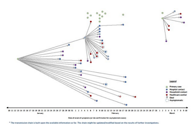
Investigations into the source of infection of the 61 cases found that 37 were health-care acquired infections, 14 were primary cases presumed to be infected from contact with dromedary camels and the remaining (10) infections occurred among close contacts outside of health care settings. As previously reported1, two human to human transmission amplification events took place at a hospital during this outbreak (one amplification event in the emergency department, and one amplification event in a cardiac intensive care unit; Figure 1).
Enlarge image

https://www.who.int/csr/don/24-April-2019-mers-saudi-arabia/en/

Figure 1. Transmission chain of laboratory-confirmed cases of MERS-CoV infection associated with Wadi Aldwasir city, Riyadh region, outbreak, 2019 (n=61)
The link below provides details of the 22 reported cases:

MERS-CoV cases reported from 14 February through 31 March 2019  xls, 116kb

From 2012 through 31 March 2019, a total of 2399 laboratory-confirmed cases of MERS-CoV and 827 associated deaths were reported globally to WHO under the International Health Regulations (IHR). The associated deaths reported to WHO were identified through follow-up with affected member states.
Public Health Response

As reported previously, the Saudi Arabian Ministry of Health (MoH) has conducted and completed a full-scale investigation of the MERS outbreak in Wadi Aldwasir including identification of all household and healthcare worker contacts of confirmed patients in all of the hospitals affected.

As of 31 March 2019, a total of 380 contacts have been identified, including 260 household contacts and 120 healthcare worker contacts. All identified contacts were monitored for 14 days from the last date of exposure as per WHO and national guidelines for MERS. All secondary cases have been reported to WHO.

Currently, all the listed contacts have been tested for MERS-CoV infection by reverse transcription polymerase chain reaction (RT-PCR) at least once and many contacts of known patients have been tested repeatedly. All secondary cases of MERS-CoV infection have been reported to WHO. The last case from Wadi Aldwasir was reported on 12 March 2019.

Within the affected health care facilities, infection prevention and control measures have been enhanced including intensive mandatory on-the-job training on infection control measures for all healthcare workers in emergency room and intensive care unit. Disinfection has been carried out in the emergency room and ICU of hospital A, which is fully operational and additional staff were mobilized to support infection control activities. Respiratory triage has been enforced in all healthcare facilities in the Riyadh region.

The MoH media department launched an awareness campaign targeting Wadi Aldawasir city with special focus on camel owners and camel related activities.

The Ministry of Agriculture is testing dromedaries in Wadi Aldwasir city and initial results have identified several PCR positive dromedaries in the city. Positive camels have been removed from the market and movement in and out of the camel market has been restricted. Camels owned by confirmed human cases were quarantined regardless of testing results. Full genome sequencing of available human and dromedary specimens have been conducted. Laboratory findings of camel testing by the Ministry of Agriculture have been reported to the World Organization for Animal Health (OIE).
WHO risk assessment

Infection with MERS-CoV can cause severe disease resulting in high morbidity and mortality. Humans are infected with MERS-CoV from direct or indirect contact with infected dromedary camels or by transmission between humans. So far, the observed non-sustained human-to-human transmission has occurred mainly in health care settings.

The notification of these additional cases does not change WHO’s overall risk assessment of MERS. WHO expects that additional cases of MERS will be reported from the Middle East, and that cases will continue to be exported to other countries by individuals who might acquire the infection after exposure to dromedary camels, dromedary camel animal products (for example, consumption of camel’s raw milk), or humans (for example, in a health care setting or household contacts).

WHO continues to monitor the epidemiological situation and conducts risk assessment based on the latest available information.
Results of the completed epidemiological investigation, as well as full genome sequencing of available dromedary and human specimens are being used by Ministry of Health officials to further evaluate the zoonotic and human-to-human transmission that has occurred in Wadi Aldwasir outbreak.
(Continue . . . )


Emer. Microb. & Inf.: Avian Flu Co-Infection in Poultry - Cambodia, 2017–2018

http://afludiary.blogspot.com/2018/02/who-genetic-characteristics-of-avian.html
Credit CDC


#14,030


Over the past 5 weeks two previously unreported avian flu subtypes -  HPAI H5N6 and LPAI H7N4 - have been detected and reported from Cambodia.
OIE: 1st occurrence of HPAI H5N6 in Cambodia

OIE: Multiple Outbreaks Of LPAI H7N4 In Cambodian Poultry
While surveillance and reporting from Cambodia is often - to put it kindly, suboptimal - in 2017 and 2018 multiple outbreaks of HPAI H5N1 were reported (see OIE Final Report) in poultry.
Cambodia hasn't reported a human avian flu case since 2014, but six years ago they saw a flurry of HPAI H5N1 human infections (see Cambodia’s H5N1 Surge & the `M’ Word).
In 2016, we looked at a seroprevalence study (see Sci Rpts: Intense Circulation Of A/H5N1 In Cambodian LBMs & Evidence Of Subclinical Human Infection) conducted during the height of the H5N1 human outbreaks in 2013 at four live bird markets in Cambodia.
Researchers not only found an incredibly high incidence of H5N1 infected poultry (35%), they found a plethora of other LPAI subtypes (HA1, HA2, HA3, HA4, HA6, HA7, HA9, HA10 and HA11) as well.
Seroprevalence studies also found evidence of sub-clinical H5N1 and H9N2 infection in LMB workers, and additional seroconversions were documented during the course of the 11 month study. 

The obvious concern with multiple co-circulating subtypes of avian flu is the potential for reassortment, and the generation of new, potentially dangerous, hybrid viruses.


While the avian flu world remained relatively stable from 1996 to 2013 - with HPAI H5N1 being our primary concern - over the past 6 years we've seen a spate of new reassortant viruses (H7N9, H5N6, H6N8, H10N8, H7N4, etc.) emerge - mostly from China or Southeast Asia.

All of which brings us to a research letter, published last week in Emerging Microbes & Infections, which describes recent AI surveillance results, and quantifies the level and types of co-infections, found in Cambodian poultry.

I've only included some excerpts from a much longer, more detailed letter. So follow the link to read it in its entirety. 
Avian influenza virus detection, temporality and co-infection in poultry in Cambodian border provinces, 2017–2018
Erik A. Karlsson , Srey Viseth Horm, Songha Tok, Sothyra Tum, Wantanee Kalpravidh, Filip Claes, show all
Pages 637-639 | Received 03 Feb 2019, Accepted 01 Apr 2019, Published online: 19 Apr 2019
Highly pathogenic avian influenza virus (AIV) has been endemic in Cambodia since 2004, and is a major agricultural and public health concern. Cambodia is a tropical, resource poor, lower-middle income country in Southeast Asia with a large socio-economic dependence on agriculture.

In 2015, 87% of Cambodian households with agricultural holdings raised poultry mainly on small, backyard farms with minimal biosafety and/or biosecurity. In conjunction with the National Animal Health and Production Institute (NaHPRI), Institut Pasteur du Cambodge (IPC) has maintained active longitudinal surveillance at key live bird markets (LBMs) in the heavily populated, southern part of the country.
Cambodian LBMs have high levels of AIV circulation, with 30–50% of ducks and 20–40% of chickens testing positive. Intense circulation of A/H5N1 and other avian influenza viruses in Cambodian live-bird markets with serological evidence of sub-clinical human infections.

Concerningly, a multitude of high and low pathogenic AIVs circulate concurrently. Previous studies suggest peak AIV circulation corresponds to the dry season (November to May) especially around Lunar New Year (LNY) celebrations when poultry consumption is highest.
        (SNIP)
Overall, 23.3% of the poultry samples screened were positive for AIV by RT-qPCR with 20.0% and 32.6% positivity in chickens and ducks, respectively. Percentages were similar for individual provinces (Supplemental Table 1). Longitudinally, total AIV detection fluctuated between 4.0% and 48.3%, with highest levels the week before or the week of festivals with increased poultry consumption.
Highest detection was associated with LNY followed closely by KNY (Figure 1(A)). Similar patterns were observed in individual provinces (Figure 1(B–D)). By subtype, 25.2%, 7.4%, 52.3%, 18.1% of AIV positive poultry samples were subtyped as A/H5, A/H7, A/H9, and “unknown,” respectively (Supplemental Table 1). 
        (SNIP)
Co-infections comprised 3.2% of all AIV positive samples (Figure 1(I)). The majority (86.7%) of co-infections were classified as A/H5 + A/H9; however, co-infections between A/H5 + A/H7 and A/H7 + A/H9 were also detected in 6.7% and 13.3% of total co-infections, respectively.
Prevalence of co-infection was similar between chickens (3.2%) and ducks (3.3%); however, co-infections in chickens were exclusively the A/H5 + A/H9 combination whereas ducks had a higher diversity with 50%, 16.7% and 33.3% of co-infections identified as A/H5 + A/H9, A/H5 + A/H7, and A/H7 + A/H9, respectively (Supplemental Figure 4(A,E)).
Co-infections were detected in both chickens (2.8%) and ducks (13.6%) in Kandal, but only in chickens (4.8%) in BM and only in ducks (2.7%) in Takeo (Supplemental Figure 4(B–D,F–H)). The greatest prevalence of co-infections were detected at week 11 of 2018 at 5.7% of total AIV positive samples (Supplemental Figure 4(I)).

As observed previously, A/H5 isolates that could be subtyped were also positive for neuraminidase (NA) subtype N1 by RT-qPCR. No novel H5Nx viruses were detected. A/H9 isolates were subtyped with N2 and A/H7 samples were identified with the N7 and N4 NA subtype by conventional PCR. All A/H7 samples are of the Eurasian lineage, and, to date, AIV similar to the A/Anhui/1/2013-lineage have not been detected in Cambodia. While some samples could not be typed due to low viral load, no positive samples were subtyped for N3, N6, N8 or N9 by RT-qPCR or conventional methods.

Overall, AIV continues to circulate within Cambodia at high levels as previously described, correlating to festival periods when poultry production and consumption is increased

However, a human infection has not been detected since 2014. Border regions display variable AIV prevalence and diversity, possibly due to poultry movement across borders. While A/H7 was detected previously in Cambodia, subtype A/H7N4 presents concern due to the human case in nearby China at a similar time period.

In addition, detection of co-infections in 3.2% of AIV positive poultry, especially with A/H9, raises concerns about reassortment and emergence of novel viruses with epizootic or pandemic potential.
Isolation, Whole Genome Sequencing, and unknown subtype determination is on-going to further characterize these viruses on a molecular and phylogenetic level. Continued, active, vigilant surveillance is vital and interventions to decrease the prevalence of AIVs in LBMs should be considered, especially during festival periods.
        (Continue . . . )

   
Note: The author's concerns about A/H7N4 in Cambodia were confirmed a month ago (see OIE announcement)

Not unexpectedly, over 90% of the co-infections reported involved the highly promiscuous H9N2 virus, which - while it has some pandemic potential on its own (see CDC IRAT Score) - is more infamous for lending its internal genes to many of the HPAI viruses of greatest concern today (see The Lancet's Poultry carrying H9N2 act as incubators for novel human avian influenza viruses).

While reassortments can occur anytime and anywhere - in any host (avian, swine, human, etc.) able to be co-infected with influenza A viruses - we normally look to China as being the most likely region to produce new HPAI viruses (see Viral Reassortants: Rocking The Cradle Of Influenza). 
That changed - at least temporarily - 18 months ago following their highly successful H5+H7 Nationwide poultry vaccination campaign, which has greatly suppressed avian flu activity across China for more than a year. 
Which makes increased surveillance in other locations - particularly in Southeast Asia - a high priority.