Showing posts with label Lungs. Show all posts

Wednesday, October 09, 2013

mBio: H7N9 Naturally Adapted For Efficient Growth in Human Lung Tissue

image


# 7844


Although we’ve not seen a human case of H7N9 in more than two months, the results of research over the summer continues to point towards this emerging avian influenza virus as being unusually well-adapted to mammals.  A few examples include:


Nature: Limited Airborne Transmission Of H7N9 Between Ferrets
BMJ: `Probable Person-to-Person Transmission’ Of H7N9
Lancet: Tropism Of H7N9 In the Human Respiratory Tract
Science: H7N9 Transmissibility Study In Ferrets


Yesterday the open-access journal mBio published another study,  where researchers conducted in vitro studies – using human (A549) lung tissue and canine (MDCK) Kidney cells – on an H7N9 isolate (A/Anhui/1/2013) collected from a fatal human infection, and two low-pathogenic avian H7 subtype viruses.


Researchers evaluated and compared the replication, tropism, and cytokine induction of all three viruses. They found  the A/Anhui/1/2013 (H7N9) virus was already remarkably well adapted to replicate and infect human lung tissue. 

Additionally, A/H7N9’s NS1 protein suppressed (compared to the other H7 viruses tested) the natural cellular production of beta interferon (IFN-β), an antiviral produced in response to the presence of pathogens like viruses and bacteria.


First, a link to the study, and some excerpts, after which I’ll have more.


The Novel Human Influenza A(H7N9) Virus Is Naturally Adapted to Efficient Growth in Human Lung Tissue

Jessica Knepper, Kristina L. Schierhorn, Anne Becher, Matthias Budt, Mario Tönnies, Torsten T. Bauer, Paul Schneider, Jens Neudecker, Jens C. Rückert, Achim D. Gruber, Norbert Suttorp, Brunhilde Schweiger, Stefan Hippenstiel, Andreas C. Hocke, Thorsten Wolff

(EXCERPTS)

ABSTRACT

. . .  The A(H7N9) patient isolate replicated similarly well as a seasonal IAV in explanted human lung tissue, whereas avian H7 subtype viruses propagated poorly. Interestingly, the avian H7 strains provoked a strong antiviral type I interferon (IFN-I) response, whereas the A(H7N9) virus induced only low IFN levels. Nevertheless, all viruses analyzed were detected predominantly in type II pneumocytes, indicating that the A(H7N9) virus does not differ in its cellular tropism from other avian or human influenza viruses. Tissue culture-based studies suggested that the low induction of the IFN-β promoter correlated with an efficient suppression by the viral NS1 protein. These findings demonstrate that the zoonotic A(H7N9) virus is unusually well adapted to efficient propagation in human alveolar tissue, which most likely contributes to the severity of lower respiratory tract disease seen in many patients.

IMPORTANCE Humans are usually not infected by avian influenza A viruses (IAV), but this large group of viruses contributes to the emergence of human pandemic strains. Transmission of virulent avian IAV to humans is therefore an alarming event that requires assessment of the biology as well as pathogenic and pandemic potentials of the viruses in clinically relevant models. Here, we demonstrate that an early virus isolate from the recent A(H7N9) outbreak in Eastern China replicated as efficiently as human-adapted IAV in explanted human lung tissue, whereas avian H7 subtype viruses were unable to propagate. Robust replication of the H7N9 strain correlated with a low induction of antiviral beta interferon (IFN-β), and cell-based studies indicated that this is due to efficient suppression of the IFN response by the viral NS1 protein. Thus, explanted human lung tissue appears to be a useful experimental model to explore the determinants facilitating cross-species transmission of the H7N9 virus to humans.

(Continue . . . )


For more detail, including methods and materials, you will want to read the entire study.


While scientists know there are many types of viruses that could potentially cause a pandemic, influenza viruses – due to their ability to spread rapidly (including from carriers who are pre-symptomatic or asymptomatic), and to cause severe disease  – are the ones that keep most researchers up at night. They have a long history of causing human misery and death.


Although the news of new cases in China has been absent since early August, concerns run high that the virus will reappear this winter, or in the spring (see  FAO Warns On Bird Flu).  With temperatures dropping, and respiratory infections rising, the Chinese public health authority has already stepped up hospital surveillance for this virus (see Chinese CDC: Be Alert For H7N9).


Last week, just before the government shutdown, the CDC issued two new guidance docs on H7N9 (see H7N9: CDC Guidance On Antiviral Chemoprophylaxis & H7N9: Updated CDC Guidance For Antiviral Treatment).


The saving grace with H7N9, at least so far, is that it hasn’t demonstrated the ability spread efficiently in the human population.


Last week, in BMC: Estimating The Transmission Potential Of H7N9, we saw an encouraging study that found the virus’s R0 (R naught) or Basic Reproductive Number, was well below 1.0 last spring,  suggesting this virus doesn’t yet possess enough `legs’ to spark a major epidemic.


The concern, of course, is that influenza viruses are constantly changing, and so what was true about the virus last spring may not hold true this winter or next spring.


So we watch and we prepare (see Pandemic Preparedness: Taking Our Cue From The Experts) – not because we are convinced that H7N9 will spark the next pandemic.  It could, after all , fade back into the woodwork or simply simmer ominously for years like its H5 cousin.


We pay attention because – just like with MERS-CoV and H5N1 -  we know that it could.

Saturday, August 17, 2013

The Cytokine Storm Revisited

image

Credit CDC


# 7577


Cytokines are a category of signaling molecules that are used extensively in cellular communication. They are often released by immune cells that have encountered a pathogen, and are designed to alert and activate other immune cells to join in the fight against the invading pathogen.


This cascade of immune cells rushing to the infection, if it races out of control, can overwhelm the patient. Their lungs can fill with fluid (which makes a terrific medium for a bacterial co-infection), and cells in the lungs (Type 1 & Type II Pneumocytes) can sustain severe damage.


Previously, in Swine Flu Sequelae and Cytokine Storm Warnings, we looked at some of the severe lung damage detected during the 2009 pandemic that was thought to be due to this overreaction of the immune system.


Last night Robert Roos, Editor of CIDRAP NEWS, wrote about a study that appeared this week in the American Journal of Pathology that looked at the lungs from 50 fatal cases of H1N1 during the 2009 pandemic, and their finding of "remarkably" high levels of cytokines in the lung tissue.


Robert does a terrific job explaining this research, so I’ll invite you to read his article at the link below. When you return, I’ll have some more background on the cytokine response.


Study shows cytokine storm in fatal 2009 H1N1 cases

Aug 16, 2013

Researchers who studied lung tissue samples from 50 people who died of pandemic H1N1 (pH1N1) influenza infections in 2009 say they found clear evidence that the intense immune response known as a cytokine storm played a role in their demise.


The scientists found that the peak levels of virus in the victims' lungs correlated with "remarkably" high levels of certain cytokines in the same tissues, according to their report, which was released ahead of print this week in the American Journal of Pathology.

(Continue . . . )


The abstract to the study is available at:

Cytokine and Chemokine Profiles in Lung Tissues from Fatal Cases of 2009 Pandemic Influenza A (H1N1

Role of the Host Immune Response in Pathogenesis

Rongbao Gao, Julu Bhatnagar, Dianna Blau, Patricia Greer, Dominique C. Rollin, Amy M. Denison, Marlene Deleon-Carnes, Wun-Ju Shieh, Suryaprakash Sambhara, Terrence M. Tumpey, Mitesh Patel, Lindy Liu, Christopher Paddock, Clifton Drew, Yuelong Shu, Jacqueline M. Katz, Sherif R. Zaki


While influenza can strike people of any age, it generally exacts its greatest toll on the elderly – those over the age of 65 - whose weaker immune systems (and comorbidities) can render them less able to fight off an infection.


Exact numbers are unknown, since influenza is only rarely cited as the primary cause of death. If a cause of death (beyond`natural causes’) is given, comorbidities like COPD, heart disease, asthma are far more likely to listed on a death certificate.


Still, estimates are that 90% of seasonal flu mortality occurs in those over the age of 65 (cite CDC Pink book).


In 2010, (see Study: Years Of Life Lost Due To 2009 Pandemic), researchers estimated the median age of death due to seasonal influenza-related illness in the United States to be 76.


In contrast, pandemic influenza strains, at least during the first few years after their introduction, often produce a dramatic `age shift’ downward in mortality. 


The CDC’s estimate of average and median age of death due to the 2009 Pandemic virus reads:


Based on two CDC investigations of confirmed 2009 H1N1-related deaths that occurred during the spring and fall of 2009, the average age of people in the U.S. who died from 2009 H1N1 from April to July of 2009 was 40. The median age of death for this time period was 43. From September to October of 2009, the average age of people in the U.S. who died from 2009 H1N1 was 41, and the median age was 45.


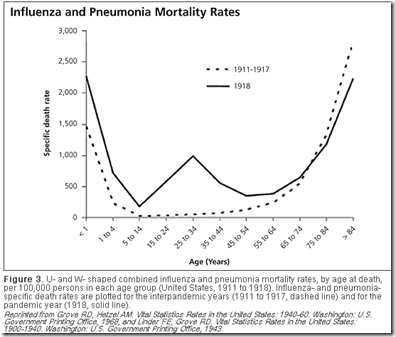
An even more pronounced `age shift’ was observed during the 1918 Spanish Flu, which exacted its heaviest toll among those aged 25 to 34 – an age group that would normally be expected to weather the illness better than any other.


image

The infamous `W shaped curve’ of the 1918 pandemic clearly shows that the death rates among those in their teens, 20s, and 30s was much higher than was normally seen in previous influenza years. Those over the age of 65, however, saw a reduction in mortality during the pandemic.


Similarly, the H5N1 avian flu virus has shown a disturbing predilection for younger victims, as illustrated by the following chart showing the ages of H5N1 cases in China over the past decade (see WHO Perspective Human infections with avian influenza A(H7N9) virus in China: preliminary assessments of the age and sex distribution).

.

image


Although not universally embraced, the Cytokine Storm theory has been adopted by many researchers as the most likely reason why the young, and healthy – those with presumably the most robust immune systems – would suffer disproportionately with some pandemic flu strains.


Somewhat surprisingly, we haven’t seen this age shift during the first wave of H7N9 avian flu cases in China this spring.  We’ll have to see if this trend continues if, and when, the virus re-emerges.


image


As you can imagine, many researchers and doctors are looking for effective ways to safely dampen runaway immune responses as a possible treatment for pandemic influenza.


This is particularly important as most of the world would have little hope of seeing a vaccine for a pandemic flu during the first year or two of its arrival.


Dr. David Fedson – former Professor of Medicine at the University of Virginia School of Medicine and formerly Director of Medical Affairs, Aventis Pasteur MSD - has long championed the idea that we should be looking at statins for pandemic flu, which he believes may help modulate the immune response.


If they can be proved effective, statins have the advantage of being cheap, easy to manufacture and distribute, and have relatively few side effects.


The problem is, while some studies on statins and pneumonia have yielded promising results, not all of the research is in agreement. Complicating matters, since many statins are now generic, there is little financial incentive for drug companies to fund expensive research.


You can read about the potential for statin therapy in the blogs below.


Study: Statins, Influenza, & Mortality

Another Study On Statins And Pneumonia

Dr. David Fedson: The Case For Using Statins In A Pandemic

Statins Revisited


There are other approaches under investigation as well. 


In 2011, in Study: Calming The Cytokine Storm, we looked at research from The Scripps Research Institute  that found a protein located on the surface of endothelial cells, called S1P1, to be largely responsible for flu-associated cytokine storms.


While a specific drug to target this protein is likely a long way off, their discovery provides new avenues for research into dampening the cytokine response.


This latest study in the American Journal of Pathology provides some of the strongest evidence we’ve seen on the impact of cytokine production in the pathogenesis of influenza.


Results that will likely spur additional interest in finding ways to moderate the cytokine storm.


For a better understanding of the pathogenesis of influenza, the innate immune system, and the role of cytokines I would invite you to read my 3-part look at the Baskin study from 2009.

Dissecting the Influenza Pathogenesis Study Pt. 1

Dissecting the Influenza Pathogenesis Study Pt. 2

Dissecting the Influenza Pathogenesis Study Pt.3

Tuesday, September 20, 2011

mBio: Lethal Synergism of H1N1 Pandemic Influenza & Bacterial Pneumonia


PHIL Image 2111

CDC PHIL - Photomicrograph of Streptococcus (Diplococcus) pneumoniae bacteria


# 5856


While the vast majority of people who contracted the H1N1 pandemic flu of 2009 recovered without incident, a very small minority saw severe – sometimes fatal – illness. 


Often during 2009 we saw reports of severe lung damage. Damage that in some cases was compared to what has been seen in H5N1 bird flu and during the great pandemic of pandemic of 1918.


A few of the stories from back then include:


In early September of 2009, in Pathology Of Fatal H1N1 Lung Infections, we looked at a report by Helen Branswell that looked early autopsy results.


Lung damage in fatal swine flu cases more bird flu than seasonal flu: expert

By Helen Branswell Medical Reporter (CP) 

TORONTO — The lungs of people who have died from swine flu look more like those of the victims of H5N1 avian influenza than those of people who succumb to regular flu, the chief of infectious diseases pathology at the U.S. Centers for Disease Control says.


Study of about 70 fatal H1N1 cases so far also reveals there may be more incidences of co-infections with bacteria than was earlier thought, Dr. Sherif Zaki told The Canadian Press in an interview.


A couple of weeks later in More On The Pathology Of Novel H1N1, we saw a report by Maggie Fox, then Health and Science Editor for Reuters, who brought us more details of this  story, including comments by Dr. Sherif Zaki of the U.S. CDC who  stated that "This is almost exactly what we see with avian flu. This looks like avian flu on steroids."


That same month, I wrote about the use of ECMO (Extracorporeal Membrane Oxygenation) in the treatment of severe lung injury in H1N1 victims in The ECMO Option.


In early December (see NIH: Post Mortem Studies Of H1N1) the NIH announced the results of a series of autopsies conducted on H1N1 victims in New York City over the summer, which are chronicled in the Archives of Pathology & Laboratory Medicine.


The NIH put together a press release, which provided highlights of the study.


FOR IMMEDIATE RELEASE
Monday, Dec. 7, 2009

Media Contact: Anne A. Oplinger
(301) 402-1663
niaidnews@niaid.nih.gov

New York Autopsies Show 2009 H1N1 Influenza Virus Damages Entire Airway

In fatal cases of 2009 H1N1 influenza, the virus can damage cells throughout the respiratory airway, much like the viruses that caused the 1918 and 1957 influenza pandemics, report researchers from the National Institutes of Health (NIH) and the New York City Office of Chief Medical Examiner. The scientists reviewed autopsy reports, hospital records and other clinical data from 34 people who died of 2009 H1N1 influenza infection between May 15 and July 9, 2009. All but two of the deaths occurred in New York City. A microscopic examination of tissues throughout the airways revealed that the virus caused damage primarily to the upper airway—the trachea and bronchial tubes—but tissue damage in the lower airway, including deep in the lungs, was present as well. Evidence of secondary bacterial infection was seen in more than half of the victims.


The team was led by James R. Gill, M.D., of the New York City Office of Chief Medical Examiner and New York University School of Medicine, and Jeffery K. Taubenberger, M.D., Ph.D., of the National Institute of Allergy and Infectious Diseases (NIAID) at NIH. The findings are reported in the Archives of Pathology & Laboratory Medicine, now available online and scheduled to appear in the February 2010 print issue.

<SNIP>

This pattern of pathology in the airway tissues is similar to that reported in autopsy findings of victims of both the 1918 and 1957 influenza pandemics,” notes Dr. Taubenberger.


While many people continued to insist that swine flu was no worse than seasonal flu, obviously something was different in the way it produced severe lung damage.  


A year into the pandemic, I summarized many of the ways that the 2009 H1N1 virus differed from seasonal flu in There’s No Flu Like A New Flu.


While the overall incidence of these complications was relatively low, those who suffered from them often experienced extremely severe illness.


All of which serves as prelude to an open access study, published today in mBio, called:


Lethal Synergism of 2009 Pandemic H1N1 Influenza Virus and Streptococcus pneumoniae Coinfection Is Associated with Loss of Murine Lung Repair Responses

John C. Kasha, Kathie-Anne Waltersb, A. Sally Davisa, Aline Sandouka, Louis M. Schwartzmana, Brett W. Jaggera, Daniel S. Chertowa, Qi Lia, Rolf E. Kuestnerb, Adrian Ozinskyb, and Jeffery K. Taubenbergera


The entire study is available, and is well worth reading, but briefly:


Scientists at NIAID and the Institute for Systems Biology (ISB) infected experimental mice with both seasonal flu and the 2009 H1N1 pandemic flu, and after 48 hours exposed some of them to Streptococcus pneumoniae, one of the main causes of pneumonia.


Mice that were exposed only to the two flu strains showed expected flu symptoms, but all survived.


Mice that were exposed to seasonal flu and S. pneumoniae experienced minor lung damage, but once again, all survived.


But all of the mice infected with the pandemic H1N1 virus, and S. pneumoniae showed severe weight loss, lung damage, and 100% mortality


Excerpts from the press release below explain what else they found:


American Society for Microbiology


2009 H1N1 pandemic flu more damaging to lungs, opens opportunities for bacterial infection

(EXCERPT)

The lung tissues of the dead mice revealed that the alveoli were severely inflamed and the surfaces of the bronchioles were wiped clean of the protective layer of cells called the epithelium. There was also increased bacterial replication in the lungs of the co-infected mice, a sign that the bacteria were thriving there.


Looking at the mouse genes that were expressed during infection revealed more details about how the pandemic influenza virus sets the stage for lethal bacterial infections. Mice infected with the pandemic flu virus and S. pneumoniae had a similar inflammatory response as the other mice, but they lack responses that would repair and regenerate their damaged epithelial cells, those protective tissues that would otherwise keep bacteria from penetrating to deeper layers of tissue.


All these factors add up to big problems in the lung: as compared with seasonal flu, infection with the pandemic strain of flu was associated with more extensive damage to the epithelium that requires more extensive tissue repair. This opens the body up to attack from bacterial invaders, including Streptococcus pneumoniae.

(Continue . . . )


So not only did this duel infection lead to greater lung damage, and increased bacterial replication, it also disabled the lung’s ability to repair itself.


Since it can take 6 months or longer to develop a vaccine for a novel influenza virus, these results may suggest a bigger role for the 23-valent Pneumonia vaccine (PPVSV) during a future pandemic. 


More than a year after the end of the 2009 pandemic, scientists are still uncovering basic information about how pandemic flu differs from seasonal flu. 


With luck, work like this will provide better ways for us to deal with an outbreak, when the next one arrives.