Wednesday, February 28, 2018

J.I.D.: NIAID's Strategic Plan To Develop A Universal Flu Vaccine

Credit NIAID


#13,180


This year's flu season has once again highlighted the pressing need for a better flu vaccine - one that isn't easily foiled by subtle changes in the virus or stymied by 60 year old egg-propagation technologies.

While progress has been made (cell culture-propagated vaccines, adjuvanted and high dose vaccines), today's vaccines far too often provide only modest protection against a virus that kills tens of thousands of Americans every year.
The holy grail - dubbed a `universal' vaccine - it is often fancifully described in the popular press as being a `one time (or every few years) shot' that would convey nearly full protection against all flu subtypes. 
The reality is, while that would be optimal, the criteria (see above) for a `universal' vaccine is a bit more realistic;  a 75%+ VE (Vaccine Effectiveness) across multiple (seasonal & novel) subtypes that would last at least a year.
Despite these reduced goals, the road to a universal flu shot is a long one, and success - at least in the near term - is far from guaranteed.  
Today the NIH's NIAID ( National Institute of Allergy and Infectious Diseases) unveils their strategic plan to accomplish this goal in a major article published in the Journal of Infectious Diseases and in a press statement.

A Universal Influenza Vaccine: The Strategic Plan for the National Institute of Allergy and Infectious Diseases

Emily Erbelding, M.D., M.P.H Catharine Irene Paules, M.D Alison Deckhut Augustine, PhD Stacy Ferguson, PhD Anthony S Fauci, M.D Paul C Roberts, Ph.D Diane Post, Ph.D Barney S Graham, MD, PhD Erik J Stemmy, Ph.D

The Journal of Infectious Diseases, jiy103, https://doi.org/10.1093/infdis/jiy103
Published: 28 February 2018

Views
PDF  
Abstract

A priority for the National Institute of Allergy and Infectious Diseases (NIAID) is development of an influenza vaccine providing durable protection against multiple influenza strains, including those that may cause a pandemic, i.e., a universal influenza vaccine. To invigorate research efforts, NIAID developed a strategic plan focused on knowledge gaps in three major research areas, as well as additional resources required to ensure progress towards a universal influenza vaccine. NIAID will use this plan as a foundation for future investments in influenza research and will support and coordinate a consortium of multidisciplinary scientists focused on accelerating progress towards this goal.
        (Continue . . . )


Media Advisory
Wednesday, February 28, 2018
NIAID unveils strategic plan for developing a universal influenza vaccine

WHAT

Developing a universal influenza vaccine — a vaccine that can provide durable protection for all age groups against multiple influenza strains, including those that might cause a pandemic — is a priority for the National Institute of Allergy and Infectious Diseases (NIAID), part of the National Institutes of Health. Writing in the Journal of Infectious Diseases, NIAID officials detail the Institute’s new strategic plan for addressing the research areas essential to creating a safe and effective universal influenza vaccine. They describe the scientific goals that will be supported to advance influenza vaccine development. The strategic plan builds upon a workshop NIAID convened in June 2017 that gathered scientists from academia, industry and government who developed criteria for defining a universal influenza vaccine, identified knowledge gaps, and delineated research strategies for addressing those gaps.  

The cornerstone of both seasonal and pandemic influenza prevention and control is the development of vaccines against specific influenza strains that pose a potentially significant risk to the public. Seasonal influenza vaccines are made anew each year to best match the strains projected to circulate in the upcoming season. However, this approach has limitations and difficulties. To reduce the public health consequences of both seasonal and pandemic influenza, vaccines must be more broadly and durably protective. Advances in influenza virology, immunology and vaccinology make the development of a universal influenza vaccine more feasible than a decade ago, according to the authors. To develop a universal influenza vaccine, NIAID will focus resources on three key areas of influenza research: improving the understanding of the transmission, natural history and pathogenesis of influenza infection; precisely characterizing how protective influenza immunity occurs and how to tailor vaccination responses to achieve it; and supporting the rational design of universal influenza vaccines, including designing new immunogens and adjuvants to boost immunity and extend the duration of protection.

The authors state that a coordinated effort of guided discovery, facilitated product development and managed progress through iterative clinical testing will be critical to achieving the goal of a universal influenza vaccine. NIAID will establish and support a consortium of scientists to meet designated goals for a universal influenza vaccine and will expand the Institute’s research resources by establishing long-term human cohorts, supporting improved animal models of influenza infection and expanding capacity for conducting human challenge studies.

The authors emphasize that broad collaboration and coordination in many disciplines and involving government, academia, philanthropies and the private sector will be vital to achieving the goal of developing a universal influenza vaccine. NIAID intends for the plan to serve as the foundation for its research investment strategy to achieve this important public health goal.
(Continue . . . )



Study: Obesity & Influenza-Like-Illness (ILI) Severity


#13,179


Early reports during the 2009 H1N1 pandemic (see H1N1 Morbidity And Previously Existing Conditions) cited unusual numbers of obese influenza patients - a previously unrecognized risk group - populating intensive care facilities around the world. 
Over the next couple of months obesity, and particularly morbid obesity, was frequently mentioned as a possible risk factor, along with asthma, diabetes, and immune disorders.   
That is, until an ACIP meeting held at the end of July, where evidence was presented that showed that the incidence of hospitalizations among those listed as obese by their BMI was practically the same as their prevalence in society.
Roughly 34% of Americans are obese, and roughly 38% of those hospitalized met that criteria.  While 6% are morbidly obese (BMI > 40), they only made up 7% of the hospitalized cases.
Since then, the debate has raged on, with some studies showing an elevated risk of severe influenza, hospitalization, and death among those who are morbidly obese, while other studies failing to find such evidence. 
Alas, science isn’t always neat, tidy, consistent or clear (see When Studies Collide (Revisited)).
In September of 2009, in Study: Half Of ICU H1N1 Patients Without Underlying Conditions, it became apparent that while pre-existing risk factors were important, they were not the sole reason behind flu victims ending up in intensive care.

In November of that year, Eurosurveillance Journal  published a Study: H1N1 Hospitalization Profiles, that similarly found :
  • The most common risk factor in admission to intensive care was chronic respiratory disease followed by chronic neurological disease, asthma and severe obesity.
  • 51% of hospitalized cases and 42% of ICU cases were not in a recognized risk group.
Over time, while studies were far from consistent, the tide began to turn, with morbid obesity increasingly cited as a risk factor for severe influenza.

In early 2011 (see Extreme Obesity: A Novel Risk Factor For A Novel Flu) the IDSA’s journal Clinical Infectious Diseases carried a study called  A Novel Risk Factor for a Novel Virus: Obesity and 2009 Pandemic Influenza A (H1N1), that found:
Extreme obesity associated with higher risk of death for 2009 H1N1 patients
[EMBARGOED FOR JAN. 5, 2011] For those infected with the 2009 pandemic influenza A (H1N1) virus, extreme obesity was a powerful risk factor for death, according to an analysis of a public health surveillance database.
In a study to be published in the February 1, 2011, issue of Clinical Infectious Diseases, researchers associated extreme obesity with a nearly three-fold increased odds of death from 2009 H1N1 influenza. Half of Californians greater than 20 years of age hospitalized with 2009 H1N1 were obese. 
 For the last few years the CDC has cited `people with extreme obesity (i.e., body-mass index is equal to or greater than 40)' on their list of High Risk groups for severe influenza.
Today, via Nature, we have a pre-release abstract from the International Journal of Obesity, whose findings I suspect are going to be viewed as controversial. 
Over a four year (2010-2014) study of ER visits for influenza-like-illness (ILI), they found that obesity was not linked to a greater risk of hospitalization - in fact - they found the opposite was true. 
Epidemiology and population health
Is weight associated with severity of acute respiratory illness?
Elizabeth E. Halvorson,Timothy R. Peters, Joseph A. Skelton, Cynthia Suerken, Beverly M. Snively & Katherine A. Poehling

International Journal of Obesity (2018)
doi:10.1038/s41366-018-0044-y
Published online: 27 February 2018

Abstract

Background/objectives

Obesity was an independent risk factor for severe disease in hospitalized adults during the 2009 pandemic H1N1 influenza season. Few studies have investigated the association between weight and severity of acute respiratory illnesses in children or in adults seeking care in the emergency department (ED) during other winter respiratory seasons.
Subjects/methods

We prospectively and systematically enrolled patients ≥2 years of age who presented to the ED or inpatient setting in a single geographic region with fever/acute respiratory illness over four consecutive winter respiratory seasons (2010–2014).
We collected demography, height and weight, and high risk co-morbid conditions. Multivariable logistic regression was used for prediction of hospital admission (primary outcome), length of stay and supplemental oxygen requirement among those hospitalized, and antibiotic prescription (secondary outcomes).
Results

We enrolled 3560 patients (N = 749 children, 2811 adults), 1405 (39%) with normal weight, 860 (24%) with overweight, and 1295 (36%) with obesity. Following multivariable logistic regression, very young or very old age (p < 0.001) and high-risk conditions (p < 0.001) predicted hospitalization.
Risk of hospitalization was decreased for adults with overweight [aOR 0.8 (95% CI 0.6–1.0)], class 1 obesity [aOR 0.7 (95% CI 0.5–1.0)], and class 2 obesity [aOR 0.6 (95% CI 0.4–0.8)] compared to normal-weight. Class 3 obesity was associated with supplemental oxygen requirement in adults [aOR 1.6 (95% CI 1.1–2.5)]. No association was seen in children.
Conclusion

Overweight and obesity were not associated with increased risk of hospitalization during winter respiratory seasons in children or adults.
While the results are interesting, this is but one study done in a single location, and was conducted after the 2009 H1N1 pandemic.  It uses `fever/acute respiratory illness' as a benchmark, and not lab-confirmed influenza. 
Being personally somewhat `gravitationally challenged', I'd very much like this to be true.
But until these results can be fully examined, and replicated by others, the best we can say right now is  . . .  of all of the studies on obesity as a risk factor for severe influenza . . .  this is without a doubt, the most recent.     

WHO Statement & Nigerian CDC Update On Lassa Fever Epidemic

http://www.ncdc.gov.ng/themes/common/files/sitreps/7b122e4047446980c5e4755437e5bee2.pdf


#13,178

As we discussed last week in Nigeria's Lassa Fever Outbreak - Epi Week 7, since just after the 1st of the year, Nigeria has seen a nearly  ten-fold increase in the number of Lassa Fever cases. 
Although endemic in West Africa, between 2013 and early 2016 Nigeria had seen a steady decline in the number of Lassa Fever cases  - and deaths - with the last significant outbreak reported in 2012.
In early 2016 that trend began to change with outbreaks starting in Nigeria (see Nigeria: Lassa Fever Outbreak With 40 Fatalities), and then flaring up in both Benin and Togo (see ECDC: Rapid Risk Assessment On Lassa Fever In Nigeria, Benin, Togo, Germany & USA) but leveling out again for the second half of 2017.


Commonly carried by multimammate rats - a local rodent that often likes to enter human dwellings - Lassa fever exposure is typically via the urine or dried feces of infected rodents, although person-to-person transmission may occur with exposure to the blood, tissue, secretions, or excretions of an infected individual (cite CDC).

First a few excerpts from this week's Nigerian CDC Epidemiological Report, followed by a statement released today by the World Health Organization.


http://www.ncdc.gov.ng/themes/common/files/sitreps/7b122e4047446980c5e4755437e5bee2.pdf

HIGHLIGHTS
  • In the reporting Week 08 (February 19-25,2018) fifty four new confirmediI cases were recorded from eight States Edo (21), Ondo (9), Nasarawa (2), Ebonyi (18), Plateau(1),Kogi(1) Imo (1)and Ekiti (1) with ten new deaths in confirmed cases from five states Ondo (2), Edo (2), Plateau (1) Ekiti(1) and Ebonyi (4 and 2 probable deaths)
  • From 1st January to 25th February 2018, a total of 1081 suspectedi cases, and 90 deaths have been reported from 18 activeiv States- (Edo, Ondo, Bauchi, Nasarawa, Ebonyi, Anambra, Benue, Kogi, Imo, Plateau, Lagos, Taraba, Delta, Osun, Rivers, FCT, Gombe and Ekiti) - Figure 1
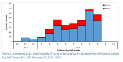
  • Since the onset of the 2018, 325 cases have been classified as: 317 confirmed cases, 8 probable cases with 72 deaths (64 in Lab confirmed and 8 in probable)
  • Case Fatality Rate in confirmed and probable cases is 22%
  • Fourteen Health Care workers have been affected in six states –Ebonyi (7), Nasarawa (1), Kogi (1), Benue (1), Ondo (1) and Edo (3) with four deaths in Ebonyi (3) and Kogi (1)
  • Predominant age group affected is age group 21-40 (Median Age = 34) - Figure 5
  • The male to female ratio for confirmed cases is 2:1
  • 69% of all confirmed cases are from Edo (43%) and Ondo (26%) states
  • National RRT team (NCDC staff and NFELTP residents) batch B continues response support in Ebonyi, Ondo and Edo States
  • Irrua Specialist Hospital has 42 cases on admission this weekend. FMC Owo has 21 isolation beds, all occupied
  • A total of 2845 contacts have been identified from 18 active states and 1897 are currently being followed up
  • WHO scaling up its support of the response at National and State levels
  • NCDC is collaborating with ALIMA and MSF in Edo, Ondo and Anambra States to support case management
  • NCDC deployed teams to four Benin Republic border states (Kebbi, Kwara, Niger and Oyo) for enhanced surveillance activities
  • National Lassa fever multi-partner multi-agency Emergency Operations Centre(EOC) continues to coordinate the response activities at all levels

This statement from http://www.afro.who.int/

Abuja, Nigeria, 28 February 2018 — Nigeria’s Lassa fever outbreak has reached record highs with 317 laboratory confirmed cases, according to figures released by the Nigeria Centre for Disease Control (NCDC) this week.
Although endemic to the West African nation, Lassa fever has never reached this case count in Nigeria before. The number of confirmed cases during the past two months exceeds the total number of confirmed cases reported in 2017.
The outbreak has affected 18 states since the first case was detected on 1 January 2018, resulting in 72 deaths caused by the acute viral haemorrhagic fever. A total of 2,845 people who have come into contact with patients have been identified and are being monitored.
The World Health Organization is supporting the NCDC-led response with a focus on strengthening coordination (including through the Global Outbreak Alert and Response Network), surveillance, contact tracing, laboratory testing, clinical management of patients, and community engagement. State health authorities are mobilizing doctors and nurses to work in Lassa fever treatment centres. 
“The ability to rapidly detect cases of infection in the community and refer them early for treatment improves patients’ chances of survival and is critical to this response,” said Dr Wondimagegnehu Alemu, WHO Representative to Nigeria.
Health facilities are particularly overstretched in the southern states of Edo, Ondo and Ebonyi. WHO is working with health authorities, national reference hospitals and the Alliance for International Medical Action (ALIMA) to rapidly expand treatment centres and better equip them to provide patient care while reducing the risks to staff. Among those infected are 14 health workers, four of whom have died. 
“Given the large number of states affected, many people will seek treatment in health facilities that are not appropriately prepared to care for Lassa fever patients and the risk of infection to healthcare workers is likely to increase,” said Dr Alemu.
Health workers are being trained in infection, prevention and control measures, such as the importance of wearing personal protective equipment (PPE) and isolating patients during treatment. WHO has provided an initial supply of PPE, other related materials and is assessing additional needs with a view to addressing them.
WHO is also supporting national response efforts in neighbouring Benin, where more than 20 suspected cases have been reported.

While primarily a regional threat, the incubation period of Lassa fever can run up to 21 days, and we've seen exported cases before. In 2014, in Minnesota: Rare Imported Case Of Lassa Fever, we saw the CDC's investigation into the 7th known imported case of Lassa into the United States.
Most of the earlier cases had been diagnosed abroad and the patients were then airlifted to the US for treatment, but two previous cases (2004 in New Jersey (MMWR) & 2010 in Pennsylvania (EID Journal)) involved travelers who arrived in the US without knowing they had been infected.
Again, in 2016, exported cases turned up in several countries, including Germany and Sweden (see Germany's RKI Statement On Lassa Fever Cluster In Cologne & WHO Lassa Fever Update - Sweden (Imported)).

In 2016 the ECDC published a Rapid Risk Assessment on the spread of Lassa Fever out of  West Africa.  While the risk of seeing Lassa Fever outside of West Africa was determined to be low, they authors wrote:
The two imported cases of Lassa fever recently reported from Togo indicate a geographical spread of the disease to areas where it had not been recognised previously. Delays in the identification of viral haemorrhagic fevers pose a risk to healthcare facilities.
Therefore, Lassa fever should be considered for any patient presenting with suggestive symptoms originating from West African countries (from Guinea to Nigeria) particularly during the dry season (November to May), a period of increased transmission, and even if a differential diagnosis such as malaria, dengue or yellow fever is laboratory-confirmed.
If you count the unprecedented West African Ebola epidemic in 2014, and last year's record setting Monkeypox outbreak in Nigeria, this year's record surge in Lassa Fever cases marks the third outbreak `anomaly' in the region in recent years.
While the world has been relatively lucky so far, and these outbreaks have been largely contained, that luck may not hold forever.
A reminder that in this increasingly interconnected and mobile world that localized outbreaks - no matter how remote - aren't guaranteed to remain such, and that without a proactive response they can very quickly turn into public health threats anywhere in the world.


Tuesday, February 27, 2018

Bulgarian NVS: Confirmed Avian Flu In 2 Illegally Transported Birds


#13,177


Yesterday the Bulgarian National Veterinary Service (NVS) announced the interception of a number of illegally transported birds and animals destined for local zoos. An except from a much longer (translated) statement posted on their website:

Illegal transport of animals in zoos in Bulgaria

26.02.2018

On 20/02/2018, the illegal transport of zoo animals for Bulgaria was stopped at checkpoint "Danube Bridge II". Animal species: alpaca, teddy bears plus birds - geese, ducks, pigeons and ducks are transported in extremely poor conditions are not met any requirements for welfare during transport and are not accompanied by veterinary documents to certify their origin and health status.

The carrier shall not transport license required knowledge for the transport of animals and documents that certify the origin im.Po his statements animals were taken from Belgium, Slovakia and Hungary.
        (Continue . . . . )


Today the Bulgarian NVS has announced that at least two birds illegally brought into the country (presumably linked to the above story) have tested positive for `avian flu', and that the Sofia Zoo has been put under quarantine.

Confirmed bird flu in two of birds destroyed in the detection of illegal transport of animals
27.02.2018
After the destruction of the 119 birds found in transport, transporting illegal animals and birds, the Regional Directorate for Food Safety - Sofia city grant for study by the National Reference Laboratory for avian influenza and Newcastle disease samples from the euthanized specimens.
Initial studies have shown the presence of bird flu in two of the birds - a swan and duck. The final results for the strain of the disease will be announced within days.
Under the Animal Veterinary Act Zoo park Sofia has been quarantined..

In this regard, the executive director of the Bulgarian Agency for Food Safety ordered the following:

Pursuant to Art. 126, para. 1 and Art. 117, para. 1, p. 1, 2 and 7 of the veterinary activity in relation to the obtained positive for the M gene of avian influenza "laboratory result of the National Referral Laboratory for" Avian "A" and Newcastle disease "to NDRVI with protocol of test ref. ADI № 02-646 / 02.27.2018, the sample taken from the confiscated animals protocol order № 1 / 20.02.2018, the outgoing and № 475 / 20.02.2018, the RIEW Montana and placed in holding reg. № 1407-044, garden- Sofia Zoo, located in the town. Sofia municipality. Sofia., Sofia-city,

N A R E W D M A:

I. Director RFSDs-c. Sofia city to organize the implementation of these measures to eradicate and contain the spread of avian influenza:

  1. Foreclosure of movement of poultry to and from Sofia Zoo garden-.
  2. Foreclosure of movement of eggs, poultry feed, litter, manure from poultry equipment used in the facility and having contact with birds from the Zoo garden- Sofia.
  3. Close and bring indoors in flocks of bird species for which it is possible to prevent contact with wild birds.
  4. Strengthening biosecurity measures and storage of all feed to feed the birds indoors.
  5. Organization of clinical examination of birds found in the Zoo garden- Sofia.
  6. Organization of taking fecal samples from all aviaries and premises where birds are kept in the facility.
  7. Imposition of ban on access by unauthorized persons to garden- Sofia Zoo, until measures under p. 1-6.
II. For the implementation of activities under item. I, t. 1-6 to submit weekly report to the Directorate "Health and welfare, and feed control" at the HQ of NVS an email. Email: ahwfc@bfsa.bg.
         (Continue . . .  )

Although it doesn't get as much press as it deserves, the illegal transport and trade in wild animals, birds, and even `bushmeat'  poses a significant danger to both public health and agricultural security all over the world.

The lengths that some people will go to move these animals, birds, and goods across international borders sometimes staggers the imagination, but for many of these items - particularly from endangered species - the profit margins can be huge. 

 A few notable examples I've blogged over the past decade include:
  • In 2015's A Quail Of A Tale the U.S. Customs Agency intercepted 26 pounds of raw quail eggs at Boston's Logan Airport in the luggage of a traveler from Vietnam, who declared the items, apparently unaware of the danger they posed.
  • In May of 2013, in All Too Frequent Flyers, we saw a Vietnamese passenger, on a flight into Dulles Airport, who was caught with 20 raw Chinese Silkie Chickens in his luggage.
  • The following month we saw a traveler (see Vienna: 5 Smuggled Birds Now Reported Positive For H5N1) attempt to smuggle 60 live birds into Austria from Bali, only to have 39 die in transit, and five test positive for H5N1.   Fortunately, no humans were infected. 
  • In 2012, in Taiwan Seizes H5N1 Infected Birds, we learned of a smuggler who was detained at Taoyuan international airport in Taiwan after arriving from Macau with dozens of infected birds. Nine people exposed to these birds were observed for 10 days, and luckily none showed signs of infection.  
  • In 2011, in Bushmeat,`Wild Flavor’ & EIDs, we looked at the illegal trade in exotic food, including bats, monkeys, large rats, crocodiles, small antelopes and pangolins.
  • And most audacious of all, in 2010 two men were indicted for attempting to smuggle dozens of song birds (strapped to their legs inside their pants) into LAX from Vietnam (see Man who smuggled live birds strapped to legs faces 20 years in prison). 

It has been estimated that as much as three-quarters of human diseases originated in other animal species, and there are undoubtedly more out there, just waiting for an opportunity to jump to a new host.
Add in the threat from importing (and releasing) invasive species, agricultural diseases like Rift Valley Fever, FMD, or any number of plant diseases, and the damage that these illegal smuggling operations can do is incalculable.
Sadly, the role of `wild flavor’ cuisine in the 2002-2003 SARS epidemic in China and the introduction of HIV to humans via the hunting of bushmeat in Africa, are lessons we have yet to fully take to heart.

JNeurosci: Another Study On The Neurocognitive Impact Of Influenza Infection

Credit CDC 2018 COCA Call On Severe Influenza


#13,176


Although rare, there is no doubt that severe influenza infection can sometimes produce acute neurological effects, which can range from minor transient confusion to more serious events like cerebral vascular accidents (strokes), GBS (Guillain-Barré syndrome), and even death.

Credit CDC 2018 COCA Call On Severe Influenza

While the exact mechanism behind these neurological complications are not known - seasonal flu viruses are generally regarded as being non-neurotropic - suggesting that these neurological symptoms may be due to neuroinflammation induced by the host's immune response. 
We have seen some evidence that some influenza viruses - particularly novel flu types - can be more neuroaffective than others. 
In 2009, a PNAS study (link below) found that the H5N1 virus was highly neurotropic in lab mice, and in the words of the authors `could initiate CNS disorders of protein aggregation including Parkinson's and Alzheimer's diseases’.
Highly pathogenic H5N1 influenza virus can enter the central nervous system and induce neuroinflammation and neurodegeneration

Haeman Jang, David Boltz, Katharine Sturm-Ramirez, Kennie R. Shepherd, Yun Jiao, Robert Webster and Richard J. Smeyne
In 2015, after the death of the first imported H5N1 case in Canada, we saw a study (see CJ ID & MM: Case Study Of A Neurotropic H5N1 Infection - Canada), where the authors wrote:
These reports suggest the H5N1 virus is becoming more neurologically virulent and adapting to mammals. Despite the trend in virulence, the mode of influenza virus transmission remains elusive to date. It is unclear how our patient acquired the H5N1 influenza virus because she did not have any known contact with animals or poultry.
Similarly, in a Scientific Reports open access study on the genetics of the H5N1 clade 2.3.2.1c virus - Highly Pathogenic Avian Influenza A(H5N1) Virus Struck Migratory Birds in China in 2015 – the authors warned: 
This suggests that the novel Sanmenxia Clade 2.3.2.1c-like H5N1 viruses possesses tropism for the nervous system in several mammal species, and could pose a significant threat to humans if these viruses develop the ability to bind human-type receptors more effectively.
Far less certain are the long-term neurological impacts of severe (or repeated) influenza infections, although we've seen studies suggesting links to Parkinson's, Schizophrenia, and even Alzheimer's.

In 2011 a study by Boise State biology professor Troy Rohn  appeared in PLOS ONE , which unexpectedly found immunohistochemical evidence of prior influenza A infection in the post-mortem brain tissues of 12 Parkinson’s patients they tested.
Immunolocalization of Influenza A Virus and Markers of Inflammation in the Human Parkinson's Disease Brain
Troy T. Rohn*, Lindsey W. Catlin
The following year, in Revisiting The Influenza-Parkinson’s Link, we looked at another study, conducted by the University of British Columbia, that found a linkage between a past history of severe bouts of influenza and the likelihood of developing Parkinson’s disease later in life.
According to their research, a severe bout of influenza doubled a person’s chances of developing the neurological condition (Severe flu increases risk of Parkinson's: UBC research).
Last summer the journal Nature published a brief communications with the daunting title of Synergistic effects of influenza and 1-methyl-4-phenyl-1,2,3,6-tetrahydropyridine (MPTP) can be eliminated by the use of influenza therapeutics: experimental evidence for the multi-hit hypothesis that raised (again) the theory that certain types of influenza infections may increase a person's chances of developing neurological disorders like Parkinson's disease (PD). 
My full review may be found at  Nature Comms: Revisiting The Influenza-Parkinson's Link.
We've also looked at the potential impact of maternal influenza infection on the developing fetus in Pregnancy, Influenza & Elevated Psychosis Risks In Adult Offspring and in Pregnancy, Influenza, & Bipolar Disorder In Offspring.
It should be noted that these studies are small and less than conclusive, and while they suggest an increase in relative risk over pregnancies without fever or viral infection – in terms of absolute riskthe odds that a mother’s fever or viral infection during pregnancy would result in a developmentally challenged child remains low.
All of which brings us to a new study, published yesterday in the Journal of Neuroscience (but which was unavailable when I tried to access it), produced by researchers at the Technical University of Braunschweig, which finds long-term neurocognitive impairment in mice following infection with specific types (H3N2 & H7N7) of influenza viruses but not in others (H1N1).

Luckily, the Society for Neuroscience has provided a brief summary (below), and following that I have a link to the the study's abstract which is available on the University's website.

Flu may impact brain health
February 26, 2018, Society for Neuroscience
Long-term effect of influenza A virus infection on glial cell density and activation status within the hippocampal subregions. The neurotropic H7N7 IAV infection induced an increased microglia density in all hippocampal subregions at 30 days pi. Credit: Hosseini et al., JNeurosci (2018)

Female mice infected with two different strains of the flu exhibit changes to the structure and function of the hippocampus that persist for one month after infection, according to new research published in JNeurosci.
Although influenza is considered to be a respiratory disease, it has been associated with neurological symptoms in some cases. However, the long-term effects of flu on the brain have not been studied.

Martin Korte and colleagues investigated three different flu strains (H1N1, H3N2, H7N7) in mice. Two of these strains, H3N2 and H7N7, caused memory impairments that were associated with structural changes to neurons in the hippocampus. The infections also activated the brain's immune cells in this region for an extended period and altered the expression of genes implicated in disorders including depression, autism and schizophrenia. These findings suggest that some strains of the flu may pose a threat to healthy brain function.
More information: Long-term neuroinflammation induced by influenza A virus infection and the impact on hippocampal neuron morphology and function, JNeurosci (2018). DOI: 10.1523/JNEUROSCI.1740-17.2018

Obviously bad news if you are a mouse, but how predictive these results will turn out to be for humans is unknown at this time.  The abstract can be found at.

Long-term neuroinflammation induced by influenza A virus infection and the impact on hippocampal neuron morphology and function

Hosseini, Shirin
English
German

Influenza A viruses (IAV) as a major threat to human and animal health today are still a leading cause of worldwide severe pandemics. Although the primary target of these viruses in mammals is the lung, an influenza infection can be associated with neurological complications. However, the long-term consequences of an IAV infection for the central nervous system remain largely elusive.
In the first part of this study, two months old female mice were infected intranasally with non-neurotropic (H1N1 and H3N2) as well as neurotropic (H7N7) IAV subtypes in order to investigate possible long-term effects on hippocampal structure and function.

(Continue . . . )
       

Whenever we talk about long-term sequelae from influenza, the mysterious decade-long epidemic of Encephalitis Lethargica (EL) that followed the 1918 pandemic always comes to mind.

More than a million people were affected with severe Parkinson's-like symptoms, and while some scientists have suggested it may have been linked to the pandemic virus, others have pointed to a post-streptococcal immune response, or believe it was an aberrant autoimmune response, and dismiss the link with the 1918 pandemic.
The cause remains a mystery.
In 2008's The relationship between encephalitis lethargica and influenza: A critical analysis Sherman McCall, Joel A Vilensky and Jeffery K Taubenberger looked at both sides of this longstanding debate.Quite interestingly, they conclude:
Empirical studies provide little evidence of influenza causation; but, as we have demonstrated, technical limitations and the shortage of appropriate material for testing limit the degree of confidence. Therefore, unless another cause of classical EL is positively identified, its return in the context of another influenza pandemic remains formally possible. Such a recurrence would provide an opportunity to establish the etiology of EL using modern methods.
While the jury is still out on the long-term neurological impact of severe (or multiple) influenza infections - and more research is needed - these studies should at least provide us with some additional motivation to avoid the flu whenever possible.

Monday, February 26, 2018

Two Studies On Impact Of Fever & Antipyretics On Flu Vaccine Immune Response In Children


#13,175


As any parent will attest, it isn't unusual for a child (or sometimes an adult) to develop a mild fever after receiving the influenza (or other) vaccine. According to the CDC, in addition to soreness and swelling at the injection site, headache and minor muscle aches may also commonly occur. 
These symptoms generally last a day or so, and are widely believed to be a sign  the body's immune response. 
The question, often asked, it whether it is better to let these symptoms run their course, or to take an antipyretic (acetaminophen of an NSAID) to reduce fever and pain.  While adults can generally tough it through without taking anything, children may experience more discomfort.

We've looked at this debate a number of times over the years, and the answers have not always been consistent.  
Also in 2014's  Start Spreading the Flus, we looked at a study that appeared in the Proceedings of the Royal Society B, that calculated the use of fever suppression meds increased the number of annual cases by approximately 5%, resulting in more than 1,000 additional flu deaths each year in North America.
Simply put, the use of antipyretics not only produces a host environment conducive to better viral replication, it can allow someone who is still contagious to feel good enough go to work, or school, and further spread the virus.
We saw this tactic used during the 2009 pandemic among airline passengers trying to evade fever screenings at airports (see Why Airport Screening Can’t Stop MERS, Ebola or Avian Flu).

While it is always a good idea to avoid any medication whenever possible - as even OTC meds can cause rare but serious side effects (see Common anti-fever medications pose kidney injury risk for children)  - this can be a tough sell to a parent with a cranky, feverish child.
So the question remains, is it better to let a mild fever after a flu vaccination (or infection) run its course, or treat it with antipyretics?
And we have two recent studies on point, which unfortunately don't exactly come up with the same answer. First, from December of 2017:

Vaccine. 2017 Dec 4;35(48 Pt B):6664-6671. doi: 10.1016/j.vaccine.2017.10.020. Epub 2017 Oct 19.

The effect of antipyretics on immune response and fever following receipt of inactivated influenza vaccine in young children.


Walter EB1, Hornik CP2, Grohskopf L3, McGee CE4, Todd CA4, Museru OI5, Harrington L4, Broder KR5.
BACKGROUND:

Antipyretics reduce fever following childhood vaccinations; after inactivated influenza vaccine (IIV) they might ameliorate fever and thereby decrease febrile seizure risk, but also possibly blunt the immune response. We assessed the effect of antipyretics on immune responses and fever following IIV in children ages 6 through 47 months.


(SNIP)


RESULTS:

Significant differences in seroconversion and post-vaccination seroprotection were not observed between children included in the different antipyretic groups and the placebo group for the vaccine antigens included in IIV over the course of the studies. Frequencies of solicited symptoms, including fever, were similar between treatment groups and the placebo group.


CONCLUSIONS:

Significant blunting of the immune response was not observed when antipyretics were administered to young children receiving IIV. Studies with larger sample sizes are needed to definitively establish the effect of antipyretics on IIV immunogenicity.

Slightly newer, and with a different take, is this Feb 19th study from  Pediatric Infectious Diseases Journal.

Pediatr Infect Dis J. 2018 Feb 19. doi: 10.1097/INF.0000000000001949. [Epub ahead of print]
Impact of Fever and Antipyretic Use on Influenza Vaccine Immune Reponses in Children.
Li-Kim-Moy J, Wood N, Jones C, Macartney K, Booy R.

Abstract

BACKGROUND:


Comparing post-vaccination fever rates in pediatric influenza vaccine clinical trials is difficult due to variability in how fever is reported. The impact of vaccine-related fever and antipyretic use on trivalent influenza vaccine (TIV) immunogenicity in children is also unclear.
METHODS:

In this pilot study, we obtained individual-level data provided by GlaxoSmithKline (GSK) from three pediatric clinical trials of GSK versus comparator TIV. We explored a primary study (NCT00764790), the largest trial involving young children (6-35 months, n=3317), and further explored key findings in the two other trials (3-17 years, NCT00980005; 6m-17y NCT00383123). We analyzed post-vaccination fever and antipyretic use, and their association with immunogenicity through use of multivariable regression.
RESULTS:

Post-vaccination fever data were re-analyzed from the primary study using the Brighton Collaboration standardized definition (vaccine-related fever ≥38°C, measured by any route, reported after each dose). Rates were substantially lower after first (2.7-3.4%) and second doses (3.3-4.1%), than those published (6.2-6.6%; combined dose data, any causality).
A pooled immunogenicity analysis combining the 3 studies (n=5902) revealed children with post-vaccination fever had significantly higher adjusted Geometric Mean Titers (GMT) than those without fever (ratio 1.21-1.39; p≤0.01). Conversely those with antipyretic use had significantly lower adjusted GMTs (ratio 0.80-0.87; p < 0.0006), dependent on virus strain.
CONCLUSIONS:

Varying analyses and reporting methods can result in substantially different reported fever rates in studies. Standardized reporting of fever is needed to facilitate comparison between studies. Fever and antipyretic use may have important associations with influenza vaccine immunogenicity in children and need further prospective investigation.
Where these studies do agree is that more, and bigger, investigations are needed in order to accurately pin down the true impact of antipyretics on a flu vaccine's immune response in children.


Saudi MOH Announces 3 More MERS-CoV Cases


#13,174


After nearly a month of intermittent reporting from the Saudi MOH, over the past two days we've seen 3 daily updates posted (23rd, 24th & 25th), informing us of 6 new MERS cases.
You can view yesterday's report, and a recap of February's missing updates, in Saudi MOH Announces 3 MERS Cases, 1 Fatal.
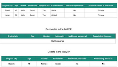
Today's report comes with a bit of a mystery, as it contains the reported death of a 15 year old female from Riyadh - one without preexisting illness -  who doesn't appear to have been previously reported as infected.
At least, not among the English language reports posted by the MOH (see Saudi MOH: Mismatching MERS Reports).
Also a bit unusual, MERS infections are rare in people under the age of 20, and are generally mild when they do occur.  According to the WHO demographic chart below, since 2012 there have been no fatalities reported among cases 19 years or younger.

http://applications.emro.who.int/docs/EMROPub_2018_EN_16780.pdf?ua=1


The details on these two updates follow, after which I'll return with a postscript.

Feb 24th:

https://www.moh.gov.sa/en/CCC/PressReleases/Pages/statistics-2018-02-24-001.aspx


Feb 25th:


Given the sporadic nature of reporting from the Saudi MOH this month it is difficult to draw any conclusions regarding current MERS activity in Saudi Arabia. 
That said, there does appear to have been an uptick in the number of primary (no known healthcare or camel exposure) MERS cases being reported in and around Riyadh.
Hopefully this weekend's back-to-back reporting of three days worth of MERS cases from the MOH is a sign that whatever barriers there have been to posting reports this month has been overcome.

Netherlands: H5 Avian Flu In A Commercial Flock In Oldekerk


#13,173


Given last year's record epizootic in Europe, and the arrival of newly reassorted HPAI H5N6 virus to the region in December, Europe has remained remarkably quiet on the avian flu front this winter (see DEFRA Avian Flu In Europe Update - Feb 14th).
Perhaps as a testament to improved biosecurity -  other than the December outbreak of H5N6 at a farm in Biddinghuizen, Netherlands - HPAI activity in Europe this winter has been pretty much limited to wild birds. 
Today, however, the government of the Netherlands is reporting on the detection of a (likely Highly pathogenic) H5 avian flu outbreak at a poultry farm in Oldekerk, in the north of the country.

Two reports.  First, the initial announcement from the Netherlands' Rijksoverheid (government) website, followed by an update today on the Wageningen Bioveterinary Research Institute website.  

Bird flu identified in poultry in Oldekerk

release | 25-02-2018 | 23:00
Olde Church (province of Groningen) on Sunday, February 25th, 2018 at a company with poultry avian influenza of the H5 type set. It is probably a highly pathogenic strain of bird flu. To prevent spread of the virus, the company is cleared. In total there are approximately 36,000 birds. In a radius of one kilometer around the holding are not other companies. The clearing are performed by the Dutch Food Safety Authority (NVWA).

In the area of ​​three kilometers around the infected holding in Oldekerk are two other companies. These companies are sampled and tested for avian influenza. Schouten Minister of Agriculture, Nature and Food Quality has immediately announced a ban on transporting poultry farms in a zone of 10 kilometers around the company Oldekerk. A movement ban applies to poultry, eggs, poultry manure and used bedding. All existing national measures, such as the indoor confinement, remain unimpaired.

Bird flu in Oldekerk 

published February 26, 2018

Olde Church (province of Groningen) on Sunday, February 25th, 2018 at a company with poultry avian influenza of the H5 type set. It is probably a highly pathogenic strain of bird flu. To prevent spread of the virus, the company is cleared. In total there are approximately 36,000 birds. In a radius of one kilometer around the holding are not other companies. The clearing are performed by the Dutch Food Safety Authority (NVWA). 


Two other companies investigated

In the area of ​​three kilometers around the infected holding in Oldekerk are two other companies. These companies are sampled and tested for avian influenza Wageningen Bioveterinary Research.

Movement ban zone 10 km

Schouten Minister of Agriculture, Nature and Food Quality (LNV) has immediately announced a ban on transporting poultry farms in a zone of 10 kilometers around the company Oldekerk. A movement ban applies to poultry, eggs, poultry manure and used bedding.
All existing national measures, such as the indoor confinement, remain unimpaired.

Sunday, February 25, 2018

Frontiers: Two Studies On The Epidemiology of Avian Influenza Viruses


#13,172


Although the debate over the role of wild and migratory birds in the spreading of both LPAI and HPAI viruses - at least across moderate to long distances - appears to be settled (see Revisiting The Migratory Bird Debate), often when we see outbreaks among tightly clustered poultry farms, other vectors appear to be in play.


MAY 2015 North American Epizootic

In the spring of 2015 we saw a number of clusters of North American farms impacted with H5N8 and/or H5N2, while surrounding areas appeared to have been `skipped over'. Furthermore, while HPAI was detected in a small number of wild birds, they were widely scattered, and wouldn't have directly accounted for the hundreds of farms affected. 
The lateral transfer of the virus from one farm to another via the movement of poultry products, vehicles, or personnel is often cited, but epidemiological investigations have often failed to find a link (see APHIS Releases Updated HPAI Epidemiology Report).
One idea, increasingly being considered, is the possibility that the virus is being dispersed – at least across short distances - `on the wind’.   It's a topic we looked at in some depth less than a month ago in `It's Raining Viruses'.

Going back even further, in December of 2012  (see Barnstorming Avian Flu Viruses?) we looked at a study in the Journal of Infectious Diseases called Genetic data provide evidence for wind-mediated transmission of highly pathogenic avian influenza that found patterns that suggested farm-to-farm spread of the 2003 H7N7 in the Netherlands due to the prevailing wind.
Another study of the same outbreak, Modelling the Wind-Borne Spread of Highly Pathogenic Avian Influenza Virus between Farms (PloS One 2012), found that wind borne transmission could have accounted for up to 24% of the transmission over distances up to 25 km. 
While the amount of virus a single wild bird might shed is fairly limited, when you have hundreds of thousands of infected chickens - all confined in a small area, producing copious amounts of waste products, and shedding feathers - it isn't difficult to envision a plume of infectious material being exhausted into the air by their ventilation system.
Given the right environmental conditions (temperature, humidity, UV light, wind speed & direction), farm-to-farm spread via airborne particles begins to make sense.  Particularly when you see some of the tight clustering of outbreaks in recent epizootics. 
All of which brings us to the first of two open-access studies today that have recently been published in Frontiers Epidemiology of Avian Influenza Viruses. This one looks at airborne detection of HPAI viruses during the 2016-17 H5N8 epizootic in France, which saw more than 400 farms affected.  

Airborne Detection of H5N8 Highly Pathogenic Avian Influenza Virus Genome in Poultry Farms, France

Axelle Scoizec1*, Eric Niqueux2, Rodolphe Thomas1, Patrick Daniel3, Audrey Schmitz2 and Sophie Le Bouquin
In southwestern France, during the winter of 2016–2017, the rapid spread of highly pathogenic avian influenza H5N8 outbreaks despite the implementation of routine control measures, raised the question about the potential role of airborne transmission in viral spread. 

As a first step to investigate the plausibility of that transmission, air samples were collected inside, outside and downwind from infected duck and chicken facilities. H5 avian influenza virus RNA was detected in all samples collected inside poultry houses, at external exhaust fans and at 5 m distance from poultry houses. For three of the five flocks studied, in the sample collected at 50–110 m distance, viral genomic RNA was detected. 

The measured viral air concentrations ranged between 4.3 and 6.4 log10 RNA copies per m3, and their geometric mean decreased from external exhaust fans to the downwind measurement point. These findings are in accordance with the possibility of airborne transmission and question the procedures for outbreak depopulation. 

(SNIP)

Our results also question the management of infected flocks. The confinement inside housing does not seem to be effective enough to prevent viral diffusion into the environment surrounding infected premises and the culling process requiring the loading of the animals into containers located outside the poultry house seems to generate an important emission of potentially infectious dust and/or aerosols into the environment. It would be essential to reduce this diffusion by rapidly implementing the depopulation using a method that reduces the air viral emission. To achieve this goal, new case management methods must require less human resource in terms of time and volume because human resources availability is the main cause of increasing time between the confirmation date and the depopulation. 

Furthermore, the methods must include a depopulation process minimizing the air viral diffusion to the surrounding environment. Methods such as emergency mass culling of poultry using a foam blanket over birds and in-house carcasses and litter composting could contribute to improve the control of influenza outbreaks (25, 26).

In conclusion, our results sustain the hypothesis of a potential airborne transmission contribution to the spread of the H5N8 HPAIV. However, more investigations would be required to support this hypothesis so as to provide evidence of virus viability in fine particles emitted from poultry outbreaks and epidemiological evidence.

(Continue . . . )

Another potential for spreading HPAI - to other farms, and back into the environment (where it can be picked up by wild or migratory birds, and spread onward) -  is the disposal of infected garbage and other waste material from poultry farms.

This is an area of vulnerability that - until recently - hasn't received much attention.  First some excerpts from the study, then I'll return with a bit  more.

Garbage Management: An Important Risk Factor for HPAI-Virus Infection in Commercial Poultry Flocks

Emily Walz1*, Eric Linskens1, iJamie Umber1, Marie Rene Culhane2,David Halvorson1, Francesca Contadini3 and Carol Cardona1

Garbage management represents a potential pathway of HPAI-virus infection for commercial poultry operations as multiple poultry premises may share a common trash collection service provider, trash collection site (e.g., shared dumpster for multiple premises) or disposal site (e.g., landfill). The types of potentially infectious or contaminated material disposed of in the garbage has not been previously described but is suspected to vary by poultry industry sector. 


A survey of representatives from the broiler, turkey, and layer sectors in the United States revealed that many potentially contaminated or infectious items are routinely disposed of in the trash on commercial poultry premises. On-farm garbage management practices, along with trash hauling and disposal practices are thus key components that must be considered to evaluate the risk of commercial poultry becoming infected with HPAI virus.

(SNIP)
  
Conclusion

This exploratory survey identified items in garbage that may contain infectious HPAI virus, some of which may carry high titers of infectious virus. Given that there is potential for HPAI virus to be associated with trash contents and garbage management practices, and taking into account the ease with which virus could be introduced into the poultry house, the potential for a commercial poultry flock becoming infected with HPAI virus due to garbage management during an outbreak should be considered.
Further research is needed to determine prevalence of garbage management practices in different production systems and across geographic regions in the United States and producers should develop appropriate mitigation measures in the event of a HPAI outbreak in commercial poultry.

(Continue . . . )

As the above study points out, garbage trucks - which often go from one farm to the next - may not only spread viruses along their route, the waste material they deposit in landfills may also contribute to the environmental spread of HPAI. 
In recent years we've seen efforts to quickly bury infected carcasses (either on site, or in landfills) in North America, Europe, Japan and South Korea. After the lessons from North America's Epizootic, the USDA's APHIS issued updated  Landfill Disposal Guidance—Recommended Waste Acceptance Practices for Landfills.
But in other places around the globe, we've seen thousands of dead birds dumped into rivers, ponds, and open ditches.  Most go unreported, but a few (of many) examples include:
All of this can make a real difference as avian influenza viruses can - under the right  environmental circumstances - survive for days or even weeks in water, soil, or in biological materials (see EID Journal: Persistence Of H5N1 In Soil).

A study reported in the August 2010 issue of Applied and Environmental Microbiology  determined that the H5N1 virus may persist on the dropped feathers from infected ducks for weeks or even months.
The bottom line is improper or sub-optimal disposal of infected bird carcasses and waste materials from poultry farms may not only be infecting nearby farms, it may also help reseed the migratory and wild bird population with new and improved HPAI viruses. 
While the risk of spread of avian viruses via airborne routes, or by improper carcass or waste disposal, remains uncertain -  the circumstantial evidence continues to mount. And given the stakes involved, the sooner we have firm answers and implement the right solutions, the better.