Showing posts with label RBD. Show all posts

Friday, January 18, 2013

EuroSurveillance: Revisiting The D222G Mutation In A/H1N1pdm09


image 

Flu Virus binding to Receptor Cells – Credit CDC 


# 6865


One of the unresolved  mysteries of the 2009 H1N1 pandemic is that while most people saw a relatively mild illness - for a small percentage of the population - the virus proved unusually severe and sometimes deadly. 


We saw the burden of the disease shift to younger adults and adolescents, a cohort that normally endures influenza infection pretty well. In Study: Years Of Life Lost Due To 2009 Pandemic, researchers calculated the mean age of death from the pandemic virus to be half that of seasonal flu, or 37.4 years.


And in September of 2011 we saw research indicating the H1N1pdm virus was more likely to exacerbate an S. pneumoniae co-infection (in mice, anyway) than was seasonal H1N1 (see mBio: Lethal Synergism of H1N1 Pandemic Influenza & Bacterial Pneumonia)


During November of 2009, news of a small change in the novel H1N1 virus came by way of the Norwegian Institute of Public Health (see Norway Reports An H1N1 Mutation) who announced the discovery of a mutation that “could possibly make the virus more prone to infect deeper in the airways and thus cause more severe disease."


The announcement of the `Norway’ or D222G (D225G in influenza H3 Numbering) mutation immediately sent researchers around the world on a hunt for similar changes in the virus, and over the following months several variations on a theme were discovered; D222N, D222E, and D222A.


The D222G mutation involves a single amino acid change in the HA1 gene at position 222 from aspartic acid (D) to glycine (G).


The pdmH1N1 virus carrying this mutation appeared to bind more readily to receptor cells (α2-3) found deep in the lungs, whereas unmutated seasonal flu strains bind preferentially to the (α2-6) receptor cells found in the upper airway.


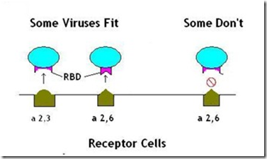
A virus’s ability to bind to specific cells is controlled by its RBD or Receptor Binding Domain; an area of its genetic code that allows it to attach to, and infect, specific types of host cells.

image

(A Very Simplified Illustration of RBDs)

Like a key into a padlock, the RBD must `fit’ in order to open the cell to infection.

Similarly, D222A changes the HA1 gene at position 222 to Alanine, while D222E changes the gene to Glutamic acid and D222N changes to Asparagine.


This D222G mutation had actually been detected months earlier, and in several other countries, but Norway was the first country to announce a possible link between that mutation and greater virulence.


In January of 2010, the World Health Organization’s  Weekly Epidemiological Record (No. 4, 2010, 85, 21–28) provided a detailed overview of what was then known about this mutation. 


While stating that more study was needed, the WHO pointed out the lack of apparent ongoing transmission of this mutation, and stated that:


`Based on currently available virological, epidemiological and clinical information, the D222G substitution does not appear to pose a major public health issue.’


Later in 2010, in Study: Receptor Binding Changes With H1N1 D222G Mutation, we saw more evidence of preferential binding to deep lung cells by viruses with the D222G mutation.


The debate over the significance (and origins) of the D222G mutation have continued since then. You can revisit some of those studies in the following blogs:


Eurosurveillance: Analysis Of Fatal H1N1 Cases In The UK)

Eurosurveillance: Debating The D222G/N Mutation In H1N1

D222G And Deep Lung Infections


All of which serves as prelude to a new study on the D222G mutation – again from Norwegian Institute of Public Health – that appeared yesterday in the journal Eurosurveillance.


Eurosurveillance, Volume 18, Issue 3, 17 January 2013

Within-patient emergence of the influenza A(H1N1)pdm09 HA1 222G variant and clear association with severe disease, Norway

R Rykkvin, A Kilander, S G Dudman, O Hungnes


Date of submission: 29 December 2012

ABSTRACT (reparagraphed for readability)

The association between a particular mutation in the HA1 subunit of the influenza virus haemagglutinin, D222G, and severe and fatal disease in cases of influenza A(H1N1)pdm09 in Norway during the 2009 pandemic was investigated using pyrosequencing.


The prevalence of the variant among fatal cases was 8/26 and among severe non-fatal cases 5/52. No D222G mutations were found among the 381 mild cases.


This difference could not be attributed to sampling differences, such as body location of sampling, or duration of illness. In cases with mutant virus where clinical specimens from different days of illness were available, transition from wild-type to mutant virus was commonly observed (4/5), indicating that the mutant virus emerged sporadically in individual patients.


In patients with paired samples from both the upper and lower respiratory tract (n=8), the same viral genotypes were detected in both locations. In most of the D222G cases (11/13), the mutant virus was found as a quasispecies.


This a long, and fairly technical report with a lot to digest, and I’m sure many of you will want to read it in its entirety.


But in short the authors present several findings, which they summarize in the discussion portion of the paper:


In the present study, we provide further epidemiological evidence of the association between the D222G mutation in HA1 of influenza A(H1N1)pdm09 virus and severe or fatal clinical course.


Furthermore, we present evidence that the mutated viruses emerge in individual patients after the onset of illness and demonstrate the presence of mutant virus in both the upper and lower respiratory tract. We also address some potential biases that could conceivably confound the analysis.


The Norwegian cases of infection with HA1 222G genotype viruses have occurred sporadically and do not cluster epidemiologically or in phylogenetic analysis.

<SNIP>


The 222G viruses appear to be rare among circulating strains, but are still quite frequent in patients with severe disease, who are not epidemiologically linked. A likely explanation is that the presence of mutant viruses in these particular individuals experiencing severe disease is due to selective upgrowth of mutant genomes during infection.


In other words, this mutation appears to occur spontaneously after a person is infected by the H1N1pdm virus, and then, only rarely. But when that happens, the patient appears more likely to experience a more severe illness.


As previously reported, It does not appear to transmit efficiently in the wild.


The concern here is that viruses can change, and co-mutations could occur that make the D222G mutation more transmissible in the future. 


We’ve seen that happen before.


In 2006 we saw a smattering of oseltamivir (Tamiflu ®) resistant seasonal H1N1 cases, almost always attributed to `spontaneous mutations’ within a patient receiving the drug.  While of concern to the patient afflicted, it appeared to be poorly transmissible.


In the 2006-2007 flu season, laboratories found no resistant strains in Europe or Japan, and in less than 1% of samples from the United States.


This resistance was caused by a mutation called H275Y, where a single amino acid substitution (histidine (H) to tyrosine (Y)) occurs at the neuraminidase position 275.


(Note: some scientists use 'N2 numbering' (H274Y) and some use 'N1 numbering' (H275Y))


The following year, during the 2007-2008 flu season, oseltamivir resistant viruses suddenly took flight, and by the spring of 2008 roughly 25% of European samples tested showed the H275Y mutation (see Increased Tamiflu Resistance In Seasonal Influenza).   


By the end of the year, resistant seasonal H1N1 was pretty much the norm around the world.


Influenza viruses are both unpredictable and constantly changing. So we watch subtle mutations like the D222G carefully, with the knowledge that the limited threat it poses today (due to its poor transmissibility) may not necessarily hold true tomorrow.

Wednesday, March 21, 2012

Study: Dual Receptor Binding H5N1 Viruses In China


RBD

(Very Simplified Illustration of RBDs)

# 6238


Human adapted influenza viruses have an RBD - Receptor Binding Domain (the area of its genetic sequence that allows it to attach to, and infect, host cells) that – like a key slipping into a padlock -`fit’ the receptor cells commonly found in the human upper respiratory tract; the alpha 2,6 receptor cell.  


Avian adapted flu viruses, like the H5N1 virus, bind preferentially to the alpha 2,3 receptor cells found in the gastrointestinal tract of birds. 


While there are some alpha 2,3 cells deep in the lungs of humans, for an influenza to be successful in a human host, it needs to a able to bind to the a 2,6 receptor cell.


A design flaw that - at least for the time being – is believed to have kept the H5N1 virus from taking off in the human population (note: there are likely other genetic barriers as well).


All of which serves as prelude to a study out of China where researchers have found at least two strains of the H5N1 virus that demonstrated the ability to bind to both a2,3 and a2,6 receptor cells.


Before anyone starts to head down to the bunker, these viruses were collected between 2003 and 2009, and despite their ability to bind to human-type receptors (at least at high viral loads), they have not managed to spark a pandemic.


The following excerpts come the abstract that appears in Biomedical and Environmental Sciences, Volume 25, Issue 1(h/t Tetano on FluTrackers for this link). 

Follow the link to read it in its entirety.


Identification of Dual Receptor-binding Specific Strains of Human H5N1 Viruses in China


Jian Fang ZHOU, Shu Mei ZOU, Zi LI, Min WANG, Jie DONG, Jun Feng GUO, He Jiang WEI, Le Ying WEN, Hong XU, Yue Long SHU

Abstract (excerpts):


<SNIP>

Results Dual binding preference to 2, 3 and 2, 6-glycans were found in two strains: A/Guangdong/1/06 (A/GD/1/06) and A/Guangxi/1/08 (A/GX/1/08). Though minor effect of short-2, 6-binding was detected in A/GX/1/08 at a low virus titer, both showed high affinity to the oligosaccharide at a high load. Notably both are of the long-2, 6-recognition, with the same topology as that of human H1N1 and H3N2 viruses.

Conclusion The findings suggest that human H5N1 virus in China likely acquired the potential human-adaptation ability. Further research and surveillance on receptor-binding specificity of H5N1 viruses are required.


While a significant step towards adaptation to human hosts, obviously something more is needed to make the H5N1 virus a pandemic threat.


Lest anyone be too comforted, this study does show that the H5N1 virus is capable of evolving towards a more `humanized’ pathogen.


Which is why the world remains at pre-pandemic Phase III on the H5N1 virus, and we continue to watch for signs of better adaptation to humans.


 image

Monday, December 13, 2010

Virology Journal: Receptor Cells In Minor Poultry Species


# 5136


Constant readers of this blog are aware that avian influenza strains bind preferentially to the kind of receptor cells commonly found in the digestive and respiratory tracts of birds; alpha 2,3 receptor cells.


Human influenzas – on the other hand - are adapted to bind to the kind of receptor cells that line the surfaces of the human respiratory system; alpha 2,6 receptor cells.


While not an absolute, flu viruses that bind to one type of receptor cell, tend not to bind to the other.


This ability to bind to a specific type of cell has often been described as the host cell being a padlock, and the virus needing a specific key (determined by the genetics of the virus’s Receptor Binding Domain: RBD) to unlock it.


image

(A Very Simplified Illustration of RBDs)


Now humans do have some avian-like alpha 2,3 receptor cells, particularly deep in the lungs.


This has been suggested as the reason that - when on rare occasions humans contract H5N1 - it is usually a deep lung infection.


It has also been postulated that H5N1’s deeper lung infections may reduce human-to-human transmission, as sneezing is a less common symptom.   


The concern is that over time, the H5N1 (or some other avian flu) virus might mutate in such a way as to be able to bind to human α2,6 receptor cells.


And while that may not be the only obstacle keeping the virus from becoming a pandemic strain, it does appear to be a major one.


The two main ways the virus could `learn’ to adapt to humans are through a mutation, or through a reassortment (a sharing of genetic material) with another flu virus.


It is possible for a simple mutation to change the binding preference of an influenza strain, to allow it to bind to a different type of receptor cell (or, to more than one type).


This is actually something we’ve observed with the novel H1N1 virus.  The D222G  or ``Norway’ mutation has been shown to convey dual receptor specificity for complex α2,3 and α2,6-linked sialic acids (see  Study: Receptor Binding Changes With H1N1 D222G Mutation).


Since pigs are known to have both types of receptor cells, they have often been cited as a potential `mixing vessel’ for influenza strains.  


Reassortant pig


Last January I wrote a blog titled Mixing Vessels For Influenza  which mentioned research done by two wildlife disease experts from the San Diego Zoo - Mark Schrenzel and Bruce Rideout – that identified the North American Striped Skunk and the Persian leopard - along with a handful of other small carnivores - as a potential host for influenza reassortment.


Additionally, we’ve seen H5N1 infections among dogs, cats, civets, raccoons, martens, and – of course – humans.   And researchers have successfully infected cattle with the H5N1 virus, along with ferrets and mice for testing.


But since avian strains mostly infect birds, the opportunities for avian viruses to encounter human adapted viruses, or to learn to unlock alpha 2,6 receptor cells, are limited. 


But perhaps, not quite as limited as we have previously believed.


Which brings us to today’s study, from the Virology Journal, that looks at types of receptor cells detected in a half dozen minor species of poultry.


The condensed version is, some types of poultry have both types of receptor cells . . . but read the entire abstract, or better yet, the entire article.


Characterization of influenza virus sialic acid receptors in minor poultry species

Brian Kimble, Gloria Ramirez Nieto and Daniel R Perez

It is commonly accepted that avian influenza viruses (AIVs) bind to terminal alpha2,3 sialic acid (SA) residues whereas human influenza viruses bind to alpha2,6 SA residues.


By a series of amino acid changes on the HA surface protein, AIVs can switch receptor specificity and recognize alpha2,6 SA positive cells, including human respiratory epithelial cells.


Animal species, like pigs and Japanese quail, that contain both alpha2,3 and alpha2,6 SA become ideal environments for receptor switching. Here, we describe the SA patterns and distributions in 6 common minor domestic poultry species: Peking duck, Toulouse geese, Chinese ring-neck pheasant, white midget turkey, bobwhite quail, and pearl guinea fowl.


Lectins specific to alpha2,3 and alpha2,6 SA (Maakia amurensis agglutinin and Sambuca nigra agglutinin, respectively) were used to detect SA by an alkaline phosphotase-based method and a fluorescent-based method.


Differences in SA moieties and their ability to bind influenza viruses were visualized by fluorescent labeling of 4 different H3N2 influenza viruses known to be specific for one receptor or the other.


The geese and ducks showed alpha2,3 SA throughout the respiratory tract and marginal alpha2,6 SA only in the colon.


The four other avian species showed both alpha2,3 and alpha2,6 SA in the respiratory tract and the intestines. Furthermore, the turkey respiratory tract showed a positive correlation between age and alpha2,6 SA levels.


The fact that these birds have both avian and human flu receptors, combined with their common presence in backyard farms and live bird markets worldwide, mark them as potential mixing bowl species and necessitates improved surveillance and additional research about the role of these birds in influenza host switching.

The complete article is available as a provisional PDF. The fully formatted PDF and HTML versions are in production.

Tuesday, November 16, 2010

Study: The Continuing Evolution Of Avian H9N2


# 5058


When it comes to avian influenzas, H5N1 gets the lion’s share of our attention, but it isn’t the only avian flu strain with the potential to jump to humans.


Below you’ll find a chart lifted and edited from CIDRAP’s excellent overview Avian Influenza (Bird Flu): Implications for Human Disease  showing non-H5N1 avian flu infections in humans over the past decade.


CIDRAP FluA


Since surveillance is – at best - haphazard (or even non-existent) in many parts of the world,  how often this really happens is unknown.  


Like the novel swine flu cases that caused such a stir last week, they probably happen a bit more often than we realize.


A couple of years ago, we saw a study in PNAS that indicated that the H7 virus might be moving more towards adapting to humans.


Contemporary North American influenza H7 viruses possess human receptor specificity: Implications for virus transmissibility


You can read more about this in a couple of blogs from 2008, H7's Coming Out Party and H7 Study Available Online At PNAS.


Today, in a similar vein, we have study appearing in the Virology Journal that suggests that the H9N2 virus may also be evolving more towards humans as well.


While most virological research studies are in vivo or in vitro, this one is in silica, or based primarily on computer analysis of existing data.


Avian influenza A (H9N2): computational molecular analysis and phylogenetic characterization of viral surface proteins isolated between 1997 and 2009 from the human population

Azeem M Butt, Samerene Siddique, Muhammad Idrees and Yigang Tong

Virology Journal 2010, 7:319 doi:10.1186/1743-422X-7-319

Published: 15 November 2010

Abstract (provisional)
Background

H9N2 avian influenza A viruses have become panzootic in Eurasia over the last decade and have caused several human infections in Asia since 1998. To study their evolution and zoonotic potential, we conducted an in silico analysis of H9N2 viruses that have infected humans between 1997 and 2009 and identified potential novel reassortments.

Results

A total of 22 hemagglutinin (HA) and neuraminidase (NA) nucleotide and deduced amino acid sequences were retrieved from the NCBI flu database.

It was identified that mature peptide sequences of HA genes isolated from humans in 2009 had glutamine at position 226 (H3) of the receptor binding site, indicating a preference to bind to the human alpha (2-6) sialic acid receptors, which is different from previously isolated viruses and studies where the presence of leucine at the same position contributes to preference for human receptors and presence of glutamine towards avian receptors.

Similarly, strains isolated in 2009 possessed new motif R-S-N-R in spite of typical R-S-S-R at the cleavage site of HA, which isn't reported before for H9N2 cases in humans. Other changes involved loss, addition, and variations in potential glycosylation sites as well as in predicted epitopes. The results of phylogenetic analysis indicated that HA and NA gene segments of H9N2 including those from current and proposed vaccine strains belong to two different Eurasian phylogenetic lineages confirming possible genetic reassortments.

Conclusions

These findings support the continuous evolution of avian H9N2 viruses towards human as host and are in favor of effective surveillance and better characterization studies to address this issue.

The complete article is available as a provisional PDF. The fully formatted PDF and HTML versions are in production.


In December 2008, after receiving the news of a baby in Hong Kong having been diagnosed with H9N2, I reran a blog featuring an interview in which world famous Hong Kong virologist Malik Peiris cautioned that the H9N2 virus may be circulating far more commonly than we believe. 


Revisiting A Malik Peiris Interview On H9N2


As it exists now, H9 poses a low threat to humans.


Sporadic reports of human infections – particularly when there is no evidence of ongoing transmission – are interesting, but not particularly alarming.


But H9, like a number of other avian viruses (H5’s, H7’s) have some pandemic potential, particularly if they can `drift’ or mutate sufficiently, or pick up genetic material from other viruses.  


This `reassortment’ could conceivably create a new, hybrid strain of influenza.


image

How likely is this to happen?


Well, that’s the big question.  No one really knows.


We just know that it is possible.


It obviously doesn’t happen often, otherwise we’d be hip deep in new, hybrid viruses all of the time.  But this is essentially the route that the 2009 H1N1 virus took to become a pandemic, and is likely the way the 1957 and 1968 pandemics came about.


And so we watch these rare human cases with great interest.  A dangerous reassortment or mutation may never occur with the H9 virus, or it could happen tomorrow.


Influenza viruses, as they say, are unpredictable.

Saturday, October 23, 2010

D222G And Deep Lung Infections


# 5002


This morning we’ve a joint study from Imperial College London and the University of Marburg that may shed some light on why at least some cases of pandemic H1N1 proved severe (or fatal) while the great majority remained mild.


The `Norway’ or D222G (D225G in influenza H3 Numbering) mutation first announced by Norwegian Scientists last November has sparked repeated speculation that it might be associated with increased virulence.


Although we’ve covered this territory a number of times over the past year, a brief (and hopefully simple) review is in order. If you are up to speed on receptor binding, and the history of the D222G variant, feel free to skip the next section.


The D222G mutation had actually been detected months earlier, and in several other countries, but Norway was the first country to announce a possible link between that mutation and greater virulence.


This mutation involves a single amino acid change in the HA1 gene at position 222 from aspartic acid (D) to glycine (G).


The pdmH1N1 virus carrying this mutation appeared to bind more readily to receptor cells (α2-3) found deep in the lungs, whereas unmutated seasonal flu strains bind preferentially to the (α2-6) receptor cells found in the upper airway.


A virus’s ability to bind to specific cells is controlled by its RBD or Receptor Binding Domain; an area of its genetic code that allows it to attach to, and infect, specific types of host cells.


image 

(A Very Simplified Illustration of RBDs)


Like a key into a padlock, the RBD must `fit’ in order to open the cell to infection.


For some deeper background you may wish to read Looking For the Sweet Spot, and a follow-up blog called Receptor Binding Domains:Take Two.


The World Health Organization’s take on this mutation has been that it is worth following, and studying, but there is no evidence (as yet) that it poses a substantial public health hazard.


In January, in a blog entitled WER Review: D222G Mutation In H1N1, I quoted the latest WHO report that stated:


`Based on currently available virological, epidemiological and clinical information, the D222G substitution does not appear to pose a major public health issue.’


This view is not universally held, however. There are some who have maintained that that the WHO is underestimating the impact of this mutation.


In March of this year, researchers from the Norwegian Institute of Public Health in Oslo reported that they found the mutation in 11 of 61 severe illness cases that they analyzed, but that it was not found in any of the 205 mild cases they looked at  (see CIDRAP Report On The H1N1 Mutation Debate).


The WHO WER Review reported that the overall prevalence of D222G was <1.8% (52 detections among >2755 HA sequences) in contrast to a rate of 7.1% in fatal cases.


The WHO paper also reported on the occurrence of  two other mutations at this amino acid position, D222E and D222N, although their significance is unclear.


While this all may sound like fairly damning evidence, it should be noted that mild cases have been detected with this D222G mutation in other studies, and many severe and fatal cases of pandemic H1N1 that have been examined did not have this mutation.


Some recent blogs on this mutation include:


Study: Receptor Binding Changes With H1N1 D222G Mutation

Eurosurveillance On Recently Isolated H1N1 Mutations

Referral: Virology Blog On D225G Mutation


Today’s study, which appears in the Journal of Virology, is called:


Altered receptor specificity and cell tropism of D222G haemagglutinin mutants from fatal cases of Pandemic A(H1N1) 2009 influenza


Yan Liu, Robert A. Childs, Tatyana Matrosovich, Stephen Wharton, Angelina S. Palma, Wengang Chai, Rodney Daniels, Victoria Gregory, Jennifer Uhlendorff, Makoto Kiso, Hans-Dieter Klenk, Alan Hay, Ten Feizi*, and Mikhail Matrosovich*


Admittedly a daunting title, but the abstract is a bit easier to follow.  I’ve re-paragraphed, and added a couple of highlights to it for readability.


Abstract


Mutations in the receptor-binding site of the haemagglutinin of pandemic influenza A(H1N1) 2009 viruses have been detected sporadically. An Asp222Gly (D222G) substitution has been associated with severe or fatal disease.


Here we show that 222G variants infected a higher proportion of ciliated cells in cultures of human airway epithelium than viruses with 222D or 222E which targeted mainly non-ciliated cells.


Carbohydrate microarray analyses showed that 222G variants bind a broader range of {alpha}2-3-linked sialyl receptor sequences of a type expressed on ciliated bronchial epithelial cells and on epithelia within the lung.


These features of 222G mutants may contribute to exacerbation of disease.


The discovery that D222G enhances the binding to ciliated cells is important because cilia are motile hair-like protuberances that line the airway and help move mucus (and debris) up and out of the lungs.


SEM micrograph of the cilia projecting from respiratory epithelium in the lungs


If you infect (and impair) the lung’s cilia, you (theoretically, at least) increase the odds of that person developing pneumonia.


In this study, researchers tested 6 different variants of the pdmH1N1 virus, including 3 (Lvi, Nor, Ham-E) with the D222G mutation.  


The `money quote’ from the study is:


The viruses with  222D  (Mol and Ham) and 222E  (Dak) showed a pattern of cell tropism typical of seasonal influenza A and B viruses  infecting predominantly non-ciliated cells known to be rich in α2-6 Sia sequence: less than 5% of infected cells were ciliated.


By contrast, the three viruses with 222G, Lvi, Nor and Ham-e, infected both ciliated and non-ciliated cells, and  20% or more of infected cells were ciliated, known to express α2-3 Sia sequences.


This change in the cell tropism, with a 5-10 fold increase in infection of ciliated cells, thus correlated with the presence of the D222G substitution in the HA, and other amino acid  differences, in particular D222E, had little or no effect.


Where then, does all of this leave us?


Well, the authors state that:


Whether the selection of the D222G mutation is a cause or a consequence of more severe lower respiratory tract infection has still to be resolved. It is evident, however, that its emergence is likely to exacerbate the severity of disease.


Luckily, this mutation has been slow to spread. 


It has been detected in less than 2% of the samples tested, and that suggests that (right now, anyway) it may be less fit for transmission than other competing strains.  


The fact that it tends to promote deep lung infections, and reduces the ability to expel mucus (and therefore cough productively), may help inhibit its spread.


A scenario not unlike what we’ve seen with the H5N1 (bird flu) virus, which as an avian virus, binds even more preferentially to α2-3 receptor cells. 


What is true today, however, may not hold true tomorrow. Influenza viruses are capable of swift and sometimes dramatic mutations. 


This research shows that even a seemingly mild strain of influenza can easily pick up virulence, and if it can retain transmissibility, could spark a serious public health hazard.


Which is why continued influenza research, the monitoring of this and other influenza strains, and the maintaining of pandemic readiness remain vital even after the pandemic of 2009 has passed.

Monday, April 19, 2010

Master Of Its Binding Domain


# 4505


With one pandemic virus still smoldering in the background, and another one – H5N1 – potentially still in the wings (sitting at pre-pandemic Level III and holding), the question on many scientist’s minds is: what would it take for bird flu to become a pandemic?


Researchers at Erasmus MC (Medical Center) in Rotterdam are among those trying to answer that question. 


In an article that appears in the current issue of the Journal of Virology, they detail their findings that a single amino acid substitution may be all that is required to make H5N1 better adapted to the human respiratory system.


First, a little background (I’m keeping this very simple. My apologies to any real scientists reading this - you may wish to avert your eyes), then the research abstract.


There are three criteria for an influenza virus to become a pandemic.


  • It must be a novel strain for which humanity has little or no immunity.  
  • It must infect humans and cause significant illness.
  • It must be able to sustain human-to-human (H2H) transmission


The H5N1 virus has met the first two criteria, but (thankfully) has not yet adapted well enough to humans to spread easily between them.


Although there may be other factors at work, the virus’s main obstacle is believed to be H5N1’s affinity for avian α2-3 linked sialic acid receptor cells rather than for the α2-6 linked sialic acid cells commonly found in the human respiratory system.


Humans do possess α2-3 receptor cells, but mostly deep in the lungs or in the human gut, areas where the virus has trouble reaching. 


For a virus to have a good shot at transmission, it needs to be able to bind to cells in the upper respiratory system.


A virus’s ability to bind to specific cells is controlled by its RBD or Receptor Binding Domain; an area of its genetic code that allows it to attach to, and infect, specific types of host cells.


RBD

(Very Simplified Illustration of RBDs)


Like a key into a padlock, the RBD must `fit’ in order to open the cell to infection.


For some deeper background you may wish to read Looking For the Sweet Spot, and a follow-up blog called Receptor Binding Domains:Take Two.


The genetic sequence of the flu virus can be represented by the letters of the amino acids that make up the viral genome. These are long chains comprised of hundreds of amino acid molecules.


A tiny sub-section of that chain might have a sequence something like:


NPECESLSTASSWSYI


As the virus inhabits a cell, and begins to replicate, it makes thousands of copies of itself which then burst out of the cell after a few hours and go on to infect other cells.


Those cells, in turn, make copies that go forth to infect more cells.


But being a single-strand RNA virus, the influenza virus tends to be sloppy in making copies of itself. Errors sometimes creep in. If in the process of replicating it mixes up just a single amino acid, we can end up with a mutated virus.


NPECESLSTASSWSYI

NPECESLSTASSWSYI
NPECKSLSTASSWSYI           < – A mutation!
NPECKSLSTASSWSYI            
NPECKSLSTASSWSYI                


Above, I’ve swapped out the amino acid Glutamic acid (E) at position 5 for Lysine (K). Assuming the result is a `biologically fit’ and competitive virus (most aren’t), then it may go on to infect other cells, and conceivably, other hosts.


Of course, that doesn’t mean it will make the virus more dangerous.  A mutation can make the virus less virulent or less transmissible. 


Or it may simply have no effect at all.


These small changes in the virus are called antigenic drift, and over time changes in the virus can accumulate to the point that last year’s vaccine is no longer effective.  They can also bring about antiviral resistance, or even increase the virulence or transmissibility of the virus.


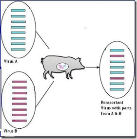
Big jumps, or mutations in the virus generally come about through a process called reassortment.  And that happens when two different flu strains inhabit the same host (human or otherwise) at the same time. 


They can swap gene segments and thereby produce a hybrid virus.


Human Reassortant


The point being that influenza viruses are constantly changing, evolving, and mutating through a variety of processes, not all of which are well understood. 


The H5N1 bird flu virus that appeared in Hong Kong in 1996 now has now mutated into well over a dozen different clades (branches of their genetic tree), and more are expected to form over time.


H5N1 clades


Which should be enough background for us to now look at the work conducted by researchers at Erasmus MC, (including such familiar names as Ab Osterhaus and Ron Fouchier).  


Here is the abstract (it’s a pay-to view article), slightly reformatted and reparagraphed for readability.


J Virol. 2010 Apr 14. [Epub ahead of print]

In vitro assessment of attachment pattern and replication efficiency of H5N1 influenza A viruses with altered receptor specificity.

Chutinimitkul S, van Riel D, Munster VJ, van den Brand JM, Rimmelzwaan GF, Kuiken T, Osterhaus AD, Fouchier RA, de Wit E.

Department of Virology and National Influenza Center, Erasmus MC, P.O. Box 2040, 3000CA, Rotterdam, The Netherlands.

Abstract

The continuous circulation of the highly pathogenic avian influenza (HPAI) H5N1 virus has been a cause of great concern.


The possibility of this virus acquiring specificity for the human influenza A virus receptor, alpha2,6-linked sialic acids (SA), and being able to transmit efficiently among humans is a constant threat to human health.


Different studies have described amino acid substitutions in hemagglutinin (HA) of clinical HPAI H5N1 isolates or that were introduced experimentally, that resulted in an increased, but not exclusive, binding of these virus strains to alpha2,6-linked SA.


Here, we have introduced all previously described amino acid substitutions and combinations thereof into a single genetic background, influenza virus A/Indonesia/5/05 HA and tested the receptor specificity of these 27 mutant viruses.


The attachment pattern to ferret and human tissues of the upper and lower respiratory tract of viruses with alpha2,6-linked SA receptor preference was then determined and compared to the attachment pattern of a human influenza A virus (H3N2).


At least three mutant viruses showed an attachment pattern to the human respiratory tract similar to that of the human H3N2 virus. Next, the replication efficiency of these mutant viruses and the effect of three different neuraminidases on virus replication were determined.


These data show that influenza virus A/Indonesia/5/05 potentially requires only a single amino acid substitution to acquire human receptor specificity, while at the same time remaining replication competent, thus suggesting that the pandemic threat posed by HPAI H5N1 is far from diminished.

PMID: 20392847 [PubMed - as supplied by publisher]


Since I haven’t seen the whole article (and honestly would be unlikely to understand it all, even if I had), I can’t really comment on the specifics of this study. 


The take-away message here is that there are a number of routes by which bird flu could become a more human-adapted pathogen.  Some may require as little as one amino acid substitution.


The unanswerable question right now is whether this substitution ever will occur in nature and spark an H5N1 pandemic.  


The best scientists can say is that it is possible.


And with a virus that’s exhibited the kind of mortality rate that we’ve seen with bird flu, that’s more than enough to keep them awake at night.

Wednesday, March 10, 2010

Study: H5N1 Can Replicate In Human Gut


# 4417


This morning a brief, but fascinating tidbit from The Journal of Infectious Diseases where scientists have demonstrated that the H5N1 bird flu virus can replicate ex vivo in the human gut.


A hat tip to Tetano on FluTrackers for posting this item. First the abstract, then a bit of discussion.


DOI: 10.1086/651457
BRIEF REPORT


Avian Influenza A(H5N1) Viruses Can Directly Infect and Replicate in Human Gut Tissues

Yuelong Shu, Chris Ka‐fai Li, Zi Li, Rongbao Gao, Qian Liang, Ye Zhang, Libo Dong, Jiangfang Zhou, Jie Dong, Dayan Wang, Leying Wen, Ming Wang, Tian Bai, Dexin Li, Xiaoping Dong, Hongjie Yu, Weizhong Yang, Yu Wang,Zijian Feng,  Andrew J. McMichael,3 and Xiao‐Ning Xu3


The human respiratory tract is a major site of avian influenza A(H5N1) infection. However, many humans infected with H5N1 present with gastrointestinal tract symptoms, suggesting that this may also be a target for the virus.


In this study, we demonstrated that the human gut expresses abundant avian H5N1 receptors, is readily infected ex vivo by the H5N1 virus, and produces infectious viral particles in organ culture.

An autopsy colonic sample from an H5N1infected patient showed evidence of viral antigen expression in the gut epithelium. Our results provide the first evidence, to our knowledge, that H5N1 can directly target human gut tissues.


If you’ve followed the H5N1 story closely over the past five years, then the findings of this study shouldn’t come as a complete surprise.  


We’ve had more than a few hints along the way. 


Influenza in humans (and in most mammals) is primarily seen as a respiratory disease.  Human adapted influenza viruses have an affinity to bind to the α2-6 receptor cells that line the upper airway and lungs in humans. 


Avian influenza viruses, however, preferentially bind to the α2-3 receptor cells that are commonly found in the gastrointestinal tract of aquatic birds, the virus’s natural host.   


Influenza in birds is a mostly a gastrointestinal illness, and the virus is often spread via feces deposited in lakes and ponds.


In order for an avian flu virus, like H5N1, to successfully `jump the species barrier’ and become easily transmissible among humans, it is believed that it must adapt its RBD (receptor binding domain) to match the  α2-6 receptor cells found in the more easily accessible upper respiratory system.


image

(Very Simplified Illustration of RBDs)


For a layman’s explanation of the science of RBDs, you might want to have a look at a couple of essays I’ve written in the past.


RBD: Looking For The Sweet Spot
Study: H1N1 Receptor Binding


Humans do have α2-3 receptor cells, however.  Just not in abundance in their upper airways, where influenza viruses can most easily bind.


These α2-3 receptor cells can be found deep in the lungs and in some human epithelial tissues, although their prevalence in the human digestive tract has been a bit of an open question.


We’ve seen rare instances of human avian flu infections that were primarily gastrointestinal in nature, raising the intriguing possibility that avian viruses can replicate outside of the human respiratory system.


One of the earliest indications that H5N1 could bind and flourish in the human gastrointestinal tract comes from this study involving the deaths of a brother and sister in Vietnam in 2004.


Fatal avian influenza A (H5N1) in a child presenting with diarrhea followed by coma.

de Jong MD, Bach VC, Phan TQ, Vo MH, Tran TT, Nguyen BH, Beld M, Le TP, Truong HK, Nguyen VV, Tran TH, Do QH, Farrar J.

Oxford University Clinical Research Unit, Hospital for Tropical Diseases, Ho Chi Minh City, Vietnam.


In southern Vietnam, a four-year-old boy presented with severe diarrhea, followed by seizures, coma, and death. The cerebrospinal fluid contained 1 white cell per cubic millimeter, normal glucose levels, and increased levels of protein (0.81 g per liter).


The diagnosis of avian influenza A (H5N1) was established by isolation of the virus from cerebrospinal fluid, fecal, throat, and serum specimens. The patient's nine-year-old sister had died from a similar syndrome two weeks earlier. In both siblings, the clinical diagnosis was acute encephalitis.


Neither patient had respiratory symptoms at presentation. These cases suggest that the spectrum of influenza H5N1 is wider than previously thought.


In June of 2007, we got a report (see Atypical Presentations of H5N1)  out of Indonesia, of a child infected with H5N1 but that presented without respiratory symptoms.


A year later, in a large review of Chinese bird flu patients (see Clinical Case Review Of 26 Chinese H5N1 Patients), we find several mentions of gastrointestinal involvement as well.


Diarrhea was present in only two H5N1 cases at admission, but developed in a quarter of cases during hospitalization. Diarrhea was a common presenting symptom among H5N1 cases in Vietnam [11], [12] and Thailand [13], but was reported infrequently among cases in Hong Kong SAR, China [9], [10], and Indonesia [4], [16].


H5N1 virus and viral RNA have been detected in feces and intestines of human H5N1 cases [12], [17], [30], [33]. Whether the gastrointestinal tract is a primary site for H5N1 virus infection is currently unknown.


And even novel H1N1 (and perhaps Influenza B) are being looked at for exhibiting unusual gastrointestinal symptoms.   Last January, in Influenza’s Gastrointestinal Connection, I wrote about a study that appeared in BMC Infectious Diseases.


Influenza virus infection among pediatric patients reporting diarrhea and influenza-like illness

The detection of influenza viral RNA and viable influenza virus from stool suggests that influenza virus may be localized in the gastrointestinal tract of children, may be associated with pediatric diarrhea and may serve as a potential mode of transmission during seasonal and epidemic influenza outbreaks.


And for my final exhibit, in the CDC’s Interim guidance on Infection Control for the pandemic H1N1 Virus, they state:


Transmission of influenza through the air over longer distances, such as from one patient room to another, is thought not to occur. All respiratory secretions and bodily fluids, including diarrheal stools, of patients with 2009 H1N1 influenza are considered to be potentially infectious.


With today’s study, another piece has been added to the influenza jigsaw puzzle.  One that may help answer a nagging question about the atypical presentation of influenza infections. 

Thursday, January 07, 2010

Mixing Vessels For Influenza


# 4228


My thanks to Helen Branswell for alerting me (via her Twitter feed) to this article in The Economist about research conducted by two scientists at the San Diego Zoo’s Institute for Conservation Research on which animal species are likely to contract different types of influenza.


Influenza and wildlife

Mix and match

Jan 7th 2010


From The Economist print edition

Which animal species are most likely to get flu?

Science Photo Library

What, me?

THE scientific value of zoos is sometimes called into question, but Mark Schrenzel and Bruce Rideout, two experts on wildlife diseases who work at San Diego Zoo’s Institute for Conservation Research, have just shown the value of having a wide range of animals to hand for study. They have been looking at which species might act as reservoirs for influenza viruses and—worse, from the human point of view—which might act as “mixing vessels” in which new strains of virus are generated.

(Continue . . . )


New strains of influenza come about from reassortment, or the mixing and matching of genetic material between two compatible flu strains in a shared host. 


While any host (human, swine, avian) could produce a reassorted virus, hosts (like pigs) that are susceptible to a wider range of viruses are thought to be more likely to serve as an efficient mixing vessel.


Reassortant pig


Human adapted influenza have an RBD - Receptor Binding Domain (the area of its genetic sequence that allows it to attach to, and infect, host cells) that `fit’ the receptor cells commonly found in the human upper respiratory tract; the alpha 2,6 receptor cell.  


Avian adapted flu viruses bind preferentially to the alpha 2,3 receptor cells found in the gastrointestinal tract of birds. 


While there are some alpha 2,3 cells deep in the lungs of humans, for an influenza to be successful in humans, it needs to bind to the a 2,6 receptor cell.  


 RBD

(Very Simplified Illustration of RBDs)


Some mammalian hosts (like pigs) have both types of receptor cells, and therefore, increase the range of influenza viruses to which they are susceptible.   And that, in turn, increases the odds that they could host two different viruses at the same time, and help facilitate a reassortment of two flu viruses into a new hybrid.


Drs. Schrenzel and Rideout at the San Diego Zoo have examined 60 different species of small mammals, and have determined that several carry the type of receptor cells (a2,3) favored by the H5N1 avian flu virus; including opossums, the Arctic Fox, and the Chinese wolf.


Additionally, we’ve seen H5N1 infections among dogs, cats, civets, raccoons, martens, and – of course – humans.   And researchers have successfully infected cattle with the H5N1 virus, along with ferrets and mice for testing.


Over the years, scientists have openly wondered about possible mammalian reservoirs of the virus, which could help explain how the virus can be reintroduced to an area after massive poultry cullings have taken place.  


The list of potential hosts is likely to continue to grow.


Schrenzel and Rideout have also identified a handful of small carnivores that carry both the a2,3 and a2,6 receptor cells, making them potential mixing vessels.   These include the Persian leopard and the North American striped skunk.


For more on mammalian and avian hosts for influenza, you wish to revisit these blogs. 


Japan: Bird Flu Antibodies Found In Raccoons
Reservoir Ducks
Reservoir Dogs (Cats, Foxes, and Raccoons)


And for more on RBDs, the following essays might be of interest.


Study: H1N1 Receptor Binding
RBD: Looking For The Sweet Spot
Receptor Binding Domains: Take Two


While we are learning more about influenza viruses, and the hosts they inhabit, we obviously have a long way to go.     The novel H1N1 and H5N1 viruses have rewritten the science text books over the past five years, and more rewrites are likely in store.

Thursday, September 10, 2009

Study: H1N1 Receptor Binding


# 3714


An interesting study in the current issue of Nature Biotechnology which helps explain why, in a small percentage of novel H1N1 infections, patients experience severe deep lung infections.  


It all has to do with how this virus binds to receptor cells.


The receptor binding domain (RBD) of an influenza virus is that area of its genetic sequence that allows it to attach to, and infect, host cells.   


Much like a key into a padlock, the RBD must `fit' the host cell in order for it to bind.


RBD

(Very Simplified Illustration of RBDs)


A little science.   I’ll try to be gentle.


Animal cell membranes are comprised of a lipid bilayer with lots of strands (or chains) of sugars (carbohydrates), proteins, and fat molecules poking through them.


These carbohydrate molecules, some being glycolipids (carbohydrate and fat) and others glyoproteins (carbohydrate and Protein), form a dense sugary coating to all animal cell membranes.


Since there are a lot of different sugars, there are a lot of combinations to be found.  The most accessible of these sugars for a virus are the ones at the tips of these chains, where a special sugar called sialic acid is often found. 


Now, not all sialic acid molecules are the same. 


Essentially, they are identified by what molecules they are attached to, and how they are attached. It gets pretty complicated, but suffice to say there are two special sialic acid configurations that are of particular interest to us when dealing with Influenza; the α-2,3 and the α-2,6 combinations.


The α-2,6 receptor cells are commonly found in the upper airway of humans, while the α-2,3 receptor cells are most often found deep in the lungs.


One of the reasons that the H5N1 (bird flu) virus hasn’t managed to transmit efficiently from human-to-human is because it is adapted to the kind of receptor cells most commonly found in the digestive tract of birds; the α-2,3  -  not the α-2,6 receptor cells found in the human upper airway.


AI (Avian Influenza) viruses must make their way deep into the lungs to infect humans.   And while it rarely happens, when it does, the resultant infection is generally far more severe than an upper airway infection.


Most human-adapted influenzas attach to the easy-to-reach  α-2,6 receptor cells commonly found in nose, throat, and pharynx.


While 99% of novel H1N1 victims seem to experience relatively mild upper airway infections, an unlucky 1% see the infection go deeper into the lungs.    Helen Branswell of the Canadian Press wrote about this phenomenon about a week ago, which I covered in a blog called Pathology Of Fatal H1N1 Lung Infections.


Lung damage in fatal swine flu cases more bird flu than seasonal flu: expert


By Helen Branswell Medical Reporter (CP) 

TORONTO — The lungs of people who have died from swine flu look more like those of the victims of H5N1 avian influenza than those of people who succumb to regular flu, the chief of infectious diseases pathology at the U.S. Centers for Disease Control says.


Today, in a study that appears in Nature Biotechnology, we learn that researchers have determined that the novel H1N1 virus has the ability to infect both upper respiratory α-2,6 receptor cells, and to a lesser extent, the deep lung α-2,3 cells.


Which makes novel H1N1 a versatile virus, indeed.


Receptor-binding specificity of pandemic influenza A (H1N1) 2009 virus determined by carbohydrate microarray - pp797 - 799

Robert A Childs, Angelina S Palma, Steve Wharton, Tatyana Matrosovich, Yan Liu, Wengang Chai, Maria A Campanero-Rhodes, Yibing Zhang, Markus Eickmann, Makoto Kiso, Alan Hay, Mikhail Matrosovich & Ten Feizi

doi:10.1038/nbt0909-797


This paper isn’t open access, but the Medical Research Council (which partnered to fund this research) has an overview of the paper, and an interview with Professor Ten Feizi, a corresponding author of the paper.


Professor Feizi, in the accompanying article, expresses the concern that:


"If the flu virus mutates in the future, it may attach to the receptors deep inside the lungs more strongly, and this could mean that more people would experience serious symptoms. We think scientists should be on the lookout for these kinds of changes in the virus so we can try to find ways of minimising the impact of such changes," added Professor Feizi.


Pandemic flu can infect cells deep in the lungs, says new research

10 September 2009

Pandemic swine flu can infect cells deeper in the lungs than seasonal flu can, according to a new study published today in Nature Biotechnology. The researchers, funded by the Medical Research Council, Wellcome Trust and Engineering Physical Sciences Research Council, say this may explain why people infected with the pandemic strain of swine-origin H1N1 influenza are more likely to suffer more severe symptoms than those infected with the seasonal strain of H1N1. They also suggest that scientists should monitor the current pandemic H1N1 influenza virus for changes in the way it infects cells that could make infections more serious.


Influenza viruses infect cells by attaching to bead-like molecules on the outside of the cell, called receptors. Different viruses attach to different receptors, and if a virus cannot find its specific receptors, it cannot get into the cell. Once inside the cell, the virus uses the cell's machinery to make thousands more viruses, which then burst out of the cell and infect neighbouring ones, establishing an infection.


Seasonal influenza viruses attach to receptors found on cells in the nose, throat and upper airway, enabling them to infect a person's respiratory tract. The research shows that pandemic H1N1 swine flu can also attach to a receptor found on cells deep inside the lungs, which can result in a more severe lung infection.


(Continue . . . )