Showing posts with label Eurosurveillance Journal. Show all posts

Thursday, May 21, 2015

Eurosurveillance: Genetic Diversity Of H5N8 Detected In Japanese Birds

image

Credit U.S. Fish and Wildlife Service


# 10,075


In January of 2014, an emerging HPAI H5N8 appeared in South Korean poultry and wild birds, and although it appears to have originated in China the previous year, it suddenly took off.  It showed up next in Japan, and subsequently showed up across much of China, in Russia, Western Europe, Taiwan, and North America.


Apparently at home in migratory birds, this well-traveled H5N8 virus has reassorted with several local LPAI viruses, producing viable, and highly pathogenic hybrids (H5N2, H5N3, H5N1, etc.) – which taken together have cost the world’s poultry industry billions of dollars over the past 15 months (see Taiwan hit by new type of H5N2, H5N8 for first time).


As with all of the avian flu viruses we’ve been watching, these influenza subtypes (i.e. H5N8 or H5N1) don’t represent a single entity, but rather a family of closely related viruses, all able to `better themselves’  through the evolutionary process. 


As a result, new variants and clades are constantly appearing either through antigenic drift or reassortment. Some fail miserably and quickly fade away, while others are competitive and `biologically fit’ enough to prosper.


We’ve heard a lot about the genetic diversity of H5N1 and the need to update candidate vaccines over the years (see WER: Development Of Candidate Vaccine Viruses For Pandemic Preparedness), but far less is known about how the H5N8 virus is evolving in the wild.


A little a year ago, in EID Journal: Describing 3 Distinct H5N8 Reassortants In Korea, we saw some early evidence of this evolutionary pattern, and last December in NARO: Miyazaki H5N8 Outbreak A Different Sub Clade we learned that a second variation on an H5N8 theme had turned up in a Japanese poultry outbreak.

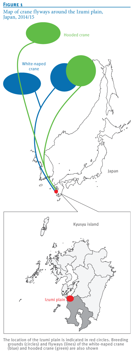
Last fall (see Japan: H5N8 Detected In Izumi Crane) H5N8 was detected among wild and migratory birds in the Izumi bird preserve on southern tip of Japan, a place famed for the yearly arrival and overwintering of thousands of rare Hooded, and White-naped cranes.

Both species spend their summers in Mongolia, Siberia, or Northwestern China - and of the roughly 10,000 hooded swans in the world - 80% overwinter in Izumi.


All of which serves as prelude to a Rapid Communications that appears in today’s Eurosurveillance that describes the discovery of at least 3 genetically distinct groups of the H5N8 virus.

Eurosurveillance, Volume 20, Issue 20, 21 May 2015

Rapid communications

Genetic diversity of highly pathogenic H5N8 avian influenza viruses at a single overwintering site of migratory birds in Japan, 2014/15

M Ozawa ()1,2,3,4, A Matsuu2,3,4, K Tokorozaki5, M Horie2,3, T Masatani2,3, H Nakagawa1, K Okuya1, T Kawabata2, S Toda5


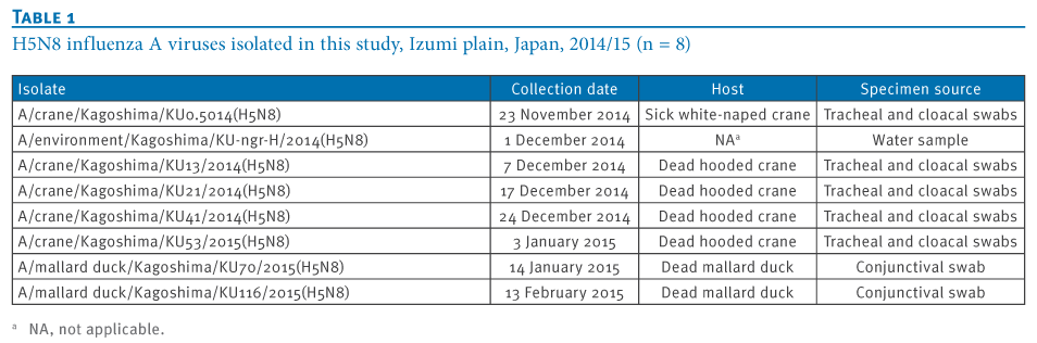
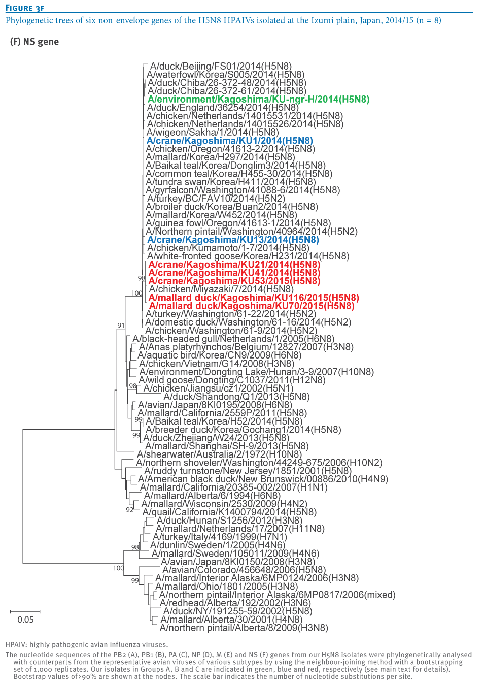
We isolated eight highly pathogenic H5N8 avian influenza viruses (H5N8 HPAIVs) in the 2014/15 winter season at an overwintering site of migratory birds in Japan. Genetic analyses revealed that these isolates were divided into three groups, indicating the co-circulation of three genetic groups of H5N8 HPAIV among these migratory birds. These results also imply the possibility of global redistribution of the H5N8 HPAIVs via the migration of these birds next winter.


In January 2014, newly discovered highly pathogenic H5N8 avian influenza viruses (H5N8 HPAIVs) caused outbreaks in poultry and wild birds in South Korea [1], although their ancestor had been isolated in China in 2013 [2]. Thereafter, these viruses have been circulating in both avian populations in South Korea [3,4] and sporadically in neighbouring countries, including China and Japan. Since November 2014, H5N8 HPAIVs have also appeared in poultry and wild birds in Europe [5,6]. Genetic analyses revealed that these isolates were closely related to the H5N8 viruses circulating in Korean birds. More recently, genetically similar HPAIVs also caused outbreaks in various avian species in North America [7]. These findings suggest that the H5N8 viruses have circulated and evolved in migratory birds.

<SNIP>

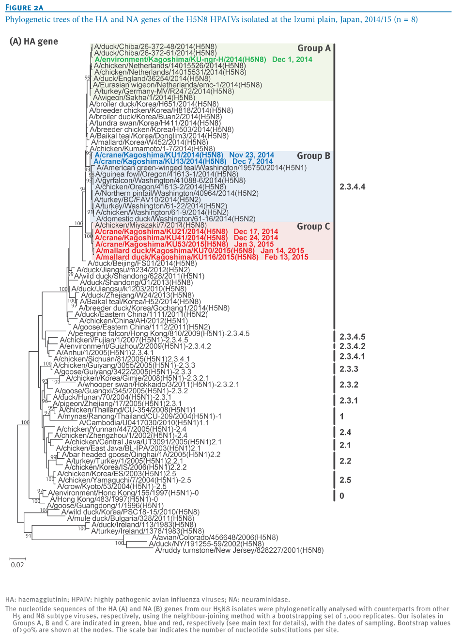
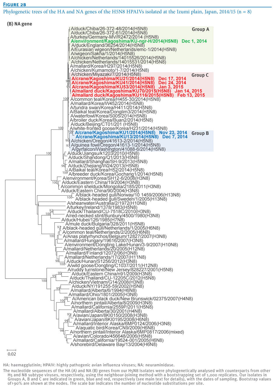
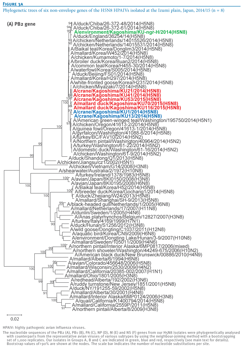
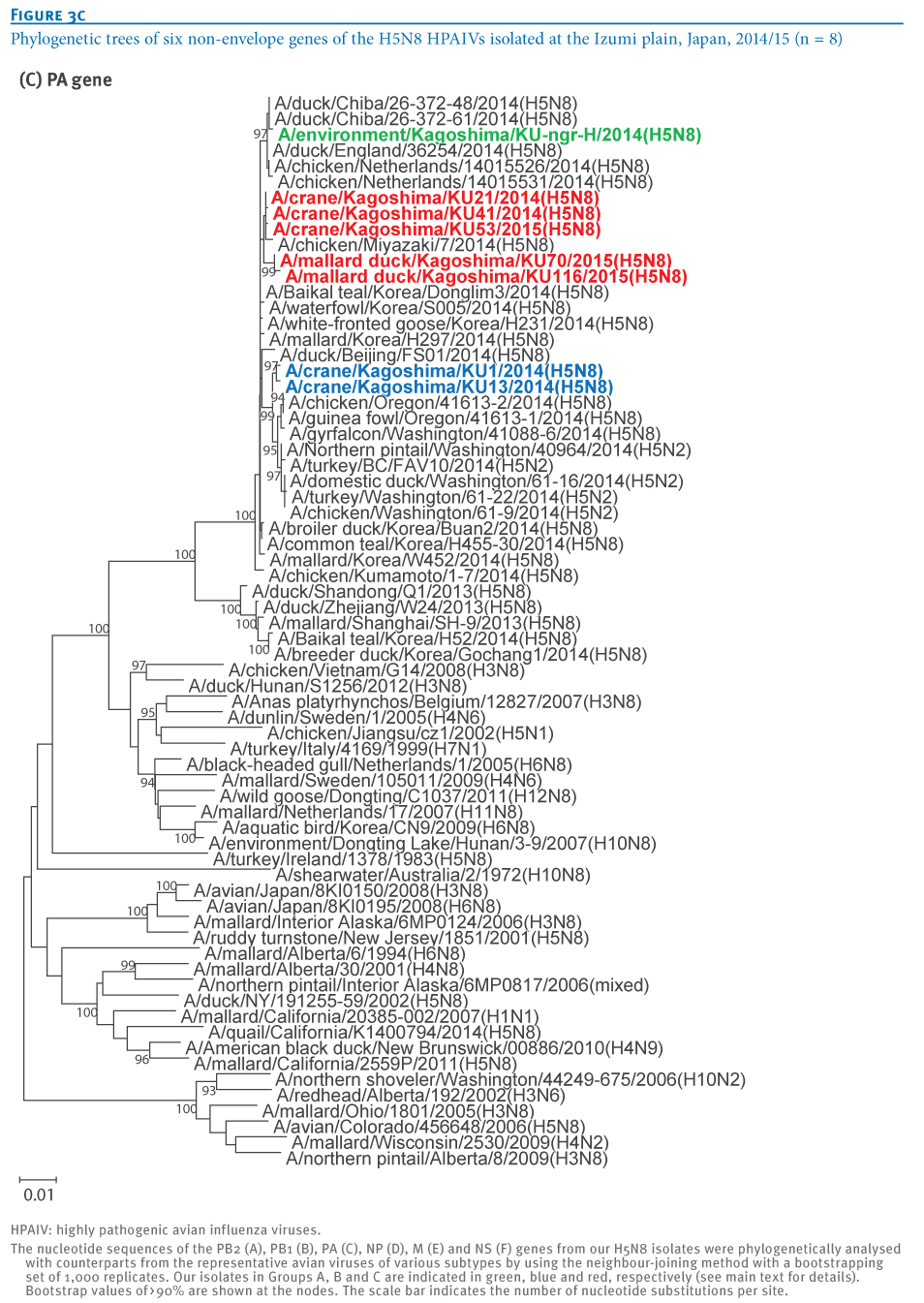
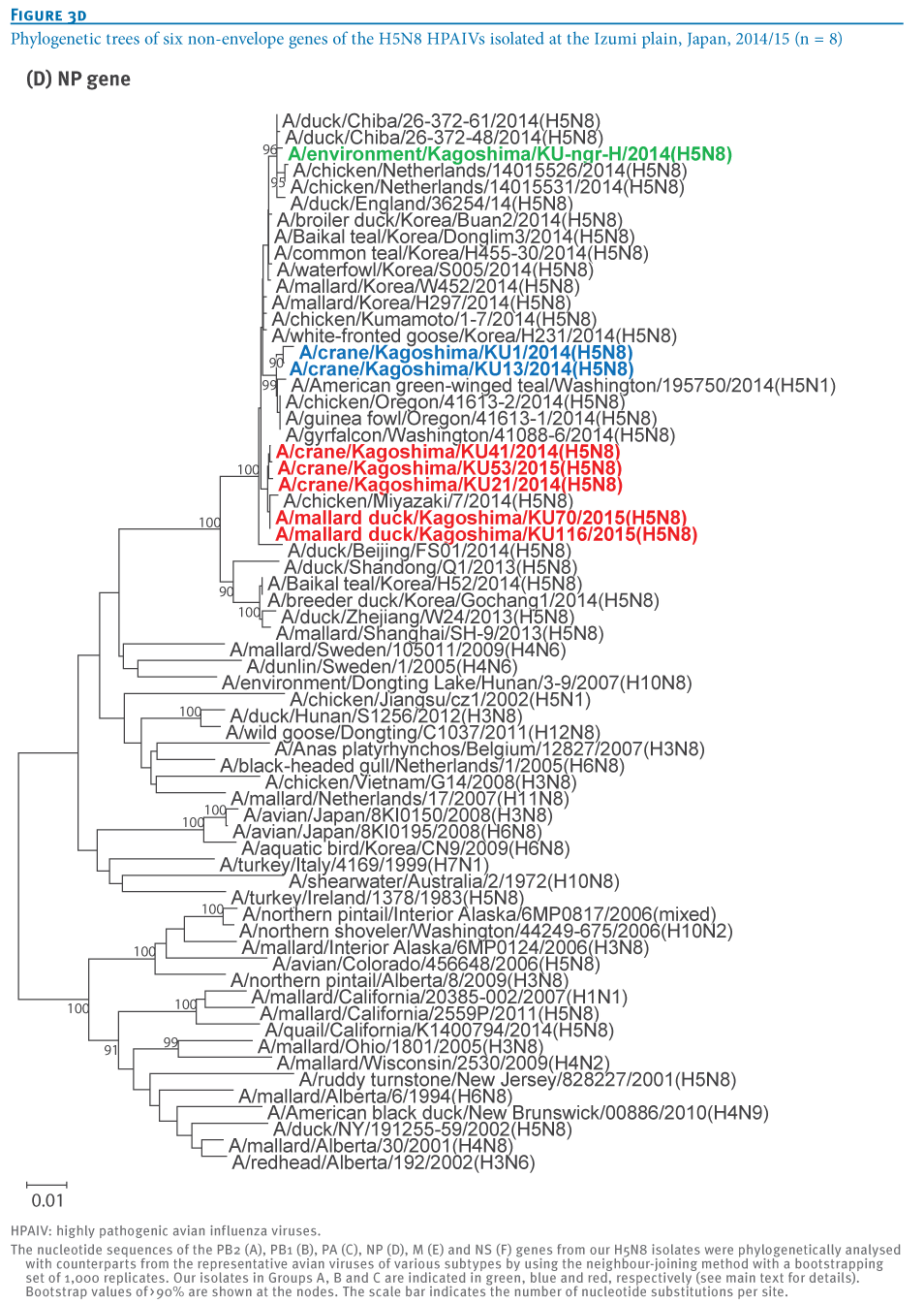
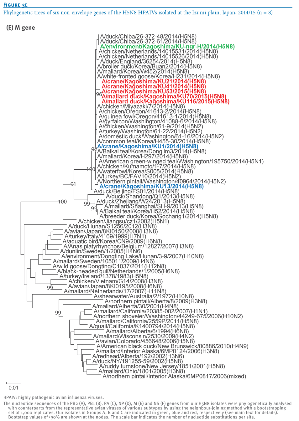
Phylogenetic analysis

To understand the genetic relationship between our isolates and related viruses, the HA and neuraminidase (NA) genes were phylogenetically analysed with counterparts from the representative avian influenza H5 (Figure 2A) and N8 (Figure 2B) subtypes, respectively.

We found that the H5 genes from our eight isolates belonged to clade 2.3.4.4 and were genetically divided into three groups. The water isolate, A/environment/Kagoshima/KU-ngr-H/2014(H5N8), fell into a phylogenetic cluster together with the European isolates and was closely related to two wild duck isolates in Japan (Group A, indicated in green in the Figures). The first and second crane isolates, A/crane/Kagoshima/KU1/2014(H5N8) and A/crane/Kagoshima/KU13/2014(H5N8), were genetically similar to the North American isolates (Group B, blue in the Figures). The HA genes of the rest of our isolates (Group C, red in the Figures), as well as a poultry isolate from Japan were clearly distinct from those of the other recent H5N8 isolates. These findings suggest that three genetically distinct groups of H5N8 HPAIVs were independently circulating among the migratory birds at the Izumi plain.

Intriguingly, the genetic grouping of our isolates matched broadly the dates of sampling; the forth to eighth isolates were categorised into Group C, while earlier isolates were categorised into Group A or B. To determine whether this virus group has genetic characteristics that become predominant among the migratory birds over the remaining virus groups, further investigation would be needed.

<SNIP>

No mutations were found that are known to confer the ability to infect mammalian hosts or to provide resistance against anti-influenza drugs to avian influenza viruses, with the exception of an asparagine at position 31 in the M2 protein, which confers resistance to the M2 ion channel blocker amantadine [11].


Conclusion


We isolated eight H5N8 HPAIVs from migratory birds and the water in their environment at the Izumi plain in southern Japan. Based on their genome sequences, these isolates were genetically divided into three groups. These results indicate the co-circulation of at least three genetic groups of H5N8 HPAIVs among the migratory birds overwintering at a single site in Japan. These H5N8 HPAIVs are most likely to be derived from wild ducks [12], rather than from cranes whose flyways were restricted to East Asian countries (Figure 1A). These findings also imply the possibility of global redistribution of the H5N8 HPAIVs via migration of these ducks next winter.


The birds that overwintered in Japan, Korea, and the Pacific Northwest last fall are now gathered in their summer breeding sites in northern China, Mongolia and Siberia -  sharing lakes, ponds, and streams and no doubt, the occasional avian virus -  and will be winging their way south again in a few short months. 


What new viral variants, clades, or reassortments we will see next fall and winter when they return is anyone’s guess.

Friday, April 03, 2015

Eurosurveillance: Emergence Of A Novel Cluster of H5N1 Clade 2.2.1.2

image

Credit Eurosurveillance 


# 9899


Over the past 5 months in Egypt we’ve witnessed the largest, and most sustained, outbreak of H5N1 among humans since the virus first emerged in China in 1996.  While numbers are murky, we are somewhere in the vicinity of 160 confirmed cases since November 1st, and roughly 50 deaths. 


Or roughly 50% higher than the greatest previous one year combined total (for 9 countries) of 115 cases set back in 2006.


Although we’ve seen a few small family clusters where H-2-H transmission may have occurred, we’ve seen no signs of increased human-to-human transmissibility of the H5N1 virus.


But obviously something has changed in Egypt. 


Last January in a CIDRAP report (see FAO Reports Mutations In H5N1 Virus From Egyptian Poultry) we learned that genetic characterizations of H5N1 viruses sampled from Egyptian poultry recently showed signs of several troubling `mammalian adaptations’, and we’ve seen repeated reports of large numbers of poultry outbreaks – even among previously vaccinated poultry (see Egypt H5N1: Poultry Losses Climbing, Prices Up 25%).


Yesterday the journal Eurosurveillance published a Rapid Communications characterizing the genetic changes in a rapidly spreading strain of H5N1 that  appears to have emerged in early 2014, and is quickly supplanting other strains of the virus in that region.


Eurosurveillance, Volume 20, Issue 13, 02 April 2015

Rapid communications

Emergence of a novel cluster of influenza A(H5N1) virus clade 2.2.1.2 with putative human health impact in Egypt, 2014/15

A S Arafa1,2, M M Naguib1,2,3, C Luttermann3, A A Selim1, W H Kilany1, N Hagag1, A Samy1, A Abdelhalim1, M K Hassan1, E M Abdelwhab1, Y Makonnen4, G Dauphin5, J Lubroth5, T C Mettenleiter3, M Beer3, C Grund3, T C Harder ()3


A distinct cluster of highly pathogenic avian influenza viruses of subtype A(H5N1) has been found to emerge within clade 2.2.1.2 in poultry in Egypt since summer 2014 and appears to have quickly become predominant. Viruses of this cluster may be associated with increased incidence of human influenza A(H5N1) infections in Egypt over the last months.


In Egypt, highly pathogenic avian influenza (HPAI) influenza A(H5N1) viruses of clade 2.2.1 and their descendants have been circulating in poultry populations since 2006, causing sporadic human infections [1]. Human influenza A(H5N1) infections in Egypt have been reported since the introduction of the virus in 2006 with 204 cases occurring until end of 2014 and a fatality rate of 35,8% in laboratory-confirmed cases reported to the World Health Organization (WHO). However, since January 2015, the incidence of human H5N1 cases in Egypt has increased dramatically: as of 21 March 2015, 116 human cases including 36 deaths have been reported to WHO [2]. This study was initiated to analyse molecular properties of H5N1 viruses that have caused outbreaks in poultry in Egypt since summer 2014 and to compare them with published sequences from H5N1 viruses obtained from recent human cases.

<SNIP>

Discussion

(Excerpt)


Our data confirm the emergence of an additional virus cluster within the Egyptian 2.2.1.2 clade of H5N1 HPAI viruses. Since November 2014, viruses of this new cluster appear to have become dominant over the previously described clade 2.2.1.2 phylotypes circulating in various poultry species. The only two publicly available sequences of viruses isolated from recent human H5N1 cases in Egypt show similar mutation patterns and fall into the same phylogenetic group. The molecular determinants that may improve the evolutionary fitness of these viruses need to be further clarified. The emergence of new clusters of H5N1 HPAI viruses in Egypt is not without precedence: In late 2007, a subclade of antigenic drift variants, later designated 2.2.1.1, emerged and expanded (clade 2.2.1.1a) in commercial poultry in Egypt but disappeared until end of 2010 [14] and, contrary to the current situation, did not replace 2.2.1 viruses. Viruses of clade 2.2.1.1 that emerged in 2007 hardly caused any human cases: according to the OpenFlu database [15]: only one of 100 H5N1 isolates from humans in Egypt belonged to clade 2.2.1.1; all others belonged to clade 2.2.1 and 2.2.1.2. In contrast, the emerging cluster identified in this study seems to be predominant across all poultry production sectors and has already caused a third of all human infections reported in Egypt since 2006 in only three months of 2015.


Given the endemic status of influenza H5N1 in poultry and the limitations of the reporting system of H5N1 HPAI virus outbreaks in poultry in Egypt, it is difficult to assess whether the altered epidemiological pattern of the emerging phylotype is due to altered biological properties in poultry or whether the increased incidence of infections in poultry merely reflects an increased viral burden across all poultry sectors in Egypt. In any case, the observed recent rise in outbreaks in poultry probably resulted in increased exposure risks for humans in contact with poultry, which may have caused an increased incidence in human cases. However, it can at this point not be excluded with certainty that the emerging phylotype of viruses may have increased zoonotic potential and may be transmitted more efficiently to humans, although this assumption cannot be drawn from the molecular evidence described here. Further studies of the pathogenicity and transmissibility of these viruses in humans, e.g. in the ferret model, are required. Concerted efforts of both veterinary and public health authorities are urgently needed to interrupt virus circulation in poultry in Egypt efficiently. This will help decrease the risk of human exposure to the virus.

(Continue . . .)


While the exact impact of these changes on the transmissibility of H5N1 to humans isn’t certain, its rapid spread among Egyptian poultry has – at the very least – increased the odds of human exposure. 


While the future course of the H5N1 virus is uncertain, the more opportunities it gets to infect humans, the more chances it will have to adapt to our physiology.  


Add in the recent emergence of H5N8, H5N6, H5N3 in Asia and novel reassortants of H5N2 and H5N1 in North America, and it is hardly surprising that the World Health Organization recently released a pointed warning that H5 Is Currently The Most Obvious Avian Flu Threat.

Thursday, November 06, 2014

Eurosurveillance: Acute Flaccid Paralysis Following EV-D68 Infection – France

image

CDC EV-D68 Fact Sheet


# 9294


In August of 2014, a seldom seen in North America non-polio enterovirus D-68 (EV-D68) appeared in America’s Midwest and quickly spread across the nation, causing a wide spectrum of respiratory illness, predominantly in young children and adolescents (see Kansas City Outbreak Identified As HEV 68).


At roughly the same time, a coincident rise in cases of neurological illness with AFP (acute flaccid paralysis) or limb weakness – often associated with a recent respiratory illness – was reported across the country.


In September the CDC  issued a HAN: Acute Neurologic Illness with Focal Limb Weakness of Unknown Etiology in Children, alerting doctors around the country to be aware of this trend, and providing information on reporting cases.


While there is a high degree of suspicion that the recent EV-D68 outbreak and the rise in paralytic cases are related, a causal link has not been established.  


The CDC’s latest EV-D68 update reads:


From mid-August to November 5, 2014, CDC or state public health laboratories have confirmed a total of 1,112 people in 47 states and the District of Columbia with respiratory illness caused by EV-D68.


While the CDC’s latest UPDATE on the unexplained neurological cases reads:

As of October 29, CDC has verified reports of 64 cases in 28 states that meet our case definition below. We are working with healthcare professionals and state and local officials to investigate all of these cases.

We are also in the process of verifying about half a dozen additional reports. These investigations take time. Therefore, the number of cases will likely increase further as we update these numbers weekly on Thursday, but the increase will not necessarily reflect changes in occurrence of cases in real time.


While it doesn’t prove causality, today the ECDC’s Eurosurveillance Journal carries a report of France’s first  EV-D68 case with Acute Flaccid Paralysis (AFP).


Eurosurveillance, Volume 19, Issue 44, 06 November 2014

Rapid communications

Acute flaccid paralysis following enterovirus D68 associated pneumonia, France, 2014

M Lang, A Mirand, N Savy, C Henquell, S Maridet, R Perignon, A Labbé, H Peigue-Lafeuille

Human enterovirus D68 (EV-D68) is known to be associated with mild to severe respiratory infections. Recent reports in the United States and Canada of acute flaccid paralysis (AFP) in children with detection of EV-D68 in respiratory samples have raised concerns about the aetiological role of this EV type in severe neurological disease. This case study is the first report of AFP following EV-D68 infection in Europe.


We report the first case of acute flaccid paralysis (AFP) following enterovirus-D68 (EV-D68) infection in Europe. The United States (US) and Canada are currently experiencing nationwide outbreaks of EV-D68 infections associated with severe respiratory diseases especially in children with underlying respiratory disease that began in mid-August 2014 [1,2]. Concomitantly, clusters of neurological illness characterised by AFP with anterior myelitis have been reported in the US and Canada [3,4]. The detection of EV-D68 in nasopharyngeal specimens of some affected children raises the question of a possible link between EV-D68 infections and severe neurological disease.

<SNIP>

Discussion

While EV-D68 has to date been almost exclusively associated with respiratory diseases, investigations are currently underway to determine its role in the acute neurological illnesses that have been reported in children in the US [3] and in Canada [4] since August 2014. Nine EV-D68-associated deaths are currently being investigated at the US Centers for Disease Control and Prevention (CDC) to confirm or refute EV-D68 as the cause of death [15]; as of 5 November, no information has been released about the death’s preceding symptoms.

The case reported here meets the definition given by CDC to identify similar neurological manifestations characterised by acute onset of focal limb weakness occurring on or after 1 August 2014 and MRI showing a spinal cord lesion largely restricted to grey matter [16]. Common features with the cases reported in the US include (i) respiratory illness preceding development of neurological symptoms, (ii) a local epidemiological context of EV-D68 detection among children admitted to hospital for respiratory infections leading to asthma crisis (data not shown) and (iii) EV-D68 detection in respiratory samples. By contrast, to our knowledge, neither meningeal syndrome nor myocarditis and acute respiratory distress syndrome had been reported in the days preceding the onset of paralysis in the US patients.

The enterovirus genome was not detected in the CSF of this patient and we cannot assert that EV-D68 was associated with meningitis. There are two case reports in the literature of EV-D68 infection associated with severe neurological disease as evidenced by detection in the CSF [17,18]. As in recent reports, the significance of EV-D68 association with AFP is hampered by the fact that it was only detected in respiratory or stool samples, in which enteroviruses can be detected many weeks after infection. However, the absence of detection in CSF does not necessarily rule out this possibility since poliovirus and EV-A71, two recognised neurotropic EVs, are not frequently recovered [19]. Further physiopathological studies may be needed to assess the neurotropism of EV-D68.

There are increasingly numerous reports of polio-like illnesses in the US (64 cases as of 30 October 2014) [15].

Surveillance of AFP cases has already been implemented as a measure in the global initiative to eradicate poliomyelitis and should allow rapid identification of similar neurological manifestations in association with EV-D68 infection [20]. However, determination of AFP aetiologies can be challenging, because of the absence of pathogen detection in the CSF. Investigation of AFP cases should include both EV screening of two stool samples collected ≥ 24 hours apart and < 14 days after symptom onset [21] and early and quick testing of diverse samples, especially upper respiratory samples, for infectious agents including EVs, to increase the chance to identify a pathogen. In the case of EV-D68 infections, the detection capabilities of the EV-D68 genome of commercial and in-house molecular methods should be assessed.


While our experience with EV-D68 goes back 50 years, the number of outbreaks that have been studied has been small. Testing has been difficult and time consuming, and treatment for EV-D68 is no different than for any other viral respiratory illness.   Therefore, we don’t really know as much about this virus as we’d like.


Other non-polio enteroviruses have a better documented track record for causing neurological complications, such as EV-71.  In recent years, EV-71 has been linked to a number of clusters of AFP  around the globe, particularly in Asia, Australia, and the Pacific (see Australia: Acute Flaccid Paralysis & EV71).


For now, the investigation into this rash of unexplained paralysis remains unresolved.

Thursday, September 11, 2014

Eurosurveillance: Stopping Ebola & R0 Calculations

image

# 9065


In somewhat the same vein as my earlier post Conventional Wisdom And Epidemic Disease Spread, the Journal Eurosurveillance has published an editorial and a paper on the transmissibility of the West African Ebola outbreak.  


Both paint a grim picture of what is to come if sufficient efforts are not mounted to stop this outbreak.


The editorial by A  J Kucharski  and  P Piot of the London School of Hygiene & Tropical Medicine, London warns that some of the near-apocalyptic case projections we discussed earlier today are not out of the question.  A couple of excerpts, but follow the link to read the article in its entirety.


Eurosurveillance, Volume 19, Issue 36, 11 September 2014

Editorials

Containing Ebola virus infection in West Africa

A J Kucharski 1, P Piot1

Ebola virus disease (EVD) is leaving a mark deeper and wider than ever before. The current outbreak now spans five countries in West Africa – Guinea, Liberia, Nigeria, Senegal and Sierra Leone – with over 4,200 cases and 2,200 deaths reported to the World Health Organization (WHO) as of 6 September 2014 (Figure 1) [1]. Unfortunately, with many cases either not reported or yet to show symptoms, the true number of infections is likely to be considerably higher. The first countries affected were among the world’s poorest, areas where long periods of civil wars have battered health services and eroded public trust. As a result, the outbreak has spread to other countries, and continues to expand. What began as a local problem has turned into an international crisis.

<SNIP>

Ebola cannot be ignored in the hope it will burn itself out. It is true that outbreaks of acute infections will generally decline once a large number people have been infected, because there are no longer enough susceptible individuals to sustain transmission. But if Ebola indeed has a reproduction number of 2 in some locations as described by Nishiura et al. [8], the susceptible pool – which likely includes most individuals – would have to shrink by at least half before the outbreak declined of its own accord [17]. Given the vast populations in affected areas and the disease’s high fatality rate, this is clearly not an acceptable scenario.

(Continue . . . )

The second study looks at the R0 of this outbreak, something we’ve looked at previously in PLoS Currents: Calculating An R0 For Ebola. As a refresher, the R0 (pronounced R-nought) or Basic Reproductive Number is essentially the number of new cases in a susceptible population likely to arise from a single infection.


image

With an R0 below 1.0, a virus (as an outbreak) begins to sputter and dies out. Above 1.0, and an outbreak can have `legs’.


Like the earlier study, today’s report finds the R0 of the Ebola outbreak to be well over 1.0. More than enough to sustain the epidemic.


Eurosurveillance, Volume 19, Issue 36, 11 September 2014

Rapid communications

Early transmission dynamics of Ebola virus disease (EVD), West Africa, March to August 2014

H Nishiura ()1, G Chowell2,3


Date of submission: 23 August 2014


The effective reproduction number, Rt, of Ebola virus disease was estimated using country-specific data reported from Guinea, Liberia and Sierra Leone to the World Health Organization from March to August, 2014. Rt for the three countries lies consistently above 1.0 since June 2014. Country-specific Rt for Liberia and Sierra Leone have lied between 1.0 and 2.0. Rt<2 indicate that control could be attained by preventing over half of the secondary transmissions per primary case.

(continue . . . )


Thursday, August 14, 2014

Eurosurveillance: Anthrax Encounters Of The 4th Kind

image

Credit CDC 


# 8954


Traditionally there are three ways to contract anthrax. 

  • Cutaneous, or through openings in the skin (the most common form of infection)
  • Gastrointestinal from the consumption of infected meat
  • And inhalational anthrax, from inhaling the spores.


Two years ago in UK: The High Cost Of Getting High we looked at the emergence of a rare 4th way in which people were getting infected with Anthrax in the UK, and across Europe; through the injection of recreational heroin.


In recent years, batches of heroin have been reported across Europe contaminated with anthrax spores, likely an unintentional contamination during the processing or transporting of the drug – and as you might expect – injecting contaminated heroin often turns out badly for the user.


The first cluster was reported in Scotland in 2009, and involved more than 100 cases. Cases resurfaced again in England and parts of Europe in 2012 (see HPA  Case of anthrax confirmed in Oxford). Today the Journal Eurosurveillance  has published a review of the literature, urging that clinicians learn to recognize the signs so that treatment can begin as early as possible.


Eurosurveillance, Volume 19, Issue 32, 14 August 2014

Review articles

Injectional anthrax - new presentation of an old disease

T Berger ()1,2, M Kassirer1, A A Aran1,3

  1. Israel Defense Force, Medical Corps, Ramat-Gan, Israel
  2. Department of Internal Medicine D, Rabin Medical Center, Beilinson Hospital, Petah-Tikva, Israel
  3. Department of Pediatric Intensive Care, The Edmond and Lily Safra Children's Hospital, The Chaim Sheba Medical Center, Tel-Hashomer, Ramat-Gan, Israel

Bacillus anthracis infection (anthrax) has three distinct clinical presentations depending on the route of exposure: cutaneous, gastrointestinal and inhalational anthrax. Each of these can lead to secondary bacteraemia and anthrax meningitis.

Since 2009, anthrax has emerged among heroin users in Europe, presenting a novel clinical manifestation, ’injectional anthrax’, which has been attributed to contaminated heroin distributed throughout Europe; before 2009 only one case was reported. During 2012 and 2013, new cases of injectional anthrax were diagnosed in Denmark, France, Germany, and the United Kingdom. Here we present a comprehensive review of the literature and information derived from different reporting systems until 31 December 2013.

Overall 70 confirmed cases were reported, with 26 fatalities (37% case fatality rate).The latest two confirmed cases occurred in March 2013. Thirteen case reports have been published, describing 18 confirmed cases. Sixteen of these presented as a severe soft tissue infection that differed clinically from cutaneous anthrax, lacked the characteristic epidemiological history of animal contact and ten cases required complimentary surgical debridement. These unfamiliar characteristics have led to delays of three to 12 days in diagnosis, inadequate treatment and a high fatality rate. Clinicians’ awareness of this recently described clinical entity is key for early and successful management of patients.

(Continue . . . )


Of note, no human-to-human transmission has been documented among injectional anthrax cases.  While inhalational anthrax doesn’t spread from human to human, there is a slight risk of transmission from drainage from opens sores that come with the cutaneous form.


Although most of the cases in today’s report presented with serious soft tissue infections – up to, and including necrotising fasciitis - none showed the classic black lesion (eschar) that gives the disease its name (anthrax is Greek for `coal’).

image

Credit CDC PHIL

Gastrointestinal, respiratory and neurological symptoms were also described, with disease progression often leading to septic shock, organ failure, and sometimes death.


The authors wrote:


Injectional anthrax presents a challenge for physicians often due to lack of evident case clusters, unfamiliar clinical presentation and severe course of disease. Unlike cutaneous anthrax, injectional anthrax is typically a systemic infection with high mortality rate (Table 4). This may be attributed to the deeper and greater inoculation of spores, higher rates of septicaemia, delayed diagnosis and to factors specific to drug addicts including delayed medical consultation, malnutrition, presence of concomitant diseases such as HIV infection and defective immune response.


For people who inject drugs (PWIDs), anthrax is admittedly pretty far down the list of the bad things that can happen to you.  Even during the biggest outbreak in Scotland,  the incidence was estimated to be only 1.96 infections per thousand addicts.


During the `bad old days’ of the 1970s, I would see 1 or 2 heroin overdoses per shift, and many of them were well beyond any help from the bolus of Narcan (naloxone) I carried when when I arrived.


Beyond simple O.D.s, viral and bacterial infections are a constant danger, and can result in illness and even death. Hepatitis C, Hepatitis B, HIV, wound botulism, Staphylococcus aureus, and even tetanus are among the nasties that drug users are at increased risk of contracting.


While the sharing of needles is the cause of many of these infections, you don’t have to indulge in that particularly unwise practice to end up with a potentially fatal infection. Reusing your own needles, or injecting into a contaminated (dirty) arm, can easily introduce bacteria into the user’s system


Proof, I suppose, that we humans seem to have an infinite capacity to find new ways to wreak havoc on ourselves.


To close this out, a little good news. According to the CDC, this 4th type of Anthrax exposure hasn’t been reported here in the United States.


Injection Anthrax

Recently, another type of anthrax infection has been identified in heroin-injecting drug users in northern Europe. This type of infection has never been reported in the United States.

Symptoms may be similar to those of cutaneous anthrax, but there may be infection deep under the skin or in the muscle where the drug was injected. Injection anthrax can spread throughout the body faster and be harder to recognize and treat. Lots of other more common bacteria can cause skin and injection site infections, so a skin or injection site infection in a drug user does not necessarily mean the person has anthrax.

Friday, June 27, 2014

Eurosurveillance: Genetic Tuning Of Avian H7N9 During Interspecies Transmission

image


# 8788


This week’s Eurosurveillance bonanza of avian flu transmission & evolution papers has more than enough good stuff to keep us busy reading for hours. All of these papers are of great interest, but one in particularly grabbed my attention, mainly due to the potential impact of its findings.


A study that looks at the continual evolution of the H7N9 virus in Mainland China.


To recap: in February of last year a new and dangerous avian influenza virus (H7N9)  jumped to humans in Eastern China, although we did not learn about it until the end of March (see More Details Emerge On Shanghai H7N9 Case). Unlike many of the other avian influenza viruses we’ve seen - this virus produced no visible signs of illness in poultry - making it particularly difficult to detect and control.


Benign in poultry, but not in humans, H7N9 often produces a severe form of pneumonia.  One that has killed roughly 30% of those known to have been infected.


The only saving grace has been this virus has not yet achieved the ability to transmit efficiently from one human to another. The vast majority of human cases appear to be the result of direct exposure to infected birds or to their environment.


In that first wave about 130 human cases emerged, tapering off only after aggressive controls on live bird markets were imposed in April and May of 2013 (see The Lancet: Poultry Market Closure Effect On H7N9 Transmission). After a quiescent summer, colder fall and winter temperatures brought with it a resurgence in the number of human cases, with the second wave roughly double the size of the first (cite).

image

Two Waves of H7N9  - Credit Hong Kong’s CHP


A month ago, in EID Journal: H7N9 As A Work In Progress, we looked at a study that found the H7N9 avian virus continues to reassort with local H9N2 viruses, making the H7N9 viruses that circulated in wave 2 genetically distinct from those that were seen during the 1st wave.


As we’ve discussed before, the genetic contributions from the avian H9N2 virus appear to be significant. 


Of the three avian flu viruses we are currently watching with the most concern – H5N1, H7N9, and H10N8 – all  share several important features (see Study: Sequence & Phylogenetic Analysis Of Emerging H9N2 influenza Viruses In China):

    • They all first appeared in  Mainland China
    • They all  have come about through viral reassortment in poultry
    • And most telling of all, while their HA and NA genes differ - they all carry the internal genes from the avian H9N2 virus


It turns out the relatively benign and ubiquitous H9N2 is actually a fairly promiscuous virus, as bits and pieces of it keep turning up in new reassortant viruses.  See PNAS: Reassortment Of H1N1 And H9N2 Avian viruses & PNAS: Reassortment Potential Of Avian H9N2 for some earlier looks at H9N2’s active social life.


Today, in research from a group of scientists working for China’s National and Provincial CDCs, we learn that the genetic diversity of the H7N9 virus is even greater than previously described, and that continual reassortment with the H9N2 virus, along with passage through a variety of host species, appears to be influencing its ongoing evolution.


A process the authors call `genetic tuning’.


Non-scientists will likely find this article tough sledding (parts certainly were for me), as it is more than a little technical.  It is, however, a fascinating paper. 


With apologies in advance to any real scientists who may be reading this - come back after the link and abstract - and I’ll do my best to hack through some of the tall grass and go over a few of the highlights.


          Eurosurveillance, Volume 19, Issue 25, 26 June 2014

Research articles

Genetic tuning of the novel avian influenza A(H7N9) virus during interspecies transmission, China, 2013

D Wang1,2, L Yang1,2, R Gao1, X Zhang3, Y Tan4, A Wu5, W Zhu1, J Zhou1, S Zou1, Xiyan Li1, Y Sun6, Y Zhang7, Y Liu8, T Liu9, Y Xiong10, J Xu11, L Chen12, Y Weng13, X Qi14, J Guo1, Xiaodan Li1, J Dong1, W Huang1, Y Zhang1, L Dong1, X Zhao1, L Liu1, J Lu1, Y Lan1, H Wei1, L Xin1, Y Chen1, C Xu1, T Chen1, Y Zhu1, T Jiang5, Z Feng15, W Yang15, Y Wang15, H Zhu16, Y Guan16, G F Gao15, D Li1, J Han1, S Wang1, G Wu1, Y Shu ()1

Date of submission: 28 July 2013


A novel avian influenza A(H7N9) virus causing human infection emerged in February 2013 in China. To elucidate the mechanism of interspecies transmission, we compared the signature amino acids of avian influenza A(H7N9) viruses from human and non-human hosts and analysed the reassortants of 146 influenza A(H7N9) viruses with full genome sequences.

We propose a genetic tuning procedure with continuous amino acid substitutions and reassorting that mediates host adaptation and interspecies transmission.

When the early influenza A(H7N9) virus, containing ancestor haemagglutinin (HA) and neuraminidase (NA) genes similar to A/Shanghai/05 virus, circulated in waterfowl and transmitted to terrestrial poultry, it acquired an NA stalk deletion at amino acid positions 69 to 73. Then, receptor binding preference was tuned to increase the affinity to human-like receptors through HA G186V and Q226L mutations in terrestrial poultry. Additional mammalian adaptations such as PB2 E627K were selected in humans.

The continual reassortation between H7N9 and H9N2 viruses resulted in multiple genotypes for further host adaptation. When we analysed a potential association of mutations and reassortants with clinical outcome, only the PB2 E627K mutation slightly increased the case fatality rate. Genetic tuning may create opportunities for further adaptation of influenza A(H7N9) and its potential to cause a pandemic.


What these researchers did was to collect specimens (as well as clinical and epidemiological information) from human H7N9 cases, along with avian and environmental samples from areas where human cases were identified. 


From this they assembled 173 influenza A(H7N9) viruses (103 human and 70 non-human) and analyzed them to try to determine how (and from where) they had evolved.


Remarkably, out of 146 H7N9 viruses with full genome sequences, they detected at least 26 seperate genotypes, mostly from the first wave in 2013. Of those 26, twenty were only detected once or twice, suggesting they were transient, and perhaps not as `biologically fit’ as some of the other genotypes.


Based on their observations, the authors propose that `a genetic tuning procedure with continuous amino acid substitutions and reassortations, mediates the host adaptation and interspecies transmission of H7N9 viruses (Figure 4)’.

image


Essentially, they describe two processes that they believe  facilitate the evolution and adaptation of the virus.  Processes that may be `tuning’ the virus in the direction of  a `human-adapted’ pathogen.


The first is ongoing reassortment with H9N2 viruses.  


Reassortment occurs when two different influenza viruses infect the same host simultaneously.  In `close quarters’ they can swap out gene segments, and if they hit the right combination, generate a successful hybrid virus. 

image


Reassortment also produces the biggest, and most abrupt changes in the virus, and is believed the mechanism behind the emergence of many pandemic viruses.  You can view a short (3 minute) video from NIAID on reassortment here.


Based on 26 distinct genotypes described in this paper, the reassortment of H7N9 appears to be a vigorous, and ongoing, process. The greatest concentration of genotypes was found in the Yangtze river delta (see map below), suggesting this may be the region where the virus first emerged.

image


The second evolutionary path occurs as these reassortant viruses passage through different species and pick up specific amino acid changes.


When a virus infects a cell, it immediately sets upon making thousands of copies of itself in order to spread the infection throughout the host. Single-stranded RNA influenza viruses are notoriously sloppy replicators, so invariably, some of these viral copies will carry small transcription errors (in the form of amino acid substitutions).


Most of these `variants’  will prove either neutral or perhaps even detrimental to the survival and propagation of the virus, but occasionally a helpful change occurs (positive selection) that increases the `biological fitness’ of the virus  – at least for the current host species. 


Viral progeny that are the best suited for their host usually win the replication wars, and soon outnumber and overrun less `fit’ variants. As a result, a better `adapted’ virus can emerge.  And if those adaptations help it jump to another host species, it is a viral win-win.


The authors point out that the `mixed bird’ environment of live markets may have helped H7N9’s evolution along, as it was able to spread stealthily, and without interruption, among a variety of species – picking up useful adaptations along the way.


As an example, avian flu viruses bind preferentially to the α2-3 receptor cells found in the gastrointestinal tract of birds.  But the H7N9 virus also binds (albeit, not as robustly) to human α2-6 human receptor cells, which are found in mammalian tracheas and upper airways (see Nature: Receptor Binding Of H7N9).


The authors speculate H7N9’s partial affinity to α2-6 receptor cells may have been picked up when it passaged through quail or pigeons, which are known to carry both types of cells.


And even once it infects man, the H7N9 virus continues to adapt and evolve, with the PB2 E627K mutation detected in a large number of human isolates.  E627K and/or D701N mutations in the PB2 protein are considered critical for mammalian adaptation of avian influenza viruses, as they allow the virus to replicate efficiently in the lower temperatures found in the upper airway.


As H7N9 reassorts and passages through different species – a process the authors call `genetic tuning’ - it continues to evolve, and reinvent itself.  Meaning that the virus we get next fall , winter, or spring may not act like the virus we saw during the first two waves. 


Obviously, I’ve just covered some of the highlights, and then, only with the broadest of strokes.  I’m certain many of my readers will want to read the entire paper.  But to close, I’ll let the authors speak to the significance of their findings.


Genetic tuning not only mediated species switching, but may also allow the virus to adapt so that it infects humans more easily and transmits among people more efficiently. Recently, Malaysia reported its first human case of influenza A(H7N9), imported from Guangdong province, China [28]. Rapid transportation and frequent travelling have made it possible to transfer the virus from China to other regions.


Overall, due to the genetic tuning procedure, the potential pandemic risk posed by the novel avian influenza A(H7N9) viruses is greater than that of any other known avian influenza viruses. A response to this threat requires the combined effort of different sectors related to human health, poultry and wild birds, as well as vigilance and co-operation of the world.

Thursday, June 26, 2014

Eurosurveillance: Papers on potential transmissibility and evolution of avian influenza A viruses

Eurosurveillance banner


# 8787


While I’ll probably return later today or tomorrow with a deeper look at some of these papers (after I’ve had a chance to actually read them), but for now I’ll just post the links to what looks like an interesting bunch of avian flu centric research.


Eurosurveillance, Volume 19, Issue 25, 26 June 2014

Table of Contents


Editorials


Epidemiological and genetic investigations of human-to-human transmission of zoonotic influenza viruses

by S Herfst, R Fouchier

Surveillance and outbreak reports


Two clustered cases of confirmed influenza A(H5N1) virus infection, Cambodia, 2011

by N Chea, SD Yi, S Rith, H Seng, V Ieng, C Penh, S Mardy, D Laurent, B Richner, T Sok, S Ly, P Kitsutani, N Asgari, MC Roces, P Buchy, A Tarantola

Transmission of avian influenza A(H7N9) virus from father to child: a report of limited person-to-person transmission, Guangzhou, China, January 2014

by XC Xiao, KB Li, ZQ Chen, B Di, ZC Yang, J Yuan, HB Luo, SL Ye, H Liu, JY Lu, Z Nie, XP Tang, M Wang , BJ Zheng

Research articles


Limited human-to-human transmission of avian influenza A(H7N9) virus, Shanghai, China, March to April 2013

by J Hu, Y Zhu, B Zhao, J Li, L Liu, K Gu, W Zhang, H Su, Z Teng, S Tang, Z Yuan, Z Feng, F Wu

Genetic tuning of the novel avian influenza A(H7N9) virus during interspecies transmission, China, 2013

by D Wang, L Yang, R Gao, X Zhang, Y Tan, A Wu, W Zhu, J Zhou, S Zou, X Li, Y Sun, Y Zhang, Y Liu, T Liu, Y Xiong, J Xu, L Chen, Y Weng, X Qi, J Guo, X Li, J Dong, W Huang, L Dong, X Zhao, L Liu, J Lu, Y Lan, H Wei, L Xin, Y Chen, C Xu, T Chen, Y Zhu, T Jiang, Z Feng, W Yang, Y Wang, H Zhu, Y Guan, GF Gao, D Li, J Han, S Wang, G Wu, Y Shu

Genesis of the novel human-infecting influenza A(H10N8) virus and potential genetic diversity of the virus in poultry, China

by W Qi, X Zhou, W Shi, L Huang, W Xia, D Liu, H Li, S Chen, F Lei, L Cao, J Wu, F He, W Song, Q Li, M Liao, M Li

Thursday, June 12, 2014

Eurosurveillance: MERS-CoV Antibodies & RNA In Camel’s Milk – Qatar

Photo: ©FAO/Ami Vitale

Credit FAO


# 8736


Today’s Eurosurveillance Journal is MERS-centric, with no less than four articles or features on this emerging coronavirus.   First, links to today’s studies, then a closer look at one focusing on MERS and camel’s milk.


Seroepidemiology of Middle East respiratory syndrome (MERS) coronavirus in Saudi Arabia (1993) and Australia (2014) and characterisation of assay specificity

by MG Hemida, RA Perera, RA Al Jassim, G Kayali, LY Siu, P Wang, KW Chu, S Perlman, MA Ali, A Alnaeem, Y Guan, LL Poon, L Saif, M Peiris

Middle East respiratory syndrome coronavirus (MERS-CoV) RNA and neutralising antibodies in milk collected according to local customs from dromedary camels, Qatar, April 2014

by CB Reusken, EA Farag, M Jonges, GJ Godeke, AM El-Sayed, SD Pas, VS Raj, KA Mohran, HA Moussa, H Ghobashy, F Alhajri, AK Ibrahim, BJ Bosch, SK Pasha, HE Al-Romaihi, M Al-Thani, SA Al-Marri, MM AlHajri, BL Haagmans, MP Koopmans

Research articles


Assessment of the Middle East respiratory syndrome coronavirus (MERS-CoV) epidemic in the Middle East and risk of international spread using a novel maximum likelihood analysis approach

by C Poletto, C Pelat, D Lévy-Bruhl, Y Yazdanpanah, PY Boëlle, V Colizza

News


The 2014 Hajj and Umrah – current recommendations

by Eurosurveillance editorial team


The study with perhaps the greatest immediate impact looks at a possible route of transmission of the virus from camels to humans.


Eurosurveillance, Volume 19, Issue 23, 12 June 2014

Rapid communications

Middle East respiratory syndrome coronavirus (MERS-CoV) RNA and neutralising antibodies in milk collected according to local customs from dromedary camels, Qatar, April 2014

C B Reusken1,2, E A Farag2,3, M Jonges2,4, G J Godeke4, A M El-Sayed3, S D Pas1, V S Raj1, K A Mohran5, H A Moussa6, H Ghobashy5, F Alhajri5, A K Ibrahim6,7, B J Bosch8, S K Pasha6, H E Al-Romaihi3, M Al-Thani3, S A Al-Marri3, M M AlHajri ()3, B L Haagmans1, M P Koopmans1,4


  • Antibodies to Middle East respiratory syndrome coronavirus (MERS-CoV) were detected in serum and milk collected according to local customs from 33 camels in Qatar, April 2014. At one location, evidence for active virus shedding in nasal secretions and/or faeces was observed for 7/12 camels; viral RNA was detected in milk of five of these seven camels. The presence of MERS-CoV RNA in milk of camels actively shedding the virus warrants measures to prevent putative food-borne transmission of MERS-CoV.

    In April 2014, serum, nasal swabs and rectal swabs were taken from 33 milking dromedary camels at two locations in Qatar (Al Shahaniya and Dukhan), areas with known Middle East respiratory syndrome coronavirus (MERS-CoV) circulation in camels [1] and data not shown. In addition, milk was collected from these animals according to local customs. Serum samples and milk were tested for the presence of MERS-CoV-specific antibodies by protein microarray, with confirmation by virus neutralisation.

    Swabs and milk were tested for the presence of MERS-CoV RNA by real-time reverse transcription (RT)-PCR testing for multiple genomic targets. Antibodies to MERS-CoV were detected in serum and milk from all camels at both locations. At the Dukhan location, none of the 21 animals tested was actively shedding viral RNA from the nose and/or in faeces and no evidence for the presence of MERS-CoV RNA in milk was observed. At the Al Shahaniya location, evidence for active virus shedding was observed for seven of the 12 camels tested. Viral RNA was detected in milk of five of the seven camels with active virus shedding.

  •  


    While important findings, there are a few caveats due.


    First, the degree of RNA detection was pretty low – too low to allow for virus isolation.  And while viral RNA was detected in the some of the milk samples, the milk was collected `according to local customs’ – which means the camel’s udders were not cleaned prior to milking (and milking takes place immediately after calves have suckled), meaning the milk could have been contaminated from outside sources.


    Of course, whether the milk is contaminated during collection or while still in the udder, becomes somewhat moot if people are being infected from consuming it. At this point, however, it isn’t clear whether there were enough infectious virus particles in the raw milk to transmit the disease.


    The low level of virus detection could also have been influenced by the rigors (both time & environmentally related) of shipment of samples to labs outside of Qatar.  It is also possible that the existence of antibodies (detected in milk samples as well) could have affected the amount of virus detected in vitro.


    More research, particularly under better controlled conditions, is needed.  The authors conclude:


    Nevertheless, it can be concluded that the presence of MERS-CoV RNA in raw milk as consumed locally might represent a source for zoonotic transmission of MERS-CoV and prudence is called for. Munster et al. showed that heat treatment (30 minutes at 63 °C) of MERS-CoV-containing camel milk reduced levels of infectious virus below detection level [24]. Boiling milk before consumption could be an easy, achievable local measure to prevent transmission and to preserve consumption of camel milk.


    As always, I would urge my readers to follow the link and read this report in its entirety, as I’ve only hit a few of the highlights. For an earlier look at the potential for milk to carry, and potentially convey, the MERS virus, you may wish to revisit:


    EID Journal: Stability Of MERS-CoV In Milk

    Thursday, May 29, 2014

    Eurosurveillance: Rapid Communications On Netherlands Imported MERS Cases

    image


    # 8680


    The journal Eurosurveillance has published a detailed rapid communications on the two related - imported (from KSA) - cases of MERS to the Netherlands reported earlier this month (see here & here).  


    There is far too much information here to excerpt efficiently, so I’ll simply post the abstract, the link, and a snippet from the discussion – but I would invite you to read it in its entirety on the Eurosurveillance site.


    Eurosurveillance, Volume 19, Issue 21, 29 May 2014

    Rapid communications

    Middle East respiratory syndrome coronavirus (MERS-CoV) infections in two returning travellers in the Netherlands, May 2014

    M Kraaij – Dirkzwager ()1, A Timen1, K Dirksen2, L Gelinck3, E Leyten3, P Groeneveld4, C Jansen3, M Jonges5, S Raj6, I Thurkow7, R van Gageldonk-Lafeber8, A van der Eijk6, M Koopmans5,6, on behalf of the MERS-CoV outbreak investigation team of the Netherlands9


    Citation style for this article: Kraaij – Dirkzwager M, Timen A, Dirksen K, Gelinck L, Leyten E, Groeneveld P, Jansen C, Jonges M, Raj S, Thurkow I, van Gageldonk-Lafeber R, van der Eijk A, Koopmans M, on behalf of the MERS-CoV outbreak investigation team of the Netherlands. Middle East respiratory syndrome coronavirus (MERS-CoV) infections in two returning travellers in the Netherlands, May 2014. Euro Surveill. 2014;19(21):pii=20817. Available online: http://www.eurosurveillance.org/ViewArticle.aspx?ArticleId=20817
    Date of submission: 21 May 2014


    Two patients, returning to the Netherlands from pilgrimage in Medina and Mecca, Kingdom of Saudi Arabia, were diagnosed with Middle East respiratory syndrome coronavirus (MERS-CoV) infection in May 2014. The source and mode of transmission have not yet been determined. Hospital-acquired infection and community-acquired infection are both possible.


    On 13 May 2014, a Dutch patient, returning to the Netherlands from pilgrimage in Medina and Mecca, Kingdom of Saudia Arabia, was diagnosed with Middle East respiratory syndrome coronavirus (MERS-CoV) infection, followed by diagnosis of a second patient, belonging to the same tour group, the day after. Here we describe the two cases and the public health response. The case definition that is used in the Netherlands is outlined in the Box.

    <BIG SNIP>

    Discussion

    There are several options for the possible source of the infection of the two Dutch cases: Case 1 could have been infected during the hospital visit of his child on 29 April, after which he infected Case 2. Alternatively, both could have been exposed to a common, as yet unknown, source in Medina. Thirdly, each case could have been infected through different sources (hospital/ community), though this seems unlikely, as the (partial) virus sequence of both cases was nearly identical.

    The resemblance in strain sequence between the Dutch cases and the case from the US is remarkable as the cases did not visit the same places in the Kingdom of Saudi Arabia. Exchange of information between the US Centers for Disease Control and Prevention and Dutch experts did not reveal any clues about mutual exposure of the Dutch and US cases.

    The current, limited scientific information does not support any conclusion on the meaning of this genetic resemblance, knowing that multiple lineages of the virus can be found in camels and people [2,12]. Continued vigilance in evaluation of contacts of imported cases, including molecular testing and serology, will hopefully lead to better insights.

    (Continue . . . )


    Thursday, April 24, 2014

    Eurosurveillance Journal: MERS- 2 Years Into The Epidemic

    image

    Credit Eurosurveillance


    # 8524


    From Eurosurveillance Journal this afternoon an editorial penned by the ECDC’s Director Marc Sprenger and  D Coulombier on the first two years of the MERS epidemic, with data current through April 23rd.


    This editorial is actually just one of three articles on MERS in this weeks edition, the others focus on the recent imported case in Greece and additional findings of the MERS virus in camels.


    All three reports are well worth reading, but the editorial below provides the broadest overview of the situation. I’ve included a few excerpts, but the entire editorial is worth reading.  I’ll have a final comment when you return.


    Eurosurveillance, Volume 19, Issue 16, 24 April 2014

    Editorials

    Middle East Respiratory Syndrome coronavirus – two years into the epidemic

    M Sprenger1, D Coulombier ()1

    1. European Centre for Disease Prevention and Control (ECDC), Stockholm, Sweden

    Euro Surveill. 2014;19(16):pii=20783. Available online: http://www.eurosurveillance.org/ViewArticle.aspx?ArticleId=20783 Date of submission: 24 April 2014


    Two years ago, on 23 April 2012, media reported a cluster of severe respiratory infection in a hospital in Jordan [1]. Only several months later did it become evident that this was the first known occurrence of the new Middle East Respiratory Symptom coronavirus (MERS-CoV) that since then continues to puzzle scientists and public health experts alike.

    <BIG SNIP>

    MERS-CoV infections present with a high case-fatality ratio, multiple transmission routes are suspected, cases are reported among healthcare workers, multiple disease foci are affecting SA, and cases have been exported. All these facts are criteria for considering declaring a public health event of international concern listed in annex II of the WHO international health regulations [18]. Two years and 345 cases after the start of this epidemic, we remain with many unanswered questions and lack serological studies and sequences from human cases.

    Currently, SA bears the main burden of managing the MERS-CoV epidemic and lately also the UAE. So far, cases detected outside the Arabian Peninsula have not resulted in sustained onward transmission. However, the recent rapid change in the epidemiological pattern of the disease should call for a change of approach to ensure a rapid understanding of the determinants of this emerging epidemic and its effective control, which will require a joint intervention from veterinary as well as human health authorities worldwide.


     (Continue . . . )


    Lastly, I quite happily note  that  Flutracker’s , 2012-2014 Case List of MoH/WHO Novel Coronavirus nCoV Announced Cases is among the article’s references and that a very kind hat tip was extended to the bloggers, journalists, flu forum newshounds, and researchers who gather and discuss emerging diseases like MERS on twitter, Facebook,  and elsewhere.

    Friday, March 07, 2014

    Eurosurveillance: Zika Virus Infection Complicated By Guillain-Barré Syndrome

    image


    # 8357


    Ten days ago in Zika, Dengue & Unusual Rates Of Guillain Barre Syndrome In French Polynesia, we looked at the history of this (normally mild) emerging mosquito-borne virus, and at a recent outbreak in the South Pacific that – quite unusually – seemed to be linked to an increase in Guillain Barré Syndrome cases as well.


    The CDC describes Guillain-Barré syndrome below:


    What is Guillain-Barré syndrome (GBS)?

    Guillain-Barré syndrome (GBS) is a rare disorder in which a person’s own immune system damages their nerve cells, causing muscle weakness and sometimes paralysis. GBS can cause symptoms that last for a few weeks. Most people recover fully from GBS, but some people have permanent nerve damage. In very rare cases, people have died of GBS, usually from difficulty breathing. In the United States, for example, an estimated 3,000 to 6,000 people develop GBS each year on average, whether or not they received a vaccination.

    What causes GBS?

    Many things can cause GBS; about two-thirds of people who develop GBS symptoms do so several days or weeks after they have been sick with diarrhea or a respiratory illness. Infection with the bacterium Campylobacter jejuni is one of the most common risk factors for GBS. People also can develop GBS after having the flu or other infections (such as cytomegalovirus and Epstein Barr virus). On very rare occasions, they may develop GBS in the days or weeks after getting a vaccination.

    Who is at risk for developing GBS?

    Anyone can develop GBS; however, it is more common among older adults. The incidence of GBS increases with age, and people older than 50 years are at greatest risk for developing GBS.


    The ECDC’s summation of the Zika situation in French Polynesia (released March 3rd) follows:


    Zika virus infection outbreak in The Pacific


    In French Polynesia, 61 new suspected cases of Zika virus infection (ZIKAV) were recorded during the last week bringing the total number of suspected cases to 8 503. One additional case of Guillain-Barré syndrome has been reported. One case of ZIKAV in a returning traveller from Tahiti was confirmed by the Norwegian Institute of Public Health. The outbreak is declining in the majority of the islands.

    This is the second documented outbreak of ZIKAV in the Pacific. It is estimated that more than 29 000 cases sought medical care with Zika-like symptoms in French Polynesia since the beginning of the outbreak in October 2013. 


    Yesterday, the journal Eurosurveillance carried a Rapid Communications describing the first case of Guillain Barre (GBS) associated with ZIKAV infection in French Polynesia.  While the exact link between ZIKAV (and/or Dengue) and GBS is undetermined, this region has reported a 20-fold increase in the neurological disorder during the recent epidemic.


    Zika virus infection complicated by Guillain-Barré syndrome – case report, French Polynesia, December 2013

    E Oehler ()1, L Watrin2, P Larre2, I Leparc-Goffart3, S Lastère4, F Valour1, L Baudouin5, H P Mallet6, D Musso7, F Ghawche2

    Zika fever, considered as an emerging disease of arboviral origin, because of its expanding geographic area, is known as a benign infection usually presenting as an influenza-like illness with cutaneous rash. So far, Zika virus infection has never led to hospitalisation. We describe the first case of Guillain–Barré syndrome (GBS) occurring immediately after a Zika virus infection, during the current Zika and type 1 and 3 dengue fever co-epidemics in French Polynesia.


    <SNIP Extensive Case Report>

    Discussion and conclusion

    During this ongoing Zika fever outbreak in French Polynesia, we report the first case of GBS developing seven days after an influenza-like illness evoking ZIKA infection. Based on IgM/IgG serological results and PNRT which, according to our experience, is reliable and specific enough to differentiate a recent ZIKA infection from cross-reactions due to former infections to DENV, we believe that this is the first case of hospitalisation because of a severe ZIKA infection.

    Since the beginning of this epidemic, and as up to 8,200 cases of ZIKA infection have already been reported of a 268,000 total population, the incidence of GBS has been multiplied by 20 in French Polynesia (data not shown), raising the assumption of a potential implication of ZIKA.

    Underlying physiopathological mechanisms of Zika-related GBS is unknown, and could be of immunological origin as described with other infectious agents [18]. There is also no explanation for the emergence of this previously undescribed complication, which could lie in a genetic evolution of the virus to a more pathogenic genotype, or a particular susceptibility in the Polynesian population.

    As suggested by DENV and ZIKA serological tests in our patient, the simultaneous epidemics of type 1 and 3 dengue fever may also be a predisposing factor for developing GBS during Zika fever, as DENV infection had also been associated with GBS [19,20]. Our patient, like part of others who also presented a GBS, harboured serological markers of resolute dengue and recent ZIKA infections. This raises the hypothesis of a sequential arboviral immune stimulation responsible for such unusual clustering of GBS cases during concurrent circulation of ZIKA and two dengue serotypes. The risk of developing GBS would be consequently underlain by a specific sequence of DENV and ZIKA infections.

    Therefore in endemic areas, clinician should be aware of the risk of diffuse demyelinating disorder in case of ZIKA infection.