Showing posts with label Study. Show all posts

Thursday, July 31, 2014

Retraction Of Major Narcolepsy – Autoimmune Link Paper

image

Photo Credit CDC – Sleep and Sleep Disorders


# 8897


In August of 2010, roughly 9 months after the first vaccines for the pandemic H1N1 virus became available, we began to see reports of an increase in narcolepsy among children and adolescents in Northern Europe who had received a specific formulation of the vaccine; GSK’s Pandemrix ® (see Finland Suspends Use of Pandemrix Vaccine). 


In February of 2011, the Finnish National Institute for Health and Welfare released an interim report on what they  called  `a probable link’ between GSK’s Pandemrix vaccination, and an increase in narcolepsy in children and adolescents 4-19 years of age.  Two findings of note were:


  • While the Pandemrix vaccine was taken by 31 million people across 47 countries, other than Finland, Sweden and Iceland, no other countries reported an increase in narcolepsy in 2010. 
  • Iceland, unlike Finland, reported an increase in narcolepsy among unvaccinated children during this same time period.


As it turns out, Iceland wasn’t alone in seeing a spike in narcolepsy after the first pandemic wave, as a Stanford study appeared in August of 2011 in the Annals of Neurology called  "Narcolepsy Onset is Seasonal and Increased Following the H1N1 Pandemic in China", among a population who never received the flu vaccine.


While the cause of narcolepsy remains a mystery, it has long been  assumed to be an auto-immune disease, and so seeing spikes following a novel flu pandemic – or even  vaccination – made sense. Narcolepsy is probably more common than most people realize, with its prevalence estimated at being between 25 and 50 per 100,000 people.


The narcolepsy-Pandemrix link story continued with a series of studies and reports that found a link between the two, but not a cause.  While still a mystery, the point became somewhat moot, since Pandemrix was no longer in use.


That is, until December of 2013 when Immunologist Elizabeth Mellins and narcolepsy researcher Emmanuel Mignot at Stanford University School of Medicine published a paper in Science Translational Medicine  (Link) that was hailed as major breakthrough (see Ed Yong’s report in  Nature Narcolepsy confirmed as autoimmune disease) in understanding the disease.

Although I’m badly over-simplifying a complex paper, the authors found that a protein found in the brain – hypocretin – which helps keep us alert and awake, was similar enough to a protein in the H1N1 virus that the body’s immune system could occasionally mistake it for the virus and send T cells to destroy hypocretin-secreting-neurons, thus inducing narcolepsy.


This concept, known as “molecular mimicry”, was an elegant explanation, and was widely reported in the academic and mainstream press.  It also suggested that other infections could trigger similar reactions. 

That is, until yesterday when the paper was retracted by the authors.


This from  Stanford Medical News.


Stanford researchers retract study examining link between narcolepsy, H1N1

Stanford researchers have retracted a 2013 study that described a possible immunological connection between narcolepsy and the H1N1 influenza virus.

Jul 30 2014

A paper published Dec. 18, 2013, in Science Translational Medicine that described a possible immunological connection between narcolepsy and the H1N1 influenza virus is being retracted at the request of the authors at the Stanford University School of Medicine.

Sleep researcher Emmanuel Mignot, MD, PhD, a professor of psychiatry and behavioral sciences, and his co-authors requested the retraction because they were unable to replicate some of the results reported in the paper.

The journal published the retraction notice online today.


While this retraction doesn’t invalidate the theory that `molecular mimicry’ plays a role in the development of narcolepsy, it is now far from `confirmed’ as earlier reports indicated.


In a perfect world, the conclusions drawn from scientific research would always be unequivocal and we would be able to automatically accept their results as being the final word on any  subject.  But no research methodology is perfect, all studies are subject to limitations, and it isn’t unusual to end up with conflicting results from different research teams.


Science is often messy and there are few `Eureka!’ moments of sudden clarity. We usually get to the truth by fits and starts – and that can often take years or even decades to sort out.  


For some earlier blogs on the vagaries of research results, you may wish to revisit:


When Studies Collide 

When Studies Collide (Revisited)

Thursday, April 03, 2014

Study: Airborne Transmission Of H7N1 in Ferrets After Serial Passage

image

Classic serial passage study, albeit with ducks instead of ferrets


# 8428


When novel flu viruses jump species, they rarely do so fully adapted to the new host. There is generally period of transition – which may be measured in days, weeks, years . . . or even decades -  before the virus can adapt to the new host species.


Sometimes the gulf between the original host and the new species is too great, and this adaptation never takes place.


Even though we don’t fully understand how it works, the concept is fairly simple.


When a flu virus infects a cell, it immediately sets upon making thousands of copies of itself.  But single-stranded RNA flu viruses are notoriously sloppy replicators, and some of these copies will invariably carry small transcription errors.   Most of these `variants’  will prove either neutral or detrimental to the survival and propagation of the virus, but occasionally a change will occur increases its biological fitness in the new host.


Those, as you might expect, are the ones that thrive and perpetuate themselves. 


Which is why we are always concerned whenever a novel flu virus jumps to humans, as each instance is another opportunity for the virus to `figure us out’.


One of the classic lab experiments used to `hurry’  this evolutionary process along is called a serial passage experiment (see graphic at top of this post), where an test subject (usually a mouse or ferret) is infected with a virus, and that virus is then collected and used to inoculate another test subject.  This process is repeated a number of times.


After 10 or so iterations, the virus is then examined for `adaptive changes’ and/or changes in behavior (ie. virulence, transmissibility).  Sometimes, after multiple passes through a series of hosts, the virus picks up mutations that favor its survival in the new species. 


This is essentially how Ron Fouchier created a `mammalian-adapted’ H5N1 virus in the laboratory in 2011, and it mimics what viruses do in the wild, albeit at an artificially enhanced speed.


For a flu virus to spark a pandemic, it basically needs to meet three criteria:


    1. It needs to be able to infect humans
    2. It needs to be pathogenic in humans (causes disease)
    3. It needs to be efficiently transmitted from human-to-human


The avian influenza viruses we’ve been watching (H5N1, H7N9, H9N2, H7N7,  etc) all appear to meet the first two criteria (although severity of disease varies greatly between subtypes), but so far item #3 remains absent.


The primarily barrier to a pandemic appears to be a lack of `airborne transmission’  between humans.


Since ferrets are highly susceptible to influenza, and exhibit a similar respiratory response to infection to humans (coughing & sneezing), they are often used for transmissibility studies. 


Infected ferrets are placed in cages adjacent to healthy ferrets, but any direct contact is prevented.  If the healthy ferrets catch the virus, its a pretty good indication of airborne transmission.


Today, we’ve a study appearing in the Journal of Virology that takes the avian H7N1 virus – passes it serially through ferrets 10 times  – and then tests the virus for both transmissibility and virulence.  The end result was an H7N1 virus that was transmissible via the airborne route (in ferrets) with no apparent loss of virulence.


Airborne Transmission of Highly Pathogenic H7N1 Influenza in Ferrets

Troy C. Sutton1, Courtney Finch, Hongxia Shao, Matthew Angel, Hongjun Chen, Ilaria Capua, Giovanni Cattoli, Isabella Monne and Daniel R. Perez

Avian H7 influenza viruses are recognized as potential pandemic viruses as personnel often become infected during poultry outbreaks. H7 infections in humans typically cause mild conjunctivitis; however, the H7N9 outbreak in the spring of 2013 has resulted in severe respiratory disease. To date, no H7 viruses have acquired the ability for sustained transmission in humans.

Airborne transmission is considered a requirement for the emergence of pandemic influenza, and advanced knowledge of the molecular changes or signature required for transmission would allow early identification of pandemic vaccine seed stocks, screening and stockpiling of antiviral compounds, and focused eradication efforts on flocks harboring threatening viruses.

Thus, we sought to determine if a highly pathogenic influenza A H7N1 (A/H7N1) vrus, with no previous history of human infection, could become airborne transmissible in ferrets.

We show that after 10 serial passages, A/H7N1 developed the ability to transmit to co-housed and airborne contact ferrets. Four amino acid mutations (PB2 T81I, NP V284M, M1 R95K, and Q211K) in the internal genes and a minimal amino acid mutation (K/R313R) in the stalk region of the HA protein were associated with airborne transmission. Furthermore, transmission was not associated with a loss of virulence.

These findings highlight the importance of the internal genes in host adaptation and suggest that natural isolates carrying these mutations be further evaluated. Our results demonstrate that a highly pathogenic avian H7 virus can become airborne transmissible in a mammalian host, and support on-going surveillance and pandemic H7 vaccine development.

Importance: The major findings of this report are that a highly pathogenic strain of H7N1 avian influenza can be adapted to become airborne transmissible in mammals without mutations altering the receptor specificity. Changes in receptor specificity have been shown to play a role in the ability of avian influenza viruses to cross the species barrier and these changes are assumed to be essential. The work herein challenges this paradigm, at least for the influenza viruses of the H7 subtype, which have recently become the focus of major attention as they have crossed to humans.


A bit surprisingly, four of the five amino acid changes that were associated with airborne transmission were found to occur in the internal genes (PB2, NP, M1) of the virus. Given the history of H9N2 donating its internal genes to novel reassortants (see Study: Sequence & Phylogenetic Analysis Of Emerging H9N2 influenza Viruses In China), these findings may help identify potential surveillance targets in that subtype, and others. 


This isn’t the first time we’ve seen evidence of airborne transmission of an H7 virus in ferrets.


In 2013, in Nature: Limited Airborne Transmission Of H7N9 Between Ferrets & Science: H7N9 Transmissibility Study In Ferrets), we saw lab experiments that showed this H7 avian flu virus could be transmitted between ferrets (albeit at low levels) via respiratory droplets.


So far, while airborne transmission has been demonstrated (in ferrets) in the lab, we’ve yet to see evidence of sustained and efficient airborne transmission of these novel avian viruses in humans.


But nature’s lab is open 24/7, new reassortant flu viruses are appearing all the time, and just because it hasn’t happened yet doesn’t mean it can’t happen sometime in the future.

Thursday, July 25, 2013

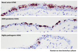
Lancet: Tropism Of H7N9 In the Human Respiratory Tract


image

ex vivo bronchus culture – Credit University Of Hong Kong


# 7513


An important new study from researchers at the University of Hong Kong appears today in The Lancet Respiratory Medicine, that finds that China’s emerging H7N9 virus infects and replicates in the cells of the human respiratory tract about as well as the 2009 H1N1 virus.


While we’ve seen a welcome reduction in human H7N9 infections over the summer, this is the latest in a series of studies that suggests this virus may have substantial pandemic potential (for more see Science: H7N9 Transmissibility Study In Ferrets).


In today’s study, researchers led by Dr. Malik Peiris inoculated in-vitro and ex-vivo cultures representing human bronchus and lung tissues with six different virus strains (H5N1, H1N1, 2 Human-derived H7N9 strains, 1 duck-derived H7N9, and the H7N7 2003 Netherlands outbreak stain)  and compared how well they infected and replicated within the cells. 


While the duck-derived H7N9 failed to replicate, both human H7N9 viruses replicated efficiently, far better than H5N1, and on a par with the 2009 H1N1 pandemic virus. 


We’ve three stops this morning for this paper, after which I’ll return with a bit more. 


First, a link to the abstract in the Lancet, then a companion piece by Jessica A Belser & Terrence M Tumpey of the CDC’s NCIRD, and finally a press release from the University of Hong Kong on this research.


Tropism and innate host responses of a novel avian influenza A H7N9 virus: an analysis of ex-vivo and in-vitro cultures of the human respiratory tract

Michael CW Chan PhD a †, Renee WY Chan PhD a †, Louisa LY Chan BSc a †, Chris KP Mok PhD a †, Kenrie PY Hui PhD a, Joanne HM Fong MSc a, Kin P Tao PhD a, Leo LM Poon DPhil a b, Prof John M Nicholls FRCPA c, Prof Y Guan PhD a b d, Prof JS Malik Peiris DPhil a b

ABSTRACT (Excerpt)
Methods

We obtained ex-vivo cultures of the human bronchus, lung, nasopharynx, and tonsil and in-vitro cultures of primary human alveolar epithelial cells and peripheral blood monocyte-derived macrophages. We compared virus tropism and induction of proinflammatory cytokine responses of two human influenza A H7N9 virus isolates, A/Shanghai/1/2013 and A/Shanghai/2/2013; a highly pathogenic avian influenza H5N1 virus; the highly pathogenic avian influenza H7N7 virus that infected human beings in the Netherlands in 2003; the 2009 pandemic influenza H1N1 virus, and a low pathogenic duck H7N9 virus that was genetically different to the human disease causing A H7N9 viruses.

Findings

Both human H7N9 viruses replicated efficiently in human bronchus and lung ex-vivo cultures, whereas duck/H7N9 virus failed to replicate in either. Both human A H7N9 viruses infected both ciliated and non-ciliated human bronchial epithelial cells and replicated to higher titres than did H5N1 (p<0·0001 to 0·0046) and A/Shanghai/1/2013 replicated to higher titres than did H7N7 (p=0·0002—0·01). Both human A H7N9 viruses predominantly infected type II alveolar epithelial cells and alveolar macrophages in the human lung and replicated to higher titres than did H5N1 (p<0·0001 to 0·0078); A/Shanghai/1/2013 replicated to higher titres than did H1N1 (p=0·0052—0·05) and H7N7 (p=0·0031—0·0151). Human H7N9 viruses were less potent inducers of proinflammatory cytokines compared with H5N1 virus.

Interpretation

Collectively, the results suggest that the novel H7N9 viruses are better adapted to infect and replicate in the human conducting and lower airways than are other avian influenza viruses, including H5N1, and pose an important pandemic threat


In a companion article, Belser and Tumpey explore these findings further (full access with free registration) in:


Tropism of H7N9 influenza viruses in the human respiratory tract

Jessica A Belser aEmail Address, Terrence M Tumpey a


While referring to the `remarkable ability of this low pathogenic avian influenza virus to cause severe human disease’  the authors caution that this study leaves a good many questions unanswered.

And our last stop is a press release from the University of Hong Kong (excerpts below).


HKU’s finding on avian influenza A (H7N9) virus well adapted to infect human respiratory tract reveals why numerous human infected cases in a short period of time

25 Jul 2013

HKU research team reveals why the avian influenza (H7N9) virus led to over 130 human cases in a relatively short period of time.  By using human respiratory tract tissues maintained in culture, the researchers discover that the avian influenza A (H7N9) virus is as efficient as the 2009 pandemic H1N1 virus, commonly known as swine influenza, in infecting the human respiratory tract.  Compared to the avian influenza A (H5N1) virus, H7N9 virus might pose an even more important pandemic threat to human. 

<SNIP>

Research findings
The researchers used human respiratory tract tissues (bronchus and lung) maintained in culture to compare infection with the avian influenza A (H7N9) virus, the 2009 pandemic influenza A (H1N1) virus (commonly known as swine influenza) and highly pathogenic avian influenza A (H5N1) virus.  They find that the H7N9 virus infects and replicates in both human bronchus as well as the H1N1 virus, and far more efficiently than the H5N1 virus.


These studies also identify the type II alveolar epithelial cells within the lung as a key target for H7N9 virus replication.  This is the key cell type supporting regeneration and repair of damaged lung tissues.  Thus infection and damage to these cells will prevent the repair processes that allow the lung to recover from injury or infection.


Taken together, this study demonstrates that the H7N9 viruses are better adapted to infect and replicate in the human airways and pose an important pandemic threat, perhaps even more so than H5N1 virus.


This latest research, along with a number of other recent studies, continue to show that the H7N9 virus has many of the qualities we would expect to find in an pandemic strain of influenza:


  • it is a novel strain to which humans appear to have little or no immunity
  • it can infect human (a2,6) receptor cells
  • it replicates efficiently in human tissue;
  • and it is capable of producing severe disease.


Yet, despite these worrisome traits in the lab, we’ve not seen sustained human-to-human transmission of the virus in the field. 


Exactly why that is, and whether or not this virus can gain the necessary changes to overcome that limitation, remain unanswered.


About the only thing we can say with any certainty is that none of the studies we’ve seen  over the summer have done anything to lower the level of concern over this emerging virus.


Which is why all eyes will be on China later this year to see if, and with how much vigor, this virus returns in the fall or winter.

Thursday, July 18, 2013

Science: H7N9 Transmissibility Study In Ferrets


image

Photo Credit Wikipedia


UPDATED: CIDRAP NEWS now has an extensive review of this paper up on their website, with expert commentary provided by Dr. Ian MacKay. 


Study: H7N9 highly transmissible by airborne route

  Highly recommended.


# 7496


While the welcomed lull in avian influenza cases in Eastern China over the past six weeks has taken H7N9 largely out of the daily headlines, history has shown that avian flu viruses often go `quiet’ during the warm summer months, only to reappear again in the fall or winter.


Complicating matters, we still don’t know the source of this virus, or exactly how it managed to spread across tens of thousands of square kilometers of Eastern China - and infect at least 132 people - in a matter of a few weeks.


So over the summer researchers have continued to examine the H7N9 virus, trying to gauge its pandemic potential should it return with colder weather this fall.


Last week we saw two studies in the Journal Nature  (see Nature: H7N9 Pathogenesis and Transmissibility In Ferrets & Mice & CIDRAP NEWS  article New studies on H7N9 raise pandemic concerns) that suggested the H7N9 virus might be better equipped to spark a pandemic than previously thought.


Researchers determined that while the virus did not appear to transmit as easily as seasonal flu via respiratory droplets between ferrets, once acquired, the virus replicated at a much higher rate than one normally sees with seasonal flu.


Today, we’ve a complex and fascinating new study by scientists from Harbin Veterinary Research Institute and the Gansu Agricultural University appearing in the Journal Science that gives us a detailed look at two critical issues; the pathogenicity and transmissibility of the virus (in mice & ferrets). 


Ferrets – whose respiratory system (and susceptibility to flu) are reasonably close to that of humans – are often used in transmissibility and pathogenicity studies of influenza. 


Today’s study tested several different H7N9 isolates (acquired from birds, and from humans) for transmissibility, and even though their HA and NA proteins were genetically quite similar, at least one H7N9 isolate transmitted readily via respiratory droplets among ferrets.


The abstract (see below), only scratches the surface of research conducted in this study.


H7N9 Influenza Viruses Are Transmissible in Ferrets by Respiratory Droplet


Science DOI: 10.1126/science.1240532


Qianyi Zhang, Jianzhong Shi, Guohua Deng, Jing Guo, Xianying Zeng, Xijun He, Huihui Kong, Chunyang Gu,  Xuyong Li, Jinxiong Liu, Guojun Wang, Yan Chen,  Liling Liu,  Libin Liang, Yuanyuan Li, Jun Fan, Jinliang Wang,  Wenhui Li, Lizheng Guan,  Qimeng Li, Huanliang Yang,  Pucheng Chen,  Li Jiang,Yuntao Guan, Xiaoguang Xin, Yongping Jiang, Guobin Tian, Xiurong Wang, Chuanling Qiao, Chengjun Li,  Zhigao Bu, Hualan Chen

Abstract (EXCERPT)

We systematically analyzed H7N9 viruses isolated from birds and humans. The viruses were genetically closely related and bound to human airway receptors; some also maintained the ability to bind to avian airway receptors. The viruses isolated from birds were nonpathogenic in chickens, ducks, and mice; however, the viruses isolated from humans caused up to 30% body weight loss in mice.


Most importantly, one virus isolated from humans was highly transmissible in ferrets by respiratory droplets. Our findings indicate nothing to reduce the concern that these viruses can transmit between humans.


While I’m sure CIDRAP NEWS will have a more detailed look later today (I’ll post a link), a few of the high points in this study include:


  • Researchers tested more than 10,700 samples taken from poultry farms, live market birds, wild bird habitats, and even poultry and swine slaughter houses and only managed to find 52 samples that were H7N9 positive (all but 2 were found in live-markets)
  • Chickens intravenously inoculated with two early strains of the virus showed no signs of illness indicating the H7N9 is largely non-pathogenic in chickens
  • Isolates taken from humans appeared to be more pathogenic in ferrets and mice than those taken from birds, suggesting adaptation of the virus may occur after the virus jumps to humans.
  • All of the human isolates had either the 627K or 701N amino acid changes in their PB2 (both associated with increased virulence and transmission in mammals) - while the avian isolates did not - again suggesting that adaptive mutations may have occurred during replication of the virus in the human host.
  • Mice inoculated with avian H7N9 isolates showed no signs of disease signs or deaths.
  • Mice inoculated with human derived H7N9 isolates experienced significant weight loss & pathogenesis.
  • In ferret transmissibility studies, four of the five viruses tested could be transmitted between ferrets in direct contact with each other, and one transmitted with high efficiency via respiratory droplets.


Being non-pathogenic in chickens, this virus has the potential to spread stealthily, and its rapid spread across multiple provinces in China suggests it transmits efficiently among poultry species.


This research suggests that the H7N9 virus already binds pretty well to human-like (a2,6) receptor cells, and it replicates efficiently in a mammalian (ferret) host.


Additionally, the H7N9 virus appears capable of accruing adaptive mutations that enhance both its virulence and transmissibility once it starts to replicate within a human host.


And perhaps most worrisome of all, at least one isolate tested already demonstrates the ability to transmit readily via respiratory droplets in ferrets.


Complicating matters, earlier this week we saw a study (see mBio: Antiviral Resistance In H7N9) suggesting that antiviral resistance may form quickly in patients infected with the H7N9 virus.


All qualities that could help make the H7N9 virus a formidable foe should we see it return in the fall.

Thursday, May 23, 2013

Branswell: Studies Show Transmissibility Of H7N9 In Ferrets

image

Credit NIAID


# 7314


Helen Branswell has the details today on a new study which appears in the Journal Science, that looks at the ability of the avian H7N9 virus – recently emerged in China – to infect and transmit among ferrets.


Ferrets are considered a reasonable mammalian substitute for humans when testing respiratory viruses because their lung physiology is similar, and because they cough and sneeze when infected, very much as humans do.


As Helen reports, researchers found signs that the H7N9 virus has already made adaptations to mammalian hosts not normally seen in `wild’ avian flu viruses, and as such, must be considered `a serious threat’.


First, I’d invite you to follow the link to read Helen’s article, then I’ll return with more.


H7N9 bird flu virus spreads among mammals, inches closer to human-to-human transmission, experts warn

Helen Branswell, Canadian Press | 13/05/23 2:50 PM ET

TORONTO — A new study shows that the H7N9 flu virus can pass between ferrets, even sometimes spreading by airborne transmission.


While the airborne spread wasn’t highly efficient, the work suggests this virus is more closely adapted to spread among mammals than other bird flu viruses.

(Continue . . .)


The study can be found at:


Published Online May 23 2013
Science DOI: 10.1126/science.1239844

Infectivity, Transmission, and Pathology of Human H7N9 Influenza in Ferrets and Pigs

H. Zhu, D. Wang, D. J. Kelvin, L. Li, Z. Zheng, S.-W. Yoon, S.-S. Wong, A. Farooqui, J. Wang, D. Banner, R. Chen, R. Zheng, J. Zhou, Y. Zhang, W. Hong, W. Dong, Q. Cai, M. H. A. Roehrl, S. S. H. Huang, A. A. Kelvin, T. Yao, B. Zhou, X. Chen, G. M. Leung, L. L. M. Poon, R. G. Webster, R. J. Webby, J. S. M. Peiris, Y. Guan,Y. Shu

The emergence of the H7N9 influenza virus in humans in Eastern China has raised concerns that a new influenza pandemic could occur. Here, we used a ferret model to evaluate the infectivity and transmissibility of A/Shanghai/2/2013 (SH2), a human H7N9 virus isolate. This virus replicated in the upper and lower respiratory tracts of the ferrets and was shed at high titers for 6 to 7 days, with ferrets showing relatively mild clinical signs.

SH2 was efficiently transmitted via direct contact, but less efficiently by airborne exposure. Pigs could be productively infected by SH2 and shed virus for 6 days but were unable to transmit the virus to other animals. Under appropriate conditions human-to-human transmission of the H7N9 virus may be possible.


Today the NIH published a press release on this study, portions of which I’ve excerpted below:


Ferrets, Pigs Susceptible to H7N9 Avian Influenza Virus

NIH-Funded Study Examined Transmissibility of Emerging Virus

WHAT:
Chinese and U.S. scientists have used virus isolated from a person who died from H7N9 avian influenza infection to determine whether the virus could infect and be transmitted between ferrets. Ferrets are often used as a mammalian model in influenza research, and efficient transmission of influenza virus between ferrets can provide clues as to how well the same process might occur in people. The research was supported, in part, by the National Institute of Allergy and Infectious Diseases, part of the National Institutes of Health.


The researchers dropped H7N9 virus into the noses of six ferrets. A day later, three uninfected ferrets were placed inside cages with the infected animals, and another three uninfected ferrets were placed in cages nearby. All the uninfected ferrets inside the cages became infected, while only one of three placed in nearby cages became infected. The team concluded that the virus can infect ferrets and be transmitted between ferrets both by direct contact and, less efficiently, by air. The scientists detected viral material in the nasal secretions of the ferrets at least one day before clinical signs of disease became apparent. The potential public health implication of this observation is that a person infected by H7N9 avian influenza virus who does not show symptoms could nevertheless spread the virus to others.


The researchers also infected pigs with the human-derived H7N9 virus. In natural settings, pigs can act as a virtual mixing bowl to combine avian- and mammalian-specific influenza strains, potentially allowing avian strains to better adapt to humans. New strains arising from such mixing have the potential to infect humans and spark a pandemic, so information about swine susceptibility to H7N9 could help scientists gauge the pandemic potential of the avian virus. Unlike the ferrets, infected pigs in this small study did not transmit virus to uninfected pigs, either through direct contact or by air. All the infected ferrets and pigs showed mild signs of illness, such as sneezing, nasal discharge, and lethargy, but none of the infected animals became seriously ill.

ARTICLE:
H Zhu et al. Infectivity, transmission and pathogenesis of human-isolated H7N9 influenza virus in ferrets and pigs. Science DOI: 10.1126/science.1239844 (2013).

Thursday, May 02, 2013

Science: A New H5N1 Reassortant Study


image

BSL-4 Lab Worker - Photo Credit –USAMRIID


# 7219


Ed Yong, writing for Nature News, has the details on a new H5N1 study that – like the Fouchier and Kawaoka studies from 2012 – involved the creation of a more transmissible bird flu virus.


A voluntary moratorium on`gain of function’ studies – those seeking to enhance the transmissibility and/or virulence of the H5N1 virus – went into effect in January of 2012 after biosecurity concerns were raised by a number of experts. (see Scientists Announced a 60 Day Moratorium On Some Types of H5N1 Research).


Objections were raised initially after the 2011 ESWI Influenza Conference in Malta, where Dutch researcher Ron Fouchier revealed that he’d created a more `transmissible’ form of the H5N1 virus (see Debra MacKenzie’s New Scientist: Five Easy Mutations).


That discovery, along with similar news coming from Yoshihiro Kawaoka, a highly respected virologist at the University of Wisconsin-Madison School of Veterinary Medicine, set alarm bells ringing in the biosecurity community.


The voluntary moratorium was later extended beyond two months to give time for international debate, and the formulation of safety rules.


The research papers by Fouchier and Kawaoka on the genetic changes needed to provide H5N1 with enhanced transmissibility have now been published, and the moratorium was lifted earlier this year.


As the following 3 minute NIAID Video explains, pandemic viruses can emerge when two (or more) flu viruses swap genetic material and create a new, hybrid strain.


The stated goal of these controversial experiments is to figure out in the lab which reassortments are likely to produce a human transmissible strain, before nature figures it out in the wild.


Today the Journal Science has published a new study where researchers in China reassorted the genes from the H5N1 and 2009 H1N1 flu viruses, creating 127 new `hybrids’. 


Of these, five showed an increased ability to transmit between guinea pigs, suggesting that they would transmit well among humans as well.


Lead author Professor Chen Hualan of the Harbin Veterinary Research Institute has been heavily involved over the past few weeks into the investigation of the H7N9 outbreak (see Study: Source Identification Of H7N9), and was the author of an EID Journal study from 2001 on H5N1 in wild birds (see EID Journal: H5N1 Branching Out).


First a link to the paper and abstract, then a jump to Ed Yong’s article.


H5N1 Hybrid Viruses Bearing 2009/H1N1 Virus Genes Transmit in Guinea Pigs by Respiratory Droplet

Ying Zhang,Qianyi Zhang, Huihui Kong, Yongping Jiang, Yuwei Gao, Guohua Deng, Jianzhong Shi, Guobin Tian, Liling Liu, Jinxiong Liu, Yuntao Guan, Zhigao Bu, Hualan Chen


  Nature | News

Scientists create hybrid flu that can go airborne

H5N1 virus with genes from H1N1 can spread through the air between mammals.

Ed Yong

02 May 2013

As the world is transfixed by a new H7N9 bird flu virus spreading through China, a study reminds us that a different avian influenza — H5N1 — still poses a pandemic threat.


A team of scientists in China has created hybrid viruses by mixing genes from H5N1 and the H1N1 strain behind the 2009 swine flu pandemic, and showed that some of the hybrids can spread through the air between guinea pigs. The results are published in Science1.


(Continue . . .)


Despite the lifting of the research moratorium, these types of experiments are still viewed  as controversial – and potentially dangerous -  by a number of experts. 


For more on this debate, you may wish to revisit:


The Furor Over H5N1 Research Continues
The Biosecurity Debate On H5N1 Research
Referral: Laurie Garrett On The Bird Flu Research Controversy
The Bird Flu Research Debate Continues

Monday, April 22, 2013

WHO H7N9 Study: Preliminary Age & Sex Distribution


image


# 7173


From the Western Pacific Surveillance and Response Journal (WPSAR) we’ve a preliminary review of the age and gender demographics of H7N9 infection among China’s first 63 cases reported through April 16th.


You’ll recall we touched on a similar analysis by CIDRAP of some of these same issues on Friday in H7N9: The Riddle Of The Ages.


Unlike the previous infection patterns we’ve seen with the H5N1 avian flu in China – which has had its biggest impact in those 15-39 years of age – the H7N9 patients have been conspicuously older.


image

Figure 1. Age group and sex distribution of reported human infections with avian influenza A(H7N9) and A(H5N1) viruses, China, as of 16 April 2013


Another notable trend : H7N9 patients have been predominantly male, by a ratio of 2 to 1. 


This skewing towards elderly males runs counter to China’s population demographics, which is heavily weighted towards young and middle-aged adults and where there are more women than men among the elderly.


Today’s study doesn’t provide us with any answers for why these trends are appearing, but it does provide us with three possible avenues of investigation:


(1) differential exposure between males and females due to gender-associated practices and norms;

(2) biological differences between males and females in the clinical course post exposure/infection; and

(3) differential healthcare-seeking/access behaviour between male and females, leading to surveillance/detection bias.


The study, which was conducted by scientists working with the World Health Organization is open access. Below you’ll find some excerpts (slightly reparagraphed for readability) - follow the link to read it in its entirety.


Human infections with avian influenza A(H7N9) virus in China: preliminary assessments of the age and sex distribution

Perspective

Yuzo Arima,a Rongqiang Zu,a Manoj Murhekar,a Sirenda Vong,b Tomoe Shimadaa and the World Health Organization Regional Office for the Western Pacific Event Management Team*

(EXCERPTS)

Since 31 March 2013, the government of China has been notifying the World Health Organization (WHO) of human infections with the avian influenza A(H7N9) virus, as mandated by the International Health Regulations (2005).


While human infections with other subgroups of H7 influenza viruses (e.g. H7N2, H7N3, and H7N7) have previously been reported, the current event in China is of historical significance as it is the first time that A(H7N9) viruses have been detected among humans and the first time that a low pathogenic avian influenza virus is being associated with human fatalities.


In this rapidly evolving situation, detailed epidemiologic and clinical data from reported cases are limited—making assessments challenging—however, some key questions have arisen from the available data. Age and sex data, as one of the first and most readily available data, may be an important proxy for gender-specific behaviours/conditions and an entry point for response.


Here, we describe the age and sex distribution of the human cases of avian influenza A(H7N9) to better inform risk assessments and potential next steps.


Between 31 March and 16 April 2013, there were 63 reported cases of avian influenza A(H7N9). The median age was 64 years (range 4–87), and 45 cases (71%) were male. Notably, 39 of the 63 cases (62%) were ≥ 60 years of age.


When stratified by age and sex, elderly men were the most affected demographic group (Figure 1).


  • < SNIP  lengthy discussion of possible factors that might influence the age/gender demographics of H7N9 infection>

At this time, it is clear that there are more questions than answers. Based on the basic age and sex distribution, we identify several critical questions and options to guide the ongoing investigation:

  • What are the societal norms and common social practices among elderly men in the affected provinces? Qualitative approaches and involvement of anthropologists/ sociologists specializing in the sociology of health of the Chinese population may be beneficial.
  • What is the age and sex distribution of severe acute respiratory illnesses and key risk factors for respiratory illness (e.g. smoking) in the underlying population in the affected provinces? While detailed case-based clinical information is pending, data from the general population may be helpful for initial assessments.
  • What is the age and sex distribution of healthcare utilization in the Chinese population in the affected provinces? Ruling out any possible selection bias will be an important initial step in understanding both the clinical and epidemiologic spectrum of infection.

In these situations, it is easy to dismiss preliminary epidemiologic assessments as being too low in numbers or with too few variables of interest.


There is a need for further case-based information, such as zoonotic exposures and underlying medical conditions. However, for public health workers engaged in rapid response to acute events, it is essential to operate as observational scientists and assess available information to help formulate the next steps.


Following age and sex distributions closely over time may detect important changes in the epidemiology of this virus and with better understanding, high-risk populations, targeted interventions (e.g. gender-specific risk communication messages), prevention and control measures (e.g. vaccination) and treatment options (e.g. antivirals) may be identified.


While this brief and rapid communication cannot offer answers, we hope that public health practitioners involved in similar responses – at various capacities around the world – may consider these key concerns and questions to help them respond to not only the current virus but also other emerging infectious threats.


Over the past 15 years the study of the H5N1 avian flu virus has produced its share of unusual and unexpected findings (see Study: What Makes Avian Flu So Deadly.  Similarly, the 2009 H1N1 pandemic virus behaved in ways that were not anticipated (see There’s No Flu Like A New Flu).


Now it is H7N9 that is challenging our preconceived notions of how influenza viruses should act. 


The mantra of flu researchers is that influenza viruses are notoriously unpredictable.  H7N9, thus far, appears to be no different.

Monday, December 31, 2012

Study: Self-Administered Vaccines In Adults

image

LAIV nasal syringe – Credit CDC PHIL 


# 6811


Beyond the difficulties of producing billions of doses of an emergency pandemic influenza vaccine, one of the logistical nightmares is how to best deploy them. The standard solutions – where vaccination centers are set up in public venues, pharmacies, and clinics – have some serious downsides.


Queuing hundreds of people together for hours during a pandemic is a good way to spread a virus further. And there are likely to be crowd control and security issues in some places as well.


During the 1976 Swine flu scare (see Deja Flu, All Over Again), I was one of thousands of health care workers around the country who were dragooned into providing pandemic flu shots. The logistics of vaccinating millions of people – even without a pandemic raging – were daunting.

image

Yes, that’s me in 1976. I and swear to this very day, that lady was making that face before I gave her the shot.


One solution –  made possible by the development of an  LAIV (Live attenuated Influenza Vaccine) – would be to have people self inoculate themselves at home. 


LAIV vaccines – unlike the standard flu shot - are delivered via a nasal spray, not a needle.


The CDC describes the major differences between the TIV (Trivalent Influenza Vaccine) shot, and the LAIV nasal spray as follows:


Major Differences Between TIV and LAIV

Influenza Prevention and Control Recommendations

Published for the 2010-11 Influenza Season; Adapted for the 2012-13 Influenza Season

Trivalent inactivated influenza vaccine (TIV) contains inactivated viruses and thus cannot cause influenza. Live-attenuated influenza vaccine (LAIV) contains live attenuated influenza viruses that have the potential to cause mild signs or symptoms related to vaccine virus infection (e.g., rhinorrhea, nasal congestion, fever, or sore throat). LAIV is administered intranasally by sprayer, whereas TIV is administered intramuscularly or intradermally by injection.

LAIV is licensed for use among nonpregnant persons aged 2-49 years; safety has not been established in persons with underlying medical conditions that confer a higher risk for influenza complications. TIV is licensed for use among persons 6 months and older, including those who are healthy and those with chronic medical conditions.


While not suitable for everyone, LAIVs could be used to inoculate a large segment of the population during an influenza pandemic, without the need of a Health care worker. 


All of which brings us to a study, recently published in the journal Vaccine (abstract slightly reparagraphed for readability), which looks at:


The safety and effectiveness of self-administration of intranasal live attenuated influenza vaccine in adults.


Ambrose CS, Wu X.


Vaccine. 2012 Dec 19. pii: S0264-410X(12)01798-7. doi: 10.1016/j.vaccine.2012.12.028.

Source

MedImmune, LLC, Gaithersburg, MD, USA. Electronic address: ambrosec@medimmune.com.

Abstract

Intranasal live attenuated influenza vaccine (LAIV) has potential for self-administration (SA) by adults and adolescents, which could save time and cost in mass vaccination settings. Participants in a study of LAIV in adults (n=4561) selected either SA or health care provider (HCP) administration and were followed for febrile illness during the influenza season.

More LAIV recipients chose SA-LAIV (72%) than HCP-LAIV (28%). Overall, 97% of SA-LAIV and 98% of HCP-LAIV recipients had no problems with vaccine administration. Four of 13 study sites enrolled more than 50 subjects in both cohorts. Overall and for these 4 sites, illness incidence was similar with SA-LAIV and HCP-LAIV.

Solicited reactogenicity events and adverse events through 7 days post vaccination were comparable for SA-LAIV and HCP-LAIV recipients; both groups exhibited increased runny nose, sore throat, and cough relative to placebo recipients. SA-LAIV and HCP-LAIV appeared similarly effective against influenza-like illness and had comparable safety profiles.


CIDRAP NEWS  summarized these findings last week in their Flu News Scan.


Study: Smooth sailing for self-administered LAIV flu vax


Self-administered intranasal live attenuated influenza vaccine (LAIV) is as safe and effective as that administered by health providers, according to recent analysis of data from a randomized, placebo-controlled study conducted in 1997 and 1998.

The study, which explored differences in administration and was funded by MedImmune, the maker of Flumist, appeared in the Dec 20 online edition of Vaccine. Self-administered flu vaccines could be useful in a pandemic mass immunization setting and could be a tool for making vaccination more efficient and less costly, the authors say.

<SNIP>

The research group found no significant difference in illness incidence between the two groups and observed comparable levels of reactions and adverse events 7 days after vaccination.
Dec 20 Vaccine
abstract


LAIVs are normally shipped  and stored at 35°F--46°F  (cite) - which makes mailing them problematic - but they could be easily distributed via drive thru windows or hand delivered to homes in neighborhoods.


LAIVs are particularly attractive during a pandemic because they can be produced in much larger quantities than Inactivated vaccines.


But there remain some questions regarding their effectiveness in adults, compared to the TIV (see Study: Nasal Flu Vaccines Generally Less Effective Than Shots In Adults).


Caveat: this study was based on seasonal flu vaccine strains used in the middle of the last decade, not on a newly emergent pandemic strain, and so the results may not be fully applicable.


Nevertheless, self-administered LAIVs have the potential to streamline vaccine delivery to a significant portion of the population during the next influenza pandemic. And with a novel flu, the quicker you can increase the levels of community immunity, the sooner the crisis will begin to abate.

Wednesday, November 28, 2012

JAMA: Waning Pertussis Vaccine Effectiveness Over Time

image
Credit CDC Pink Book


# 6744


Pertussis, or `whooping cough’ was once a major childhood disease in the United States, with more than a million cases reported between 1940 and 1945, resulting in thousands of deaths. 


All that began to change with the introduction of a whole cell Pertussis vaccine in the 1940s.  By 1960, the number of Pertussis cases in the United States had dropped to about 15,000 cases a year – a 95% reduction over 20 years.


Rates dropped another 2/3rds over the next 10 years (see chart above).


The vaccine, while highly effective, was blamed for a variety of adverse reactions, prompting a move away from whole cell pertussis vaccines to safer – but less broadly protective  - acellular vaccines in the 1990s


The following chart illustrates the rise in pertussis cases, which began in earnest in the early 1990s.  The most recent years (2010-2011) are not shown, but they were among the most active in decades.


image


While still far below the rates seen in the 1940s, the recent rise in Pertussis cases has raised a good deal of concern these past couple of years.  A few blogs on it include:


The UK’s Whooping Cough Outbreak
Referral: McKenna On Pertussis Vaccines
California Reports 9th Pertussis Fatality of 2010


The reasons behind these increases are complex, and not completely understood, but some factors may include:


  • lower vaccination uptakes
  • the move away from whole cell pertussis vaccines to safer acellular vaccines in the 1990s
  • evolutionary changes in the Bordetella pertussis bacteria.


Although infants are still at greatest risk from this illness, children aged 7 to 10 years old have experienced an unexpected rise in infections over the past decade.


A recent study published in the NEJM suggested that protection from the newer acellular pertussis vaccine – introduced in the early 1990s – may wane sooner than previously suspected. 


Waning Protection after Fifth Dose of Acellular Pertussis Vaccine in Children

Nicola P. Klein, M.D., Ph.D., Joan Bartlett, M.P.H., M.P.P., Ali Rowhani-Rahbar, M.D., M.P.H., Ph.D., Bruce Fireman, M.A., and Roger Baxter, M.D.

N Engl J Med 2012; 367:1012-1019 September 13, 2012DOI: 10.1056/NEJMoa1200850


Today we can add another study, this time appearing in JAMA, that finds that the protective effect of the Pertussis vaccine begins to wane after the fifth dose (normally given at age 5) is received, leaving 7 to 10 year olds at greater risk of infection.


Association of Childhood Pertussis With Receipt of 5 Doses of Pertussis Vaccine by Time Since Last Vaccine Dose, California, 2010

Lara K. Misegades, PhD, MS; Kathleen Winter, MPH; Kathleen Harriman, PhD, MPH, RN; John Talarico, DO, MPH; Nancy E. Messonnier, MD; Thomas A. Clark, MD, MPH; Stacey W. Martin, MSc


JAMA. 2012;308(20):2126-2132. doi:10.1001/jama.2012.14939

ABSTRACT (Excerpt)

Conclusion  Among children in 15 California counties, children with pertussis, compared with controls, had lower odds of having received the 5-dose DTaP series; as time since last DTaP dose increased, the odds increased, which is consistent with a progressive decrease in estimated vaccine effectiveness each year after the final dose of pertussis vaccine.


Pertussis remains a poorly controlled vaccine-preventable disease in the United States, despite a well-established childhood vaccination program and high coverage. Although infants have substantially higher rates of pertussis compared with other age groups, data from the National Notifiable Diseases Surveillance System reflect a recent increase in the number of reported pertussis cases among children aged 7 to 10 years. In 2010, this age group had the second highest incidence of pertussis in the United States.


The changing epidemiology raises important questions about possible waning protection from the childhood acellular pertussis vaccine series.

(Continue . . . )


This is a lengthy report, heavy on statistics.  For a less imposing summary, we have a nice write up today in Family Practice News:


Pertussis Vaccine Loses Efficacy Each Year After Fifth Dose


By: MARY ANN MOON, Family Practice News Digital Network

The odds of a child developing pertussis increase as the interval since he or she received the fifth and final dose of the DTaP vaccine increases, according to a report published in the November 28, 2012 issue of JAMA.


This pattern indicates a progressive waning of vaccine effectiveness every year after completion of the vaccine series, which would explain the recently noted surge in pertussis cases among 7- to 10-year-olds in at least 34 states, said Lara K. Misegades, Ph.D., of the Meningitis and Vaccine Preventable Disease Branch of the Centers for Disease Control and Prevention and her associates.

(Continue . . . )


As more confirmatory data comes in and is analyzed, the question then becomes what to do about this decline in protection for those aged 7 to 10 years of age.  The authors, in their conclusion, grant that their are no easy answers.


The increasing incidence of pertussis, changing epidemiology, and demonstrated decline in the estimated DTaP VE over time have raised concerns about the current US pertussis vaccine program and may prompt consideration of alternative schedules.


Options include delaying administration of the fifth DTaP dose or administering the Tdap booster at earlier than 11 years of age. However, a recommendation to delay the fifth DTaP dose until 6 years of age or later may unintentionally increase the burden of disease between the fourth and fifth doses of the childhood series, and implementation would likely be programmatically challenging because many states' school entry immunization requirements for pertussis are built around the current DTaP schedule.


Alternatively, shifting the Tdap booster to 10 years of age or earlier may have the unwanted effect of reducing coverage, as there is no established routine health care visit for children before the adolescent vaccine platform visit at 11 to 12 years of age.32


Given the options for adjustments to the pertussis vaccine schedule, these issues will require careful and ongoing review of the epidemiology and vaccine program nationwide. Ultimately, improved control of pertussis may require a vaccine that provides longer duration of protection or differently affects transmission in the community.

Tuesday, November 06, 2012

Study: MRSA In Waste Water Treatment Plants


image

Photo Credit USGS – Wastewater: The Primary Treatment Process

1. Screening 2. Pumping 3. Aerating 4. Removing sludge 5. Removing Scum 6. killing bacteria


# 6699


While it may not seem (or even be) glamorous, there are scientists who spend considerable time examining the input and output of wastewater (sewage) treatment plants. Given the potential environmental impact of the discharges from these facilities, it is important work.


The subject of wastewater treatment plants - and the substances that their processes can fail to remove during treatment - has come up before in this blog.


In 2007, with governments around the world stockpiling large quantities of Tamiflu (Oseltamivir) in anticipation of a pandemic, concerns turned to what would happen if millions of doses were dispensed, consumed, and excrete by humans into the waste water system in a short period of time.


In The Law of Unintended Consequences we looked at a report in Environmental Health Perspectives (EHP) called Potential Risks Associated with the Proposed Widespread Use of Tamiflu that found enough of excreted oseltamivir carboxylate (OC) would survive treatment, and be ejected into the environment, to raise concerns.


In October of 2009 and we saw another report (see Everything Old Is News Again), based on studies done the previous year in Kyoto, Japan – that showed elevated levels of the OC Metabolite in wastewater discharge. 


In 2011, in Pandemics & The Law Of Unintended Consequences we saw yet another study that looked at potential problems inherent in the massive distribution and consumption of antibiotics and antivirals during a pandemic.


WWTPs (Wastewater Treatment Plants) depend upon microbial activity in order to breakdown or `digest’ sewage.


Antibiotics in the sewage – at elevated levels such as might be seen during a pandemic – could inhibit microbial activity, resulting in the failure of WWTPs and the discharge of under-treated wastewater into the environment.


Today, a new study, this time on MRSA (methicillin-resistant Staphylococcus aureus) and how it fares in the wastewater treatment process. As many WWTPs provide reclaimed water for irrigation use, the concern is that MRSA shed in feces might make it through the plant and into the environment.


The University of Maryland-led study study appears as an open access article that appears in Environmental Health Perspectives. 

Methicillin-Resistant Staphylococcus aureus (MRSA) Detected at Four U.S. Wastewater Treatment Plants

November 1, 2012 Research 

Rachel E. Rosenberg Goldstein, Shirley A. Micallef, Shawn G. Gibbs, Johnnie A. Davis, Xin He, Ashish George, Lara M. Kleinfelter, Nicole A. Schreiber, Sampa Mukherjee, Amir Sapkota, Sam W. Joseph, and Amy R. Sapkota

Abstract

Background: The incidence of community-acquired methicillin-resistant Staphylococcus aureus (CA-MRSA) infections is increasing in the United States, and it is possible that municipal waste­water could be a reservoir of this micro­organism. To date, no U.S. studies have evaluated the occurrence of MRSA in waste­water.


Objective: We examined the occurrence of MRSA and methicillin-susceptible S. aureus (MSSA) at U.S. waste­water treatment plants.


Methods: We collected waste­water samples from two Mid-Atlantic and two Midwest waste­water treatment plants between October 2009 and October 2010. Samples were analyzed for MRSA and MSSA using membrane filtration. Isolates were confirmed using biochemical tests and PCR (polymerase chain reaction). Antimicrobial susceptibility testing was performed by Sensititre® micro­broth dilution. Staphylococcal cassette chromosome mec (SCCmec) typing, Panton-Valentine leucocidin (PVL) screening, and pulsed field gel electrophoresis (PFGE) were performed to further characterize the strains. Data were analyzed by two-sample proportion tests and analysis of variance.


Results: We detected MRSA (n = 240) and MSSA (n = 119) in 22 of 44 (50%) and 24 of 44 (55%) waste­water samples, respectively. The odds of samples being MRSA-positive decreased as treatment progressed: 10 of 12 (83%) influent samples were MRSA-positive, while only one of 12 (8%) effluent samples was MRSA-positive. Ninety-three percent and 29% of unique MRSA and MSSA isolates, respectively, were multi­drug resistant. SCCmec types II and IV, the pvl gene, and USA types 100, 300, and 700 (PFGE strain types commonly found in the United States) were identified among the MRSA isolates.


Conclusions: Our findings raise potential public health concerns for waste­water treatment plant workers and individuals exposed to reclaimed waste­water. Because of increasing use of reclaimed waste­water, further study is needed to evaluate the risk of exposure to antibiotic-resistant bacteria in treated waste­water.


Environ Health Perspect 120:1551–1558 (2012). http://dx.doi.org/10.1289/ehp.1205436 [Online 6 September 2012]

Full PDF FILE


The full report runs 8 pages, and can be download here.


A summation of this research is available from the University of Maryland’s School of Public Health. I’ve cut to the chase with the excerpts  posted below, so follow the link to read the full story.


The School of Public Health News

November 5, 2012
NEWS RELEASE
Contact: Kelly Blake,
kellyb@umd.edu, 301-405-9418

Superbug MRSA Identified in U.S. Wastewater Treatment Plants

University of Maryland-led study is first to document environmental source of the antibiotic-resistant bacteria in the United States

<SNIP>

They found that MRSA, as well as a related pathogen, methicillin-susceptible Staphylococcus aureus (MSSA),were present at all four WWTPs, with MRSA in half of all samples and MSSA in 55 percent.MRSA was present in 83 percent of the influent-- the raw sewage--at all plants, butthe percentage of MRSA- and MSSA-positive samples decreased as treatment progressed. Only one WWTP had the bacteria in the treated water leaving the plant, and this was at a plant that does not regularly use chlorination, a tertiary step in wastewater treatment.


Ninety-three percent of the MRSA strains that were isolated from the wastewater and 29 percent of MSSA strains were resistant to two or more classes of antibiotics, including several that the U.S. Food and Drug Administration has specifically approved for treating MRSA infections. At two WWTPs, MRSA strains showed resistance to more antibiotics and greater prevalence of a gene associated with virulence at subsequent treatment stages, until tertiary chlorination treatment appeared to eliminate all MRSA. This suggests that while WWTPs effectively reduce MRSA and MSSA from influent to effluent, they may select for increased antibiotic resistance and virulence, particularly at those facilities that do not employ tertiary treatment (via chlorination).


“Our findings raise potential public health concerns for wastewater treatment plant workers and individuals exposed to reclaimed wastewater,” says Rachel Rosenberg Goldstein, environmental health doctoral student in the School of Public Health and the study’s first author. “Because of increasing use of reclaimed wastewater, further research is needed to evaluate the risk of exposure to antibiotic-resistant bacteria in treated wastewater.”

(Continue . . . )


Most WWTPs are designed to efficiently remove solids and to disinfect discharge water, but do less well when it comes to removing chemicals.


Although only four plants were studied in this project, the results suggest that plants that fail to chlorinate routinely may be at greater risk of letting antibiotic resistant bacteria back into the environment. 


Whether it is antiviral or antibiotic metabolites, remnants from  illicit drug use, or resistant organisms themselves, what comes out of these treatment plants can affect our environment, and our population.


On a planet with 7 billion people, figuring out how to safely dispose of, recycle, or treat human waste has become a major challenge.


Shortcomings in WWTP systems – even seemingly small ones - can pose serious public health risks, including potentially creating and spreading resistant bacterial organisms.


As the authors of this study concluded, more research is now needed to evaluate and quantify the risk.