Showing posts with label PLoS. Show all posts

Wednesday, November 13, 2013

PLoS Currents: State Of Knowledge & Data Gaps On MERS-CoV

image


# 7968


Last night on twitter World Health Organization spokesperson Gregory Hartl described it as `What we know, what we don’t know. Most comprehensive paper yet . . ‘  on the MERS coronavirus.  And that’s a good assessment, as the 30 page PDF put together by the WHO MERS Research Group does a good job of laying out of what we’ve learned regarding this emerging coronavirus (through Oct 22nd, 2013), and provides an excellent reference.


While much has been learned over the past year, there remain notable gaps in our understanding of this virus, including:

  • how the virus is spilling over into the human population?
  • from what animal source did it emerge?  
  • How many people have really been infected?
  • What role do mild, or asymptomatic cases play in the transmission of the virus?
  • What is the most effective treatment for this infection?
  • Is the virus spreading outside of the Middle East?


Although I’ve excerpted the Abstract,  I would urge everyone download, and read this paper.  Then keep it handy as a reference going forward.  It is available online, or as a downloadable PDF, at the link below:


State of Knowledge and Data Gaps of Middle East Respiratory Syndrome Coronavirus (MERS-CoV) in Humans

November 12, 2013 · Research

Abstract


Background: Between September 2012 and 22 October 2013, 144 laboratory-confirmed and 17 probable MERS-CoV cases from nine countries were notified to WHO.


Methods: We summarize what is known about the epidemiology, virology, phylogeny and emergence of MERS-CoV to inform public health policies.


Results: The median age of patients (n=161) was 50 years (range 14 months to 94 years), 64.5% were male and 63.4% experienced severe respiratory disease. 76.0% of patients were reported to have ≥1 underlying medical condition and fatal cases, compared to recovered or asymptomatic cases were more likely to have an underlying condition (86.8% vs. 42.4%, p<0.001). Analysis of genetic sequence data suggests multiple independent introductions into human populations and modelled estimates using epidemiologic and genetic data suggest R0 is <1, though the upper range of estimates may exceed 1. Index/sporadic cases (cases with no epidemiologic-link to other cases) were more likely to be older (median 59.0 years vs. 43.0 years, p<0.001) compared to secondary cases, although these proportions have declined over time. 80.9% vs. 67.2% of index/sporadic and secondary cases, respectively, reported ≥1 underlying condition. Clinical presentation ranges from asymptomatic to severe pneumonia with acute respiratory distress syndrome and multi-organ failure. Nearly all symptomatic patients presented with respiratory symptoms and 1/3 of patients also had gastrointestinal symptoms.


Conclusions: Sustained human-to-human transmission of MERS-CoV has not been observed. Outbreaks have been extinguished without overly aggressive isolation and quarantine suggesting that transmission of virus may be stopped with implementation of appropriate infection control measures.

Friday, September 06, 2013

PLoS Pathogen’s Pearls: Emergence Of MERS-CoV


image


# 7736


PLoS Pathogens’s Pearls are a series of open access, short educational articles centered on pathogens research geared primarily for graduate students and post-docs. 


These provide an excellent, and not terribly difficult to digest, educational resource a variety of topics.


Yesterday PloS Pathogens  published a new one on MERS-CoV, that addresses the following topics.


Emergence of the Middle East Respiratory Syndrome Coronavirus

Christopher M. Coleman, Matthew B. Frieman

PLOS Pathogens: published 05 Sep 2013 | info:doi/10.1371/journal.ppat.1003595


Under conclusions and questions, the authors write:


Many unanswered questions remain on this newly identified virus:

  1. What is the environmental reservoir of MERS-CoV? Is it transmitted from bats to camels, goats, or cats? Is the virus linked to date palm harvesting? How did it spread to people from the environment?
  2. Are there associated comorbidities that predispose someone to contracting MERS? With the age of infected patients skewed toward older males, is there a genetic link to infection? Are the patients generally immunosuppressed?
  3. What is the seroprevalence of MERS-CoV in the general population? Has MERS-CoV been circulating for many years between animals and people and only now mutated enough to be able to cause disease in people? Or is this a new spillover event that has not been seen by humans until now?
  4. Why doesn't MERS-CoV grow in mouse cells or cause disease in mice? Is it because the viral spike protein doesn't bind mouse DPP4 at all, is it because there are other host factors necessary for entry and replication in mouse cells, or is it due to location and amounts of receptor expression?
  5. How do the MERS-CoV proteins contribute to disease? Are there any specific functions of the proteins that allow for enhanced pathogenesis?
  6. Since this virus is similar to bat coronaviruses identified in China, Africa, and Europe, why haven't other bat coronaviruses spilled over into people, causing serious disease (with the exception of SARS-CoV [7] and, potentially, hCoV-229E [23])? What is it about MERS-CoV and the conditions in the Middle East that have contributed to viral infection and the high mortality rate?

With the spread of MERS-CoV through the Middle East, one thing is certain at this point: The emergence of the novel SARS coronavirus in 2003 from a zoonotic source in China and its spread around the world is not an isolated incident of coronavirus spread. Continued spillover events will occur from animals to humans in the future. The sooner we understand these current microbial threats, the more people we can save from infection and possible death. If we can identify these microbes in our environment before they infect us, we can better protect ourselves against future infections.


Well worth reviewing.

Thursday, February 14, 2013

PLoS One: Obesity, Viral Pneumonitis & The 2009 H1N1 Pandemic


image

Photo Credit CDC PHIL


# 6936


Very early in the 2009 H1N1 pandemic we began to see reports (see H1N1 Morbidity And Previously Existing Conditions) of unusual numbers of obese influenza patients populating intensive care facilities around the world, raising concerns that obesity might be a significant pandemic risk factor.


In her address to the world announcing the declaration of the H1N1 pandemic on June 11th, (see WHO: Chan Statement On Raising Pandemic Level), Margaret Chan listed obesity as one of the pre-existing conditions that lent themselves to severe outcomes with this virus, saying:


Many, though not all, severe cases have occurred in people with underlying chronic conditions. Based on limited, preliminary data, conditions most frequently seen include respiratory diseases, notably asthma, cardiovascular disease, diabetes, autoimmune disorders, and obesity.”


Over the next couple of months obesity, and particularly morbid obesity, was frequently mentioned as a possible risk factor, along with asthma, diabetes, and immune disorders.   


That is, until an ACIP meeting held at the end of July, where evidence was presented that showed that the incidence of hospitalizations among those listed as obese by their BMI was practically the same as their prevalence in society.


Roughly 34% of Americans are obese, and roughly 38% of those hospitalized met that criteria.    While 6% are morbidly obese (BMI > 40), they only made up 7% of the hospitalized cases.


At the time the CDC’s Dr. Anne Schuchat stated that the jury was still out on the morbidly obese, but there was no clear evidence that obesity – without some comorbid condition like diabetes – created a greater risk of complications from pandemic H1N1.


Of course, this was very early in the game, and data was sparse.


In September, in Study: Half Of ICU H1N1 Patients Without Underlying Conditions, it became apparent that while pre-existing risk factors were important, they were not the sole reason behind flu victims ending up in intensive care.


In November, Eurosurveillance Journal  published a Study: H1N1 Hospitalization Profiles, that similarly found :


  • The most common risk factor in admission to intensive care was chronic respiratory disease followed by chronic neurological disease, asthma and severe obesity.
  • 51% of hospitalized cases and 42% of ICU cases were not in a recognized risk group.


This back-and-forth reporting on the significance of obesity as a risk factor continued, which I covered in blogs including:


NIH: Post Mortem Studies Of H1N1
Study: Quantifying H1N1 Risk Factors
Morbid Obesity And H1N1 Flu


In March of 2010, the CDC (in response to a recently published PLoS One study), posted the following:


What has been learned from the 2009 H1N1 pandemic about obesity and risk of serious influenza disease death?

During the 2009 H1N1 pandemic, early reports from the United States and abroad suggested that obesity was more frequent among persons hospitalized with 2009 H1N1 disease or who died following 2009 H1N1 infection.


Since that time, a number of studies have suggested that many 2009 H1N1patients tend to be morbidly obese. The study “Morbid Obesity as a Risk Factor for hospitalization and Death due to 2009 Pandemic Influenza A (H1N1) Disease,” published in PLoS ONE, sought to determine whether or not obesity or morbid obesity were in fact independent risk factors for serious 2009 H1N1-related complications, including death.  This study found that morbidly obesity persons have a higher risk of hospitalization for 2009 H1N1 infection compared to persons with normal weight. Data from this study also suggest that the risk of death following H1N1 infection may be higher for morbidly obese individuals.


As more data was gathered analyzed, it was becoming apparent that morbid obesity (BMI > 40) was associated with a greater risk from pandemic flu. 


In early 2011 (see Extreme Obesity: A Novel Risk Factor For A Novel Flu) the IDSA’s journal Clinical Infectious Diseases carried a study called  A Novel Risk Factor for a Novel Virus: Obesity and 2009 Pandemic Influenza A (H1N1), that found:


Extreme obesity associated with higher risk of death for 2009 H1N1 patients

[EMBARGOED FOR JAN. 5, 2011] For those infected with the 2009 pandemic influenza A (H1N1) virus, extreme obesity was a powerful risk factor for death, according to an analysis of a public health surveillance database. In a study to be published in the February 1, 2011, issue of Clinical Infectious Diseases, researchers associated extreme obesity with a nearly three-fold increased odds of death from 2009 H1N1 influenza. Half of Californians greater than 20 years of age hospitalized with 2009 H1N1 were obese. 


All of which serves as prelude to a new study that appears in PLoS One today called:


Viral Pneumonitis Is Increased in Obese Patients during the First Wave of Pandemic A(H1N1) 2009 Virus

Jen Kok, Christopher C. Blyth, Hong Foo, Michael J. Bailey, David V. Pilcher, Steven A. Webb, Ian M. Seppelt, Dominic E. Dwyer, Jonathan R. Iredell

Introduction

There is conflicting data as to whether obesity is an independent risk factor for mortality in severe pandemic (H1N1) 2009 influenza (A(H1N1)pdm09). It is postulated that excess inflammation and cytokine production in obese patients following severe influenza infection leads to viral pneumonitis and/or acute respiratory distress syndrome.

Methods

Demographic, laboratory and clinical data prospectively collected from obese and non-obese patients admitted to nine adult Australian intensive care units (ICU) during the first A(H1N1)pdm09 wave, supplemented with retrospectively collected data, were compared.

Results

Of 173 patients, 100 (57.8%), 73 (42.2%) and 23 (13.3%) had body mass index (BMI) <30 kg/m2, ≥30 kg/m2 (obese) and ≥40 kg/m2 (morbidly obese) respectively.

Compared to non-obese patients, obese patients were younger (mean age 43.4 vs. 48.4 years, p = 0.035) and more likely to develop pneumonitis (61% vs. 44%, p = 0.029).

Extracorporeal membrane oxygenation use was greater in morbidly obese compared to non-obese patients (17.4% vs. 4.7%, p = 0.04). Higher mortality rates were observed in non-obese compared to obese patients, but not after adjusting for severity of disease.

C-reactive protein (CRP) levels and hospital length of stay (LOS) were similar. Amongst ICU survivors, obese patients had longer ICU LOS (median 11.9 vs. 6.8 days, p = 0.017). Similar trends were observed when only patients infected with A(H1N1)pdm09 were examined.

Conclusions

Among patients admitted to ICU during the first wave of A(H1N1)pdm09, obese and morbidly obese patients with severe infection were more likely to develop pneumonitis compared to non-obese patients, but mortality rates were not increased. CRP is not an accurate marker of pneumonitis.


Interestingly, what these researchers found was a mixed bag.  While obesity was linked to a higher incidence of pneumonitis, somewhat surprisingly it was not linked to a higher rate of mortality.


The authors write:


In the present study, obesity was an independent predictor of pneumonitis after adjusting for age and chronic lung disease. Furthermore, clinicians may have intervened with more advanced levels of respiratory support in the obese patients pre-emptively and more readily prior to even more significant respiratory failure.


Although our obese patients were more likely to develop pneumonitis, they were also more likely to recover once the acute lung insult resolved.


The duration of mechanical ventilation was similar between obese and non-obese patients, comparable to the experience of critically ill patients with respiratory failure prior to the 2009 pandemic


The entire research article is very much worth reading, but the authors end by writing:


In conclusion, obese patients with severe A(H1N1)pdm09 infection from the first pandemic wave in Australia were more likely to develop pneumonitis compared to non-obese patients, but mortality rates were similar between the two groups after adjusting for severity of disease.


Although there is on-going debate as to whether obesity is a risk factor for severe A(H1N1)pdm09 infection, annual influenza vaccination should be prioritized in this group given the increased risk of serious complications from seasonal influenza infection


Nearly 4 years after the initial outbreak of novel H1N1, research on this most-studied pandemic of all time continues. The massive amount of data collected during the 2009-2012 pandemic has not yet been fully explored, and will undoubtedly fuel many more studies for years to come.

Friday, February 08, 2013

PLoS Pathogens: A New Influenza C Virus Detected In Swine

image

Credit Wikipedia


# 6917


When we talk about influenza viruses, Influenza A and Influenza B pretty much capture all of the headlines.  Less well known are the Influenza C viruses - which while less common - circulate both in humans and in swine.


For those looking for a detailed virology lesson, Vincent Racaniello wrote about the three types of influenza viruses in 2009 on his Virology Blog in a piece called The A, B, and C of influenza virus.


Although most adults have encountered the influenza C virus at least once in their life, it tends to produce only mild upper respiratory tract infections. The CDC  describes influenza B & C viruses this way.


Influenza Type B

Influenza B viruses are usually found only in humans. Unlike influenza A viruses, these viruses are not classified according to subtype. Influenza B viruses can cause morbidity and mortality among humans, but in general are associated with less severe epidemics than influenza A viruses. Although influenza type B viruses can cause human epidemics, they have not caused pandemics.

Influenza Type C

Influenza type C viruses cause mild illness in humans and do not cause epidemics or pandemics. These viruses are not classified according to subtype.


Our knowledge of influenza C is somewhat limited, but last December, Lisa Schnirring at CIDRAP NEWS wrote this report on a study of Influenza C viruses.


Study finds influenza C in kids hospitalized with pneumonia

Lisa Schnirring * Staff Writer

Dec 7, 2012 (CIDRAP News) – Influenza C generally isn't thought to be a cause clinically significant disease, but a study in Italian children who were seen in the emergency department for pneumonia found the virus in five children, with a disease severity that resembled influenza A.


The study evaluated data from four flu seasons from 2008-09 to 2011-12 at a pediatric clinic in Milan and appeared today in an early online edition of Influenza and Other Respiratory Viruses.

(Continue . . .)


Influenza B and C are considered unlikely to have much in the way of pandemic potential because they lack the multiple sub-types found with influenza A viruses (17 hemagglutinin and 9 neuraminidase subtypes) that allow for viral reassortment.


Reassortant pig[6]

Two Influenza A viruses can swap genetic segments to produce new hybrid (reassortant) viruses.


But of course, one should never say `never’. . . .


From PloS Pathogens  today we get the first study to suggest that influenza C viruses might have the ability to reassort and evolve at a rate that could pose a greater threat than previously believed.


The study is called:


Isolation of a Novel Swine Influenza Virus from Oklahoma in 2011 Which Is Distantly Related to Human Influenza C Viruses

Ben M. Hause , Mariette Ducatez, Emily A. Collin, Zhiguang Ran, Runxia Liu, Zizhang Sheng, Anibal Armien, Bryan Kaplan, Suvobrata Chakravarty, Adam D. Hoppe, Richard J. Webby, Randy R. Simonson, Feng Li

EXCERPTS (slightly reparagraphed)

Abstract

Of the Orthomyxoviridae family of viruses, only influenza A viruses are thought to exist as multiple subtypes and has non-human maintenance hosts.


In April 2011, nasal swabs were collected for virus isolation from pigs exhibiting influenza-like illness. Subsequent electron microscopic, biochemical, and genetic studies identified an orthomyxovirus with seven RNA segments exhibiting approximately 50% overall amino acid identity to human influenza C virus.


Based on its genetic organizational similarities to influenza C viruses this virus has been provisionally designated C/Oklahoma/1334/2011 (C/OK). Phylogenetic analysis of the predicted viral proteins found that the divergence between C/OK and human influenza C viruses was similar to that observed between influenza A and B viruses.


No cross reactivity was observed between C/OK and human influenza C viruses using hemagglutination inhibition (HI) assays.


Additionally, screening of pig and human serum samples found that 9.5% and 1.3%, respectively, of individuals had measurable HI antibody titers to C/OK virus. C/OK virus was able to infect both ferrets and pigs and transmit to naive animals by direct contact.


Cell culture studies showed that C/OK virus displayed a broader cellular tropism than a human influenza C virus. The observed difference in cellular tropism was further supported by structural analysis showing that hemagglutinin esterase (HE) proteins between two viruses have conserved enzymatic but divergent receptor-binding sites.


These results suggest that C/OK virus represents a new subtype of influenza C viruses that currently circulates in pigs that has not been recognized previously. The presence of multiple subtypes of co-circulating influenza C viruses raises the possibility of reassortment and antigenic shift as mechanisms of influenza C virus evolution.

Author Summary

Influenza C viruses infect most humans during childhood. Unlike influenza A viruses, influenza C viruses exhibit little genetic variability and evolve at a comparably slower rate. Influenza A viruses exist as multiple subtypes and cause disease in numerous mammals.


In contrast, influenza C viruses are comprised of a single subtype in its primary human host. Here we characterize a novel swine influenza virus, C/swine/Oklahoma/1334/2011 (C/OK), having only modest genetic similarity to human influenza C viruses. No cross-reaction was observed between C/OK and human influenza C viruses.


Antibodies that cross react with C/OK were identified in a significant number of swine but not human sera samples, suggesting that C/OK circulates in pigs. Additionally, we show that C/OK is capable of infecting and transmitting by direct contact in both pigs and ferrets.


These results suggest that C/OK represents a new subtype of influenza C viruses. This is significant, as co-circulation of multiple subtypes of influenza allows for rapid viral evolution through antigenic shift, a property previously only shown for influenza A viruses.


The ability of C/OK to infect ferrets along with the absence of antibodies to C/OK in humans, suggests that such viruses may become a potential threat to human health.


This is a fascinating paper, well worth exploring in its entirety. 


How much of a threat influenza C viruses really pose is something that needs to be established, but once again we are reminded that our understanding of the world of influenza viruses – while continually expanding – remains far from complete.

Tuesday, February 05, 2013

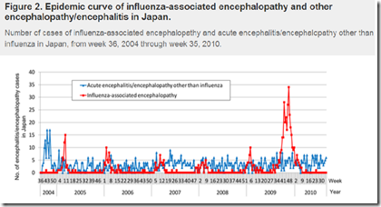
PLoS One: Influenza-Associated Encephalopathy


image

6-fold spike in encephalopathy during 2009 Pandemic Credit PLoS One


# 6908


While not viewed as having been a particularly severe pandemic, the 2009 H1N1 virus nonetheless caused serious, and sometimes fatal illness in a small percentage of patients.


And it did so in some pretty remarkable ways.


Seasonal influenza is primarily seen as a `harvester’ of the aged and infirmed, robbing its victims of the last few months or years of life, and only rarely does it seriously impact young adults and children.


Yet, in  Study: Years Of Life Lost Due To 2009 Pandemic, we saw the mean age of death from the 2009 H1N1 virus to be half that of seasonal flu, or 37.4 years


Severe lung injury, not normally seen with seasonal influenza, was also reported in a very small number of patients as well. We saw scattered reports during the summer and fall of 2009 (see Pathology Of Fatal H1N1 Lung Infections), but it wasn’t until December that the NIH: Post Mortem Studies Of H1N1 showed:


New York Autopsies Show 2009 H1N1 Influenza Virus Damages Entire Airway

In fatal cases of 2009 H1N1 influenza, the virus can damage cells throughout the respiratory airway, much like the viruses that caused the 1918 and 1957 influenza pandemics, report researchers from the National Institutes of Health (NIH) and the New York City Office of Chief Medical Examiner.

(Continue . . .)


It was also widely reported (see Pregnancy & Flu: A Bad Combination) during the 2009 pandemic that pregnant women were up to 6 times more likely to be hospitalized with influenza than were non-pregnant women.


While many of these complications made headlines, somewhat less well publicized was the neurological impact this virus had on young children.


Barely 90 days after the novel H1N1 virus emerged, the CDC’s MMWR reported on 4 pediatric patients with the novel H1N1 virus who presented with neurological symptoms including unexplained seizures and altered mental status:


Neurologic Complications Associated with Novel Influenza A (H1N1) Virus Infection in Children --- Dallas, Texas, May 2009


Additional reports came in, particularly from Japan, indicating an unusual number cases of Influenza-related encephalopathy (IAE) among children (see Japan: Influenza Related Encephalopathy).


Encephalopathy isn’t a distinct disease, but rather refers to a syndrome of diffuse brain dysfunctions, which may be associated with a variety of causes (including viral, bacterial, trauma, prions, and toxic chemicals).

The overriding hallmark of encephalopathy is an altered mental state, although depending and severity of encephalopathy, common neurological symptoms such as progressive memory loss and changes in cognitive abilitypersonality changes, inability to concentrate, lethargy, seizures and loss of consciousness may be seen.


Over the past 15 years Influenza has been increasingly recognized as a rare cause of encephalopathy. For reasons not really known, is reported most often among children and adolescents in Japan and Taiwan.


In January of 2010, the CDC’s EID Journal carried a report called Neurologic Manifestations of Pandemic (H1N1) 2009 Virus Infection and in September, the Annals of Neurology carried a study called Heightened Neurologic Complications in Children with Pandemic H1N1 Influenza that found:

The study looked at 303 children hospitalized with the pandemic H1N1 virus, of which 18 developed neurological symptoms.


They compared these cases to records of 234 children admitted to the hospital in previous years due to seasonal influenza.


The most common neurological complications exhibited with novel H1N1 were seizures (12 patients or 67%), with seven exhibiting status epilepticus, a potentially life-threatening condition involving continuous or recurrent seizures that can last for a half hour or longer.


The mean age of children admitted with neurological symptoms from H1N1 was more than twice the age (6.5 years) than usually seen with seasonal flu (2.4 years).


All of which serves as prelude to a research article that recently appeared in PloS One, that examined the rate of influenza-related encephalopathy in Japan across 6 flu seasons, including the 2009 pandemic.


National Surveillance of Influenza-Associated Encephalopathy in Japan over Six Years, before and during the 2009–2010 Influenza Pandemic

Yoshiaki Gu mail, Tomoe Shimada, Yoshinori Yasui, Yuki Tada, Mitsuo Kaku, Nobuhiko Okabe


Abstract (reparagraphed)

Influenza-associated encephalopathy (IAE) is a serious complication of influenza and is reported most frequently in Japan. This paper presents an assessment of the epidemiological characteristics of influenza A (H1N1) 2009-associated encephalopathy in comparison to seasonal IAE, based on Japanese national surveillance data of influenza-like illness (ILI) and IAE during flu seasons from 2004–2005 through 2009–2010.


In each season before the pandemic, 34–55 IAE cases (mean 47.8; 95% confidence interval: 36.1–59.4) were reported, and these cases increased drastically to 331 during the 2009 pandemic (6.9-fold the previous seasons).


Of the 331 IAE cases, 322 cases were reported as influenza A (H1N1) 2009-associated encephalopathy. The peaks of IAE were consistent with the peaks of the influenza epidemics and pandemics.


A total of 570 cases of IAE (seasonal A, 170; seasonal B, 50; influenza A (H1N1) 2009, 322; unknown, 28) were reported over six seasons. The case fatality rate (CFR) ranged from 4.8 to 18.2% before the pandemic seasons and 3.6% in the 2009 pandemic season. The CFR of pandemic-IAE was 3.7%, which is lower than that of influenza A−/B-associated encephalopathy (12.9%, p<0.001; 14.0%, p = 0.002; respectively).


The median age of IAE was 7 years during the pandemic, which is higher than that of influenza A−/B-associated encephalopathy (4, p<0.001; 4.5, p = 0.006; respectively).


However, the number of pandemic-IAE cases per estimated ILI outpatients peaked in the 0–4-year age group and data both before and during the pandemic season showed a U-shape pattern. This suggests that the high incidence of influenza infection in the 0–4 year age group may lead to a high incidence of IAE in the same age group in a future influenza season.


Further studies should include epidemiologic case definitions and clinical details of IAE to gain a more accurate understanding of the epidemiologic status of IAE.


This is a detailed, and interesting, analysis that finds that in addition to a 6 fold increase in cases during the pandemic, that (like the Annals of Neurology Study mentioned above) the median age of reported encephalopathy was twice that normally seen during other flu seasons.


The CFR (Case Fatality Ratio) of encephalopathy cases during the pandemic dropped, however, which the authors ascribe to `improved quality of diagnosis and treatment’.


Some of the 6-fold increase in IAE cases during the 2009 pandemic can probably be linked to increased public awareness and medical surveillance.  Cases of mild IAE might have been transient, and not picked up by medical authorities during other flu seasons. 


But when added to the other reports we’ve seen, the 2009 H1N1 virus does appear to have had some unusual properties.  The authors conclude by writing:


In summary, national surveillance of AE in Japan revealed a steep increase in IAE cases during the 2009–2010 pandemic season. This was likely due to the large number of children infected with influenza during the pandemic, but social attention and information bias might have affected epidemiology data. Results of the present study revealed a relatively low CFR despite a large number of reported IAE cases during the pandemic. One of the characteristic findings was the age distribution of the reported IAE cases. Further studies should include strict epidemiologic case definitions, clinical details including medication history, and epidemiological information of IAE for a more accurate understanding of the epidemiologic status of IAE.


Nearly 4 years since the emergence of the 2009 H1N1 pandemic, A/H1N109pdm is probably the most studied flu virus in history. While it did not end up being a particularly deadly pandemic virus, it has consistently yielded surprises.


  • This was the first seasonal flu shown to infect both dogs and cats (see EID Journal: Pandemic H1N1 Infection In Cats) and showed up in such diverse animal hosts as turkeys and ferrets.
  • Novel H1N1 also appears to differ from seasonal flu in how it is transmitted, at least according to researchers in Hong Kong who discovered that the novel H1N1 virus – unlike seasonal flu – easily infects and replicates in the conjunctival tissues of the eye  (see I Only Have Eyes For Flu).
  • And rather than completely supplanting the existing seasonal influenza A strains as we saw in the 1918, 1957 and 1968 pandemics, the 2009 pandemic virus now co-circulates with H3N2 (it did replace seasonal H1N1).


While some of these studies may seem a bit esoteric and limited in scope or impact, the knowledge we gain from them can hopefully prove an advantage to us the next time the world faces an emerging viral threat.

Monday, January 14, 2013

The Risks Of Chikungunya Outbreaks In The United States


image

Credit Wikipedia


# 6855


Chikungunya, up until about a decade ago, was a little known mosquito-borne disease first described in Tanganyika in the early 1950s. For the next five decades it was sporadically seen across eastern and central Africa.


That is, until 2005, when Chikungunya made a surprise jump to the Indian Ocean island of RĂ©union. There, it infected nearly 1/3rd of the island’s 770,000 residents (see 2006 EID article Chikungunya Disease Outbreak, Reunion Island) in just a matter of months.


Chikungunya typically produces a fever, severe muscle and joint pain, and headaches. The symptoms usually go away after a few weeks, but some patients can sustain permanent disability, and some deaths have been reported.


In the eight years since that  jump, `Chik’ has spread further across the Indian Ocean, Southeast Asia, and even briefly into northern Italy.


While the virus isn't normally found in Europe, the vector, the Aedes mosquito, is.  All it took was one infected traveler to arrive infected with the virus to start the chain of transmission.


image


I told the story several years ago in It's A Smaller World After All, but the short version is that a traveler, returning from India, brought the virus to Italy in 2007 which led to more than 290 cases reported in the province of Ravenna, which is in northeast Italy.


The concern is that the same sort of introduction could happen elsewhere in Europe, or here in the United States, just as we saw with West Nile Virus in 1999 (see DVBID: Final West Nile Report For 2012) and with Dengue Fever in 2010 (see MMWR: Dengue Fever In Key West)


The two primary mosquito vectors of Chikungunya are the Aedes aegypti and Aedes albopictus (cite WHO FAQ) both of which can be found across many regions of the Americas.

image

Aedes albopictus (Asian Tiger) Mosquito - Wikipedia

Dark blue: Native range
Dark green: introduced (as of December 2007)


The risk is considered great enough that early last year, the CDC and PAHO (Pan American Health Organization) put together a 161-page guide on preparing for the arrival of Chikungunya to the Americas (see Preparedness and Response for Chikungunya Virus Introduction in the Americas).


All of which serves as prelude to a report that appeared last month in PloS Neglected Tropical Diseases called:


Modeling Dynamic Introduction of Chikungunya Virus in the United States

Abstract (reparagraphed for readability)

Chikungunya is a mosquito-borne viral infection of humans that previously was confined to regions in central Africa. However, during this century, the virus has shown surprising potential for geographic expansion as it invaded other countries including more temperate regions.


With no vaccine and no specific treatment, the main control strategy for Chikungunya remains preventive control of mosquito populations. In consideration for the risk of Chikungunya introduction to the US, we developed a model for disease introduction based on virus introduction by one individual. Our study combines a climate-based mosquito population dynamics stochastic model with an epidemiological model to identify temporal windows that have epidemic risk. We ran this model with temperature data from different locations to study the geographic sensitivity of epidemic potential.


We found that in locations with marked seasonal variation in temperature there also was a season of epidemic risk matching the period of the year in which mosquito populations survive and grow. In these locations controlling mosquito population sizes might be an efficient strategy.


But, in other locations where the temperature supports mosquito development all year the epidemic risk is high and (practically) constant. In these locations, mosquito population control alone might not be an efficient disease control strategy and other approaches should be implemented to complement it.


Our results strongly suggest that, in the event of an introduction and establishment of Chikungunya in the US, endemic and epidemic regions would emerge initially, primarily defined by environmental factors controlling annual mosquito population cycles. These regions should be identified to plan different intervention measures.

In addition, reducing vector: human ratios can lower the probability and magnitude of outbreaks for regions with strong seasonal temperature patterns. This is the first model to consider Chikungunya risk in the US and can be applied to other vector borne diseases.

(Continue . . . )


More background on this modeling study is available from the Cornell University Press Office.


Chances seen rising for chikungunya outbreaks in NYC, Atlanta, Miami

ITHACA, N.Y. – Global travel and climate warming could be creating the right conditions for outbreaks of a new virus in this country, according to a new Cornell University computer model.


The model predicts that outbreaks of chikungunya, a painful virus transported by travelers and spread by the invasive Asian tiger mosquito, could occur in 2013 in New York City during August and September, in Atlanta from June through September, and year-round in Miami. The probability of a disease outbreak is correlated with temperature, as warmer weather allows the Asian tiger mosquito to breed faster and grow in numbers, according to the study published in the November issue of PLOS Neglected Tropical Diseases.


According to the simulation, there is a high probability of a chikungunya outbreak if a single infected person arrives in New York in July or August and is bitten by an Asian tiger mosquito. The risks are the same, but with wider time frames, for transmission in Atlanta and Miami, according to the paper.


Asian tiger mosquitoes were introduced to the United States in Texas in the 1980s; they are established up the East Coast into New Jersey and are rising in numbers in New York City. The aggressive mosquito outcompetes local varieties and transmits more than 20 pathogens, including chikungunya and dengue, said Laura Harrington, associate professor of entomology and the study’s senior author.


“The virus is moving in people, and resident mosquito populations are picking it up,” Harrington said.


The model estimates that with typical regional temperatures, a chikungunya outbreak in New York would infect about one in 5,000 people, said Diego Ruiz-Moreno, a postdoctoral associate and the paper’s lead author


“However, this number would increase drastically as temperatures rise due to climate change,” Ruiz-Moreno said.

(Continue . . . )


While this study focused on Chikungunya, much the same could be said about the potential for seeing Dengue, Malaria, or even Yellow Fever making inroads in the United States and Europe. 


In March of 2010 the journal  Eurosurveillance carried a series of articles on vector borne diseases and their potential to impact those living in Europe. One of the articles, Yellow fever and dengue: a threat to Europe? by P. Reiter, had these sobering comments about the future of vector-borne illnesses in Europe.


The history of dengue and yellow fever in Europe is evidence that conditions are already suitable for transmission. The establishment of Ae. albopictus has made this possible, and the possibility will increase as the species expands northwards, or if Ae. aegypti is re-established.


The epidemic of chikungunya in northern Italy in 2007 [8,49] confirms that Ae. albopictus is capable of supporting epidemic transmission, although laboratory studies indicate that the strain of virus involved was particularly adapted to this species [50,51].


Nevertheless, it is not unreasonable to assume that climatic conditions that permit malaria transmission will also support transmission of yellow fever and dengue, in which case transmission could extend into northern Europe [52].


Reason enough that if you live in - or are visiting  - a mosquito prone area, to remember to follow the `5 D’s’  (courtesy Florida Department of Health).


image

Saturday, December 29, 2012

Study: Statins & Cerebral Malaria

image

Photo Credit CDC


# 6808


Increasingly, statins – common cholesterol lowering drugs – are being looked at for their inflammation-reducing properties in the treatment of other diseases.


Long time readers of this blog will recall that Dr. David Fedson - former Professor of Medicine at the University of Virginia School of Medicine and formerly Director of Medical Affairs, Aventis Pasteur MSD – has advocated research into the potential role of low-cost statins during an influenza pandemic (see Lancet: David Fedson On Statins For Pandemic Influenza).


For more on statins, and how they might be used against pandemic influenza, you may wish to revisit:


Study: Statins, Influenza, & Mortality

Another Study On Statins And Pneumonia

Dr. David Fedson: The Case For Using Statins In A Pandemic

Statins Revisited


A couple of years ago we saw a video presentation at the 2010 ICAAC Conference called A Role for Statins in Infectious Disease? #ICAAC) (excerpt below).


Statins are well-known as a class of drugs that are used to help lower cholesterol but recent evidence suggests they might be good for more than your heart. They may play a role in preventing and treating certain bacterial infections including pneumonia and sepsis. Presenters at ICAAC discuss the latest research on the potential of these drugs.

  • Reimar Thomsen, Aarhus University Hospital, Aalborg, Denmark
  • Matthew Falagas, Alfa Institute of Biomedical Sciences, Athens, Greece
  • Nasia Safdar, University of Wisconsin, Madison, WI, United States


These presenters suggest that statins may directly affect viruses and fungi, as well as help dampen the body’s inflammatory response. One study discussed found a 30% reduction in 30-day pneumonia mortality among patients already on statins.


The caveat being that much of the evidence for statins efficacy comes from in vitro studies, or observational studies that can sometimes be influenced by what is known as the `healthy user bias’.  


Simply put, patients who are already on statins when they develop pneumonia, sepsis, or influenza may be more likely to have a healthy lifestyle than those not on statins, potentially skewing the results.


Still, the results to date have been intriguing, if not totally convincing.


Which brings us to a a new study, appearing in PloS Pathogens, that looks at the potential role of statins in the treatment of cerebral Malaria.


According to the WHO:

There were about 219 million cases of malaria in 2010 and an estimated 660 000 deaths. Africa is the most affected continent: about 90% of all malaria deaths occur there.


Between 2000 and 2010, malaria mortality rates fell by 26% around the world. In the WHO African Region the decrease was 33%. During this period, an estimated 1.1 million malaria deaths were averted globally, primarily as a result of a scale-up of interventions.


Rarely mentioned in all of these figures are the (often life-long) neurological sequelae that cerebral malaria may produce, particularly among children.


These may include blindness, epilepsy, decreased motor skills, hearing impairment, aphasia (loss of speech), and behavioral problems, as noted in the following BMC Research Note.


Severe neurological sequelae and behaviour problems after cerebral malaria in Ugandan children

Richard Idro, Angelina Kakooza-Mwesige, Stephen Balyejjussa, Grace Mirembe, Christine Mugasha, Joshua Tugumisirize and Justus Byarugaba

Conclusions

In addition to previously described neurological and cognitive sequelae, severe behaviour problems may follow cerebral malaria in children. The observed differences in patterns of sequelae may be due to different pathogenic mechanisms, brain regions affected or extent of injury. Cerebral malaria may be used as a new model to study the pathogenesis of ADHD.


The PloS Pathogens study, which looks at the potential use of statins for cerebral malaria in a murine (mouse) model, involved infecting lab mice with the malaria parasite, and then treating half of them with just chloroquine, and the other half with chloroquine and Lovastatin. 


Mice that received the combination treatment saw a significantly reduced rate of post-infection cognitive dysfunction.


Statins Decrease Neuroinflammation and Prevent Cognitive Impairment after Cerebral Malaria

Patricia A. Reis mail, Vanessa Estato, Tathiany I. da Silva, Joana C. d'Avila, Luciana D. Siqueira, Edson F. Assis, Patricia T. Bozza, Fernando A. Bozza, Eduardo V. Tibiriça, Guy A. Zimmerman, Hugo C. Castro-Faria-Neto

Author Summary

Cerebral malaria (CM) is the direst consequence of Plasmodium falciparum infection. Cognitive impairment is a common sequela in children surviving CM. Identification of adjunctive therapies that reduce the complications of CM in survivors is a priority. Statins have been suggested for the treatment of neuroinflammatory disorders due to their pleiotropic effects.


Here, we examined the effects of lovastatin on neuroinflammation in experimental CM, and its effect on the prevention of cognitive impairment. Lovastatin reduced adhesion and rolling of leukocytes in brain vessels, inhibited blood-brain barrier disruption, and reversed decreases in cerebral capillary density. Lovastatin also inhibited ICAM-1 and CD11b mRNA expression while increasing HMOX-1 mRNA levels. Proinflammatory cytokines and markers of oxidative stress were lower in the brains of infected mice treated with lovastatin.


Lovastatin administered together with antimalarial drugs during the acute phase of the disease-protected survivors from impairment in both contextual and aversive memory 15 days after infection. Similar results were observed in a model of bacterial sepsis.


Our findings support the possibility that statins may be valuable pharmacologic tools in treatment of patients with neuroinflammation associated with severe systemic inflammatory syndromes. Clinical trials with statins in CM and sepsis should be speedily considered to examine this point.


Of course, what works in mice isn’t guaranteed to work in humans.  The authors caution:


These models may provide important insights into the pathogenesis of cognitive dysfunction associated with cerebral malaria and related disorders that may be relevant to human conditions [7]. While differences between murine models of CM and the human syndrome are often emphasized [10], [11], there are also important similarities [3], [7], [12][14]. Nevertheless, caution must be exerted when translating experimental findings to the clinical scenario.


The VOA has a nice write up of this study (see Mice Study Indicates Cholesterol Drug Might Help Treat Serious Malaria Cases), including an interview with one of the authors, who recommends that:


Zimmerman recommends lovastatin be added to treatments for malaria as well as for sepsis, a systemic blood infection commonly known as blood poisoning that sickens and threatens the lives of more people worldwide than cerebral malaria.


The problem with statins is that these are are cheap, generic drugs.  They provide little financial incentive for their manufacturers to mount expensive human trials in order to prove their effectiveness against malaria, pneumonia, sepsis, or influenza.


So, while the evidence continues to suggest benefits to using statins for `off label’ purposes,  real proof of their effectiveness may be a long time in coming.

Wednesday, December 12, 2012

PLoS One: Influenza Viral Shedding & Asymptomatic Infections

image

Photo Credit PHIL (Public Health Image Library)


# 6776


Despite major advances in the study of influenza viruses, there remain significant gaps in our understanding of just how they work once they infect a human (or any other animal) host. 


Basic questions, such as `How long are we infectious?, or `How common are asymptomatic infections?  remain only partially answered.


Complicating matters, variations in individual host’s immune responses, and different strains of flu may produce varying results. Meaning that most studies can only add incrementally to our knowledge, rather than completely answering these questions.


Previously, we’ve seen evidence of asymptomatic and `presymptomatic’ shedding of influenza viruses.


In 2011, in EID Journal: Pre-Symptomatic Influenza Transmission we looked at three clusters of suspected pre-symptomatic transmission of the 2009 H1N1 virus in Japan.


And in Pre-Symptomatic Transmission Of H1N1 Influenza In the Ferret Model, researchers inoculated ferrets with the 2009 H1N1 flu, and then placed them near uninfected ferrets (some in direct contact, others in adjacent cages) at different stages after infection.


They then tested the exposed ferrets to see when, and under what circumstances, they became infected. They found that ferrets became infectious just 24 hours after becoming infected, and nearly 24 hours before showing the earliest outward signs of infection (fever).


The importance of all of this is, if presymptomatic and asymptomatic carriers of a flu virus are able to efficiently transmit the illness on to others, then strategies that seek to identify and isolate flu cases would have only limited success in containing a pandemic.


Similarly, understanding how long a person sheds the virus after becoming infected is crucial, so we can know when it is (relatively) safe for flu victims to return to work or school without endangering others.


The CDC’s general take on this topic is:


The Flu Is Contagious

Most healthy adults may be able to infect others beginning 1 day before symptoms develop and up to 5 to 7 days after becoming sick. Children may pass the virus for longer than 7 days. Symptoms start 1 to 4 days after the virus enters the body. That means that you may be able to pass on the flu to someone else before you know you are sick, as well as while you are sick. Some persons can be infected with the flu virus but have no symptoms. During this time, those persons may still spread the virus to others.


Yesterday, a new study appeared in PloS One, conducted in Germany over 4 flu seasons (2007-2011) and involving 4 flu strains - seasonal (A(H3N2), A(H1N1), influenza B, and pandemic (A(H1N1)pdm09 - that looks at many of these transmission issues. 


Comparison of Shedding Characteristics of Seasonal Influenza Virus (Sub)Types and Influenza A(H1N1)pdm09; Germany, 2007–2011

Thorsten Suess, Cornelius Remschmidt, Susanne B. Schink, Brunhilde Schweiger, Alla Heider, Jeanette Milde, Andreas Nitsche, Kati Schroeder, Joerg Doellinger, Christian Braun, Walter Haas, Gérard Krause, Udo Buchholz

Background

Influenza viral shedding studies provide fundamental information for preventive strategies and modelling exercises. We conducted a prospective household study to investigate viral shedding in seasonal and pandemic influenza between 2007 and 2011 in Berlin and Munich, Germany.

Methods

Study physicians recruited index patients and their household members. Serial nasal specimens were obtained from all household members over at least eight days and tested quantitatively by qRT-PCR for the influenza virus (sub)type of the index patient. A subset of samples was also tested by viral culture. Symptoms were recorded daily.

Results

We recruited 122 index patients and 320 household contacts, of which 67 became secondary household cases. Among all 189 influenza cases, 12 were infected with seasonal/prepandemic influenza A(H1N1), 19 with A(H3N2), 60 with influenza B, and 98 with A(H1N1)pdm09. Nine (14%) of 65 non-vaccinated secondary cases were asymptomatic/subclinical (0 (0%) of 21 children, 9 (21%) of 44 adults; p = 0.03).


Viral load among patients with influenza-like illness (ILI) peaked on illness days 1, 2 or 3 for all (sub)types and declined steadily until days 7–9. Clinical symptom scores roughly paralleled viral shedding dynamics.


On the first day prior to symptom onset 30% (12/40) of specimens were positive. Viral load in 6 asymptomatic/subclinical patients was similar to that in ILI-patients. Duration of infectiousness as measured by viral culture lasted approximately until illness days 4–6. Viral load did not seem to be influenced by antiviral therapy, age or vaccination status.

Conclusion

Asymptomatic/subclinical infections occur infrequently, but may be associated with substantial amounts of viral shedding. Presymptomatic shedding may arise in one third of cases, and shedding characteristics appear to be independent of (seasonal or pandemic) (sub)type, age, antiviral therapy or vaccination; however the power to find moderate differences was limited.


While this was a relatively small study, and their findings don’t always align perfectly with others we’ve seen (for instance, children didn’t appear contagious any longer than adults), it does provide us with some interesting data.


  • First, nearly 1/3rd of cases began shedding virus while pre-symptomatic
  • Second, viral loads in (six studied) asymptomatic cases were similar to that to patients exhibiting ILI (influenza-like-illness) symptoms.
  • Third, viral load among symptomatic patients peaked on illness days 1, 2 or 3 and declined steadily until days 7–9


Some other gems (bolding mine) excerpted from this open access article include:


  • Overall 63% of non-vaccinated secondary household cases had an ILI-syndrome and the proportion of asymptomatic/subclinical secondary cases was 14%.
  • Frequency distribution of clinical symptoms did not differ between A(H1N1)pdm09 cases and non-pandemic influenza cases.
  • Interestingly, 21% of adult secondary cases were asymptomatic/subclinical, while all children that contracted influenza were symptomatic.
  • Based on the population of ambulatory patients investigated we found no evidence that the amount of shedding is particularly higher in children, nor that duration of viral shedding is significantly longer in children compared to adults.


The authors conclude by saying:


In summary, our study addresses several important questions on clinical manifestation, duration of infectiousness, viral shedding patterns, including shedding before symptom onset and in asymptomatic/subclinical patients, as well as the effect of vaccination and antiviral therapy on viral shedding.


Important single results include the finding that children do not seem to be infected asymptomatically, that shedding one day before symptom onset may occur in one third of influenza patients, that asymptomatic/subclinical influenza patients occur rarely, but viral load (and probably infectiousness) may be substantial, and vaccinated influenza patients do not show different shedding patterns compared to non-vaccinated cases with ILI.


Overall results do not show marked differences between seasonal influenza (sub)types and influenza A(H1N1)pdm09.

Thursday, December 06, 2012

An Unwanted Lagniappe From The Kitchen


image

Norovirus – Credit CDC PHIL 


LagniappeA little something extra given to a customer. 


# 6766


The CDC estimates that norovirus (aka the `Winter Vomiting Bug’ or less accurately, `Stomach Flu’) causes more than 20 million cases of gastroenteritis each year in the United States. Anyone who has endured it can attest that a bout with norovirus is misery incarnate.


The CDC maintains an extensive Norovirus webpage where they describe the illness, how it is spread, and how to avoid infection. Among known causes of foodborne illness, norovirus is the largest culprit.


image


The CDC’s NOROVIRUS: FOR FOOD HANDLERS page warns:


The virus can easily contaminate food because it is very tiny and infective. It only takes a very small amount of virus particles (fewer than 100) to make someone sick.


Food can get contaminated with norovirus when:

  • infected people who have stool or vomit on their hands touch the food,
  • it is placed on counters or surfaces that have infectious stool or vomit on them, or
  • tiny drops of vomit from an infected person spray through the air and land on the food.


All of which makes the thorough cleaning of plates and utensils used in restaurants a priority. 


According to a PLoS One  study, published yesterday, the prescribed methods of cleaning these items in restaurants does a good job against common bacteria – like Escherichia coli K-12 and Listeria innocua – but not so well in sanitizing against norovirus.


The open access study is called:


Efficacies of Sodium Hypochlorite and Quaternary Ammonium Sanitizers for Reduction of Norovirus and Selected Bacteria during Ware-Washing Operations

Lizanel Feliciano, Jianrong Li*, Jaesung Lee, Melvin A. Pascall*


Abstract

Cross-contamination of ready-to-eat (RTE) foods with pathogens on contaminated tableware and food preparation utensils is an important factor associated with foodborne illnesses. To prevent this, restaurants and food service establishments are required to achieve a minimum microbial reduction of 5 logs from these surfaces.


This study evaluated the sanitization efficacies of ware-washing protocols (manual and mechanical) used in restaurants to clean tableware items. Ceramic plates, drinking glasses and stainless steel forks were used as the food contact surfaces. These were contaminated with cream cheese and reduced-fat milk inoculated with murine norovirus (MNV-1), Escherichia coli K-12 and Listeria innocua.


The sanitizing solutions tested were sodium hypochlorite (chlorine), quaternary ammonium (QAC) and tap water (control). During the study, the survivability and response to the experimental conditions of the bacterial species was compared with that of MNV-1.


The results showed that current ware-washing protocols used to remove bacteria from tableware items were not sufficient to achieve a 5 log reduction in MNV-1 titer. After washing, a maximum of 3 log reduction in the virus were obtained. It was concluded that MNV-1 appeared to be more resistant to both the washing process and the sanitizers when compared with E. coli K-12 and L. innocua.


(Continue . . . )


Essentially, these researchers took silverware, ceramic plates, and glassware – inoculated them with norovirus, E. coli, and Listeria – and ran them through standard dishwasher or hand washing protocols with either a bleach solution or quaternary ammonium compound (QAC) solution.


After that, they tested them for residual pathogens.


Although commercial dishwashers did a better job than did handwashing, with both, significant contamination from the norovirus remained behind.


The authors write:


Conclusions

From the results of our study, it could be concluded that QAC and sodium hypochlorite sanitizers normally used to inactivate bacteria in manual and mechanical ware-washing operations were unable to produce the same level of virus inactivation under similar conditions, irrespective of the nature of the tableware item tested.

Further studies are needed to develop more effective ware-washing protocols for the removal of viruses from food contact surfaces/tableware items. Also, the combination of different detergents and sanitizing solutions (especially those containing surfactant agents) should be evaluated since they may help to enhance the removal and inactivation of non-enveloped viruses.


Obviously, people who are sick should not be handling food, but people can shed the virus even after they stop showing symptoms.


So one of the keys to prevention is good hand hygiene.

Unfortunately, unlike with many other bacteria and viruses, alcohol gel doesn’t do a particularly good job of killing the virus, something we discussed last year in  CMAJ: Hand Sanitizers May Be `Suboptimal’ For Preventing Norovirus.


Which makes a good old fashion hand scrubbing with soap and water the best preventative.


The CDC recommends the following steps to protect yourself from the virus.

image


And of course, the obvious question is . . .if norovirus escapes routine dishwashing protocols, what about other viruses like Hepatitis A and influenza?


Reportedly, these researchers will be looking at those viruses next.