Showing posts with label Resistance. Show all posts

Saturday, April 18, 2015

Early Signs Of Artemisinin-Resistant Malaria In Africa

Plasmodium falciparum in human blood – credit Wikipedia


# 9955


One of the realities of our never-ending battle against infectious diseases is that these organisms are able to evolve, and adapt, at an amazing rate, and that the antivirals, antibiotics, and other drugs in our arsenal of therapeutic drugs can – over time – lose some or all of their effectiveness.


Antibiotic resistance has been the greatest concern, and in recent years we’ve seen some antiviral drugs falter badly (Amantadine, a stalwart against influenza for decades, became unusable in 2005), often due to poor stewardship.


Anti-malarial drugs are not immune to this sort of evolutionary obsolescence, and any loss in effectiveness would put millions of people at risk of serious illness or even death.  Spread by mosquitoes, and caused by a parasite  – Plasmodium – Malaria is common in much of the world including Sub-Saharan Africa, Asia and the Americas.  


There are four microscopic protozoan parasites in the genus Plasmodium (P. vivax, P. falciparum, P. malariae and P. ovale) that cause malaria in humans around the world.  Of these Plasmodium falciparum is generally the most serious.


The parasites multiply in the liver and infect red blood cells, resulting in recurrent fevers and headaches - and in severe cases - coma and death.  Despite advances, Malaria remains an extremely serious problem in Africa, where 1 in 5 childhood deaths is due to the disease. According to the WHO’s 10 Facts on Malaria:


About 3.2 billion people – almost half of the world's population – are at risk of malaria. In 2013, there were about 198 million malaria cases (with an uncertainty range of 124 million to 283 million) and an estimated 584 000 malaria deaths (with an uncertainty range of 367 000 to 755 000). Increased prevention and control measures have led to a reduction in malaria mortality rates by 47% globally since 2000 and by 54% in the WHO African Region.


According to the WHO, the best available treatment - particularly for P. falciparum malaria - is artemisinin-based combination therapy (ACT).  Earlier treatment options such as choloroquine - the cheapest and for years the most commonly used drug - and the combination of sulfadoxine-pyrimethamine  have slowly lost their effectiveness. .


Of concern, since about 2007 evidence of resistance to the newer drug regimen ACT  has been showing up on the Cambodian-Thai border, and more recently in Myanmar (see MYANMAR: Anti-malarial drug resistance "hotspots" identified).


For now, ACT seems to be working in Africa, but the following press release from the London School of Hygiene & Tropical Medicine, suggests that success may be in danger. Researchers report finding Plasmodium falciparum malaria parasites with a mutation to the gene Ap2mu were less sensitive to artemisinin.


The abstract and full text to the dauntingly titled study - The Mu Subunit of Plasmodium falciparum Clathrin-Associated Adaptor Protein 2 Modulates In Vitro Parasite Response to Artemisinin and Quinine – is available from Antimicrobial Agents and Chemotherapy


Fortunately we also have the following  press release to go along with it.

New genetic mutation could signal start of malaria drug resistance in Africa

London School of Hygiene & Tropical Medicine

Early indicators of the malaria parasite in Africa developing resistance to the most effective drug available have been confirmed, according to new research published in Antimicrobial Agents and Chemotherapy.

Researchers at the London School of Hygiene & Tropical Medicine found Plasmodium falciparum malaria parasites with a mutation to the gene Ap2mu were less sensitive to the antimalarial drug artemisinin.

A study in 2013, also led by the School, suggested an initial link between a mutation in the ap2mu gene and low levels of malaria parasites remaining in the blood of Kenyan children after they had been treated.[1] However, further research was needed to confirm if these genetic characteristics represented an early step towards resistance.

In the new study, researchers genetically altered the malaria parasite in the laboratory to mutate ap2mu in the same way that had been observed in Kenya. They found the altered parasite was significantly less susceptible, requiring 32% more drug to be killed by artemisinin. The genetically altered parasite was also 42.4% less susceptible to the traditional antimalarial drug, quinine.

Earlier this year a different research group discovered mutations in the gene kelch13 which were linked to reduced susceptibility to artemisinin combination treatment in South East Asia.[2] Historically, resistance to antimalarial medicines has emerged in South East Asia and then spread to Africa. But these new findings suggest a different route to drug resistance may be developing independently in Africa.

Lead researcher Dr Colin Sutherland, Reader in Parasitology at the London School of Hygiene & Tropical Medicine, said: "Our findings could be a sign of much worse things to come for malaria in Africa. The malaria parasite is constantly evolving to evade our control efforts. We've already moved away from using quinine to treat cases as the malaria parasite has become more resistant to it, but if further drug resistance were to develop against our most valuable malaria drug, artemisinin, we would be facing a grave situation.

"We now know that the gene ap2mu is an important factor in determining how well our drugs kill malaria parasites. We will be conducting laboratory and field studies to more accurately measure the impact of mutations in the ap2mu gene. We hope our findings will help understand resistance of malaria to drugs, and potentially be an important tool for monitoring malaria treatment in the future."

The World Health Organization estimates more than half a million people die from malaria every year, mostly children under five. Plasmodium falciparum is the most deadly form of the malaria parasite.


If nature weren’t capable enough on its own, helping these parasites along in their arms race has been the fact that a large percentage of the anti-malarial drugs used in Asia and sub-Saharan Africa are either fake, or are of inferior quality (see Lancet: 1/3rd Of Malaria Drugs Fake Or Sub-Standard).


Using drugs that contain too little of their intended active ingredient can contribute to pathogens developing increased and widespread resistance over time.


This is a big enough problem that the CDC maintains a webpage devoted to Counterfeit and Substandard Antimalarial Drugs: Information for Travelers.


With World Malaria Day set for April 25th, we can expect to hear a good deal more about this devastating and often deadly disease over the next week.

Tuesday, July 22, 2014

Hong Kong: Two Hospital Clusters Of MDR Acinetobacter Infections

image

Credit CDC PHIL


# 8856


Because it is such an international city, and boasts one of the most diligent (and open) public health agencies in the world (Centre For Health Protection), Hong Kong has become a terrific  barometer for the growth of multiple drug resistant infections from around the world.


One of the toughest bacteria that hospitals must deal with is called multidrug-resistant (MDR) Acinetobacter baumannii, which in recent years has made headlines as the cause of difficult to treat wound infections among our troops serving in the Middle East.


Acinetobacter (of which there are many varieties, but A. baumannii is most often linked to human infection ) are ubiquitous in nature, and can be found in soil, water, animals and humans. A very hardy species, they can survive for extended period of time on inanimate surfaces, making them difficult to control in a health care setting (see AJIC report Hospital cleaning protocol ineffective against A. baumannii)..


And like with MRSA, many people can be colonized, but not show signs of infection.  Often very serious infections develop among those who are very ill, wounded, or immunocompromised.


Today Hong Kong’s CHP has published two reports on hospital clusters of MDR  Acinetobacter infection.


Cluster of Multi-drug Resistant Acinetobacter cases in Queen Elizabeth Hospital

The following is issued on behalf of the Hospital Authority:


The spokesperson for Queen Elizabeth Hospital (QEH) made the following announcement today (July 22):


Five male patients (aged 35 to 80) of a Ventilator Ward have been confirmed as having Multi-drug Resistant Acinetobacter (MDRA) since July 14. Two of them are infected cases and are still hospitalised at QEH. The remaining three patients were confirmed to be MDRA carriers without clinical symptoms. Out of these cases, two are still hospitalised under medical surveillance and isolation at QEH. The remaining patient has been transferred to Hong Kong Buddhist Hospital. All of the five patients are in stable condition.

(Continue. . . )

Cluster of Multi-drug Resistant Acinetobacter cases in Caritas Medical Centre

The following is issued on behalf of the Hospital Authority:


     The spokesperson of Caritas Medical Centre made the following announcement today (July 22):


Three patients (aged 37 to 88) of a male Medicine and Geriatrics Ward have been confirmed as having Multi-drug Resistant Acinetobacter (MDRA) since July 17. Two of them are infected cases. The patients are still hospitalised under medical surveillance and isolation. Two of them are in stable condition, while the other one is in serious condition.

(Continue . . .)


Just yesterday, Hong Kong reported a Case of NDM-5 Carbapenemase-producing Enterobacteriaceae under CHP investigation in a 30-year-old woman with a urinary tract infection.  


NDM-5 is a novel variant of the NDM-1 enzyme which first made headlines four years ago when  The Lancet published a study (see NDM-1: A New Acronym To Memorize)  by Walsh, Toleman, Livermore, et al.  on the emergence and growing prevalence of the antibiotic resistant enzyme on the Indian sub-continent.

While still relatively rare – at least in the United States and Europe – this growing rogues gallery of new, multi-drug resistant organisms continues to gain traction around the world, threatening an early demise for much of our current antibiotic arsenal. 


In early 2012 World Health Director-General Margaret Chan expressed a dire warning about our dwindling antibiotic arsenal (see Chan: World Faces A `Post-Antibiotic Era’) – a sentiment echoed a year later by CDC Director Thomas Frieden during the release of a major US report on the threat (see McKenna On CDC Antibiotic Resistance Report).


Dark, if not Inevitable conclusions, backed up by a long list of reports and studies showing the inexorable erosion the effectiveness of our current antibiotics to deal with rapidly evolving bacteria.   Some of these reports I’ve covered in the past include:


EID Journal: Acquisition of Drug Resistant Genes Through International Travel

AAP/CDC: New Guidance On For Antibiotics For Children

The Lancet: Antibiotic Resistance - The Need For Global Solutions

UK CMO: Antimicrobial Resistance Poses `Catastrophic Threat’

MMWR Vital Signs: Carbapenem-Resistant Enterobacteriaceae (CRE)


For a more complete look at the complex issues of antibiotic resistance, and the dearth of new drugs on the horizon, I can think of no resource better than Maryn McKenna’s superb book (and recent winner of the 2013 June Roth Memorial Book Award, American Society of Journalists and Authors) Superbug: The Fatal Menace of MRSA.

Superbug (MRSA) Book

And while I dabble in writing about the issues of antibiotic resistance, undoubtedly the best coverage can be found on Maryn’s Superbug blog.

Monday, July 21, 2014

WWTPs As `Mixing Vessels’ For Resistant Bacteria

image

Photo Credit USGS – Wastewater: The Primary Treatment Process

1. Screening 2. Pumping 3. Aerating 4. Removing sludge 5. Removing Scum 6. killing bacteria


# 8853


While it is not a scientifically recognized law, as is Boyle’s or Torricelli’s, all too often the Law of Unintended Consequences (LUC) seems equally immutable and pervasive in the universe.  For every action, we often see an unanticipated, and usually negative, reaction (almost invariably referred to as `bad LUC)


And man’s expanding technology, quite naturally, serves to amplify the impact of these consequences. 


While futurists worry about nanotechnology, or artificial intelligence ultimately leading to our demise 50 years from now, the world is facing a more immediate threat.  And it is, as you’ve already guessed, one of our own making.


Antibiotic resistance is probably the greatest public health threat facing mankind,  and soon - many health experts fear - we may face a post-antibiotic future.  One where simple wounds could kill once more, and elective surgeries could become too dangerous to perform.


In early 2012 World Health Director-General Margaret Chan expressed a dire warning about our dwindling antibiotic arsenal (see Chan: World Faces A `Post-Antibiotic Era’) – a sentiment echoed a year later by CDC Director Thomas Frieden during the release of a major US report on the threat (see McKenna On CDC Antibiotic Resistance Report).


Last April,  WHO: Antibiotic Resistance – Serious, World-Wide Threat we looked at an in-depth report showing just how close we are to finally seeing this grim future realized, while last year, we looked at a report from the  UK CMO: Antimicrobial Resistance Poses `Catastrophic Threat’.


While usually ascribed to too many people not finishing their antibiotic prescriptions, or the over prescribing of antibiotics for non-bacterial infections, there are many other reasons behind  rise of antibiotic resistance.


One of the less obvious ones that we’ve discussed in the past concerns Waste Water Treatment Plants (WWTPs), where sewage is gathered, and processed.  Undeniably a crucial part of our modern infrastructure, these plants literally make it possible for people to live in large cities, but in recent years they have also been implicated in aiding and abetting the creation of antibiotic resistant bacteria.


Over the weekend another study – this time by the UK’s University of Warwick – has made headlines (see Drug-resistant bacteria: Sewage-treatment plants described as giant 'mixing vessels' after scientists discover mutated microbes in British river), for which you’ll find the link below.


A little crib sheet for those non-scientists with a desire to read the full report:


  • Beta-lactamases are enzymes that confer resistance to β-Lactam antibiotics (penicillins, cephamycins & Carbapenems)  with  blaCTX-M-15,  perhaps the most common around the globe.
  • Carbapenems are class of broad spectrum antibiotics that includes imipenem, meropenem, doripenem, and ertapenem, that are often the drug of last resort for treating difficult bacterial infections. 
  • Enterobacteriaceae comprise a large family of Gram-negative bacteria that range from harmless strains to pathogenic invaders, and includes such familiar names as Salmonella, Escherichia coli, Klebsiella and Shigella.


The entire 7-page PDF is available online, and I’ll have more when you return.


Waste water effluent contributes to the dissemination of CTX-M-15 in the natural environment

G. C. A. Amos ,P.M.Hawkey , W. H. Gaze and E. M. Wellington

ABSTRACT (EXCERPT)


Results: We report the first examples of blaCTX-M-15 in UK river sediment; the prevalence of blaCTX-M-15 was dramatically increased downstream of the WWTP. Ten novel genetic contexts for this gene were identified, carried in pathogens such as Escherichia coliST131 as well as indigenous aquatic bacteria such as Aeromonas media.The blaCTX-M-15 gene was readily transferable to other Gram-negative bacteria. We also report the first finding of an  imipenem-resistant E. coli in a UK river.


Conclusions: The high diversity and host range of novel genetic contexts proves that evolution of novel combinations of resistance genes is occurring at high frequency and has to date been significantly underestimated. We have identified a worrying reservoir of highly resistant enteric bacteria in the environment that poses a threat to human and animal health

(Continue . . . )


The inability of waste-water plants to kill all of the bacteria during their processing, combined with the pooling of resistant  & non-resistant bacteria together, provides an opportunity for new, resistant bacteria to form, and to then enter the environment.


The lead author of this report, Professor Elizabeth Wellington of the University of Warwick, is quoted in The Independent article saying:


“The problem is we use river water to irrigate crops, people swim or canoe in rivers, and both wildlife and food animals come into contact with river water. These bacteria also spread during flooding, and with more flooding and heavy rain this could get worse.


Stricter regulations and higher levels of sewage treatment, with an emphasis on preventing untreated sewage being discharged during a storm, are needed to halt the rise of antibiotic resistance in the environment, Professor Wellington said.


We’re on the brink of Armageddon and this is contributing to it. Antibiotics could just stop working”


Complicating matters, WWTPs  are also called upon to deal with drugs and chemicals either dumped into the system, or excreted from humans in their waste.  In recent years we’ve seen a number of reports on detectable levels of drugs in rivers and streams that passed relatively intact through treatment facilities, including antibiotics and antiviral meds.


Since Wastewater Treatment Plants depend upon microbial activity in order to breakdown or `digest’ sewage, large quantities of antibiotics in the sewage could inhibit microbial activity, resulting in the failure of WWTPs and the discharge of under-treated wastewater into the environment


In 2007, I looked at the issue of what might happen if millions of people simultaneously began taking Tamiflu ® during a pandemic and releasing it into our environment, prompted by a study conducted at the Centre for Ecology and Hydrology in Oxford, England. 


More recently, investigators looking at the levels chemicals in rivers downstream from a pharmaceutical manufacturing hub in India, found staggering amounts of antibiotics along with signs of resistant bacteria in 2011.


That story was  well covered by Maryn McKenna on her Superbug Blog (see Drug residues and drug resistance in water: Not good).


While I’m sure most of us would like to simply `flush and forget it’, the truth is wastewater infrastructures around the world are continually called upon to deal with new, and sometimes difficult challenges, and in many places the technology simply isn’t currently up to the task. 


All of which makes me wonder if when Nietzsche’s said `That which does not kill me, makes me stronger’, he wasn’t really talking about bacteria and viruses.

Wednesday, April 30, 2014

WHO: Antibiotic Resistance – Serious, World-Wide Threat

image

WHO Report 257 pg PDF


# 8552


For years we’ve heard dire warnings that our limited arsenal of antibiotics was in danger of being rendered useless against a growing army of resistant bacteria, and we could be facing a `post-antibiotic’ world.   Today, the World Health Organization released an in-depth report showing just how close we are to finally seeing that grim future realized.


First, an excerpt from the press release, and then some links to some other reportage on the situation, after which I’ll be back with a bit more.


 WHO’s first global report on antibiotic resistance reveals serious, worldwide threat to public health

New WHO report provides the most comprehensive picture of antibiotic resistance to date, with data from 114 countries

News release

30 April 2014 | Geneva - A new report by WHO–its first to look at antimicrobial resistance, including antibiotic resistance, globally–reveals that this serious threat is no longer a prediction for the future, it is happening right now in every region of the world and has the potential to affect anyone, of any age, in any country. Antibiotic resistance–when bacteria change so antibiotics no longer work in people who need them to treat infections–is now a major threat to public health.

“Without urgent, coordinated action by many stakeholders, the world is headed for a post-antibiotic era, in which common infections and minor injuries which have been treatable for decades can once again kill,” says Dr Keiji Fukuda, WHO’s Assistant Director-General for Health Security. “Effective antibiotics have been one of the pillars allowing us to live longer, live healthier, and benefit from modern medicine. Unless we take significant actions to improve efforts to prevent infections and also change how we produce, prescribe and use antibiotics, the world will lose more and more of these global public health goods and the implications will be devastating.”

Key findings of the report

The report, "Antimicrobial resistance: global report on surveillance", notes that resistance is occurring across many different infectious agents but the report focuses on antibiotic resistance in seven different bacteria responsible for common, serious diseases such as bloodstream infections (sepsis), diarrhoea, pneumonia, urinary tract infections and gonorrhoea. The results are cause for high concern, documenting resistance to antibiotics, especially “last resort” antibiotics, in all regions of the world.

Key findings from the report include:

  • Resistance to the treatment of last resort for life-threatening infections caused by a common intestinal bacteria, Klebsiella pneumoniae–carbapenem antibiotics–has spread to all regions of the world. K. pneumoniae is a major cause of hospital-acquired infections such as pneumonia, bloodstream infections, infections in newborns and intensive-care unit patients. In some countries, because of resistance, carbapenem antibiotics would not work in more than half of people treated for K. pneumoniae infections.
  • Resistance to one of the most widely used antibacterial medicines for the treatment of urinary tract infections caused by E. coli–fluoroquinolones–is very widespread. In the 1980s, when these drugs were first introduced, resistance was virtually zero. Today, there are countries in many parts of the world where this treatment is now ineffective in more than half of patients.
  • Treatment failure to the last resort of treatment for gonorrhoea–third generation cephalosporins–has been confirmed in Austria, Australia, Canada, France, Japan, Norway, Slovenia, South Africa, Sweden and the United Kingdom. More than 1 million people are infected with gonorrhoea around the world every day.
  • Antibiotic resistance causes people to be sick for longer and increases the risk of death. For example, people with MRSA (methicillin-resistant Staphylococcus aureus) are estimated to be 64% more likely to die than people with a non-resistant form of the infection. Resistance also increases the cost of health care with lengthier stays in hospital and more intensive care required.

            (Continue . . .)


Some related articles on today’s announcement include:


In early 2012 World Health Director-General Margaret Chan expressed a dire warning about our dwindling antibiotic arsenal (see Chan: World Faces A `Post-Antibiotic Era’) – a sentiment echoed a year later by CDC Director Thomas Frieden during the release of a major US report on the threat (see McKenna On CDC Antibiotic Resistance Report).


Inevitable conclusions backed up by a long list of reports and studies showing the inexorable erosion the effectiveness of our current antibiotics to deal with rapidly evolving bacteria.   Some of these reports I’ve covered in the past include:


EID Journal: Acquisition of Drug Resistant Genes Through International Travel

AAP/CDC: New Guidance On For Antibiotics For Children

The Lancet: Antibiotic Resistance - The Need For Global Solutions

UK CMO: Antimicrobial Resistance Poses `Catastrophic Threat’

MMWR Vital Signs: Carbapenem-Resistant Enterobacteriaceae (CRE)

And for a far more complete discussion of antimicrobial resistance issues, I can think of no better primer than Maryn McKenna’s book SUPERBUG: The Fatal Menace of MRSA.

Superbug (MRSA) Book

Superbug (MRSA) Book

Meanwhile, Maryn’s SUPERBUG Blog, continues to provide the best day-to-day coverage of these issues, and I expect she’ll post something on today’s WHO report later today.

Friday, April 04, 2014

PLoS Pathogens: Fitness Advantage From Permissive NA Mutations In Oseltamivir Resistant pH1N1

image


# 8429


Hopefully today’s blog won’t be as tedious as the title might first suggest.


Oseltamivir (aka Tamiflu ®) – an NAI (Neuraminidase Inhibiting) antiviral drug – is our primary pharmaceutical weapon against influenza.   While it doesn’t `cure’  the flu - when started early enough (preferentially within 48 hrs of onset of symptoms) - it can reduce both the severity and duration of infection (see Effectiveness Of NAI Antivirals In Reducing Mortality In Hospitalized H1N1pdm09 Cases).


Up until the middle of the last decade, we had another class of antiviral drugs - M2 ion channel blockers (e.g. Amantadine, Rimantadine) –  which were first developed in the late 1950s. But excessive use over the years (including in agricultural settings) eventually led to widespread resistance.


The replacement antivirals introduced during the last decade include Oseltamivir (Tamiflu), Zanamivir (Relenza), and Peramivir. Of these, Oseltamivir is by far the most widely used, and has been stockpiled by many governments for use in the event of a pandemic.


While occasional instances of Oseltamivir resistance was recorded prior to 2007, in nearly every case, it developed after a person was placed on the drug (`spontaneous mutations’).  While of concern to the patient being treated, it occurred in only about 1% of treated cases, and studies suggested that these resistant strains were `less biologically fit’, and were therefore believed to be unlikely to spread.


Which of course, is exactly what they did do.  Between 2007 and 2008, the incidence of resistant seasonal H1N1 viruses literally exploded around the globe. 


So much so, that by the end of 2008, nearly all of the H1N1 samples tested in the United States were resistant to oseltamivir and the CDC was forced to issue major new guidance for the use of antivirals (see CIDRAP article With H1N1 resistance, CDC changes advice on flu drugs).

This resistance was primarily due to an H275Y mutation - where a single amino acid substitution (histidine (H) to tyrosine (Y)) occurs at the neuraminidase position 275 (Note: some scientists use 'N2 numbering' (H274Y)). 


While this mutation had been seen before, obviously something had changed between 2006 and 2008 to allow the resistant form of the virus to spread so quickly.


In 2010  Bloom, Gong & Baltimore discussed these `enabling’ changes in the Journal Science in  a report called Permissive Secondary Mutations Enable the Evolution of Influenza Oseltamivir Resistance.

ABSTACT

The His274→Tyr274 (H274Y) mutation confers oseltamivir resistance on N1 influenza neuraminidase but had long been thought to compromise viral fitness. However, beginning in 2007–2008, viruses containing H274Y rapidly became predominant among human seasonal H1N1 isolates. We show that H274Y decreases the amount of neuraminidase that reaches the cell surface and that this defect can be counteracted by secondary mutations that also restore viral fitness.

Two such mutations occurred in seasonal H1N1 shortly before the widespread appearance of H274Y. The evolution of oseltamivir resistance was therefore enabled by “permissive” mutations that allowed the virus to tolerate subsequent occurrences of H274Y. An understanding of this process may provide a basis for predicting the evolution of oseltamivir resistance in other influenza strains.


In 2011 Abed,  Pizzorno,  Bouhy &  Boivin identified several `permissive’ neuraminidase mutations that occurred just prior to the spread of resistant H1N1 - that when combined with H275Y -  `enabled’ its efficient transmission (see PLoS Pathogens Role of Permissive Neuraminidase Mutations in Influenza A/Brisbane/59/2007-like (H1N1) Viruses).


This pervasive spread of resistant H1N1 would have been a much bigger deal had it not been for the arrival of the 2009 H1N1 pandemic virus, which effectively supplanted the old (resistant) H1N1, and replaced it with a new – but fortunately, still susceptible to NAIs – H1N1 virus.


Fast forward five years, and the (now seasonal, formerly pandemic) pH1N1 virus remains overwhelmingly susceptible to Oseltamivir and other NAI antiviral drugs, although we have seen a few signs of `creeping resistance’


Reassuringly, the latest FluView report (week 12) indicated that of 4524 viruses tested this flu season in the United States, only 54 (1.2%) showed signs of NA Inhibitor resistance.

image


But we have seen a few worrisome clusters of NAI resistant flu (see Eurosurveillance: Community Cluster Of Antiviral Resistant pH1N1 in Japan & NEJM: Oseltamivir Resistant H1N1 in Australia), which has raised concerns that we could see a repeat of the 2007-2008 rise in antiviral resistance in our current H1N1 strain.


All of which serves as prelude to a new study that appears in PloS Pathogens, that looks at the potential of pH1N1 following the same course as its predecessor.  Their assessment is not particularly rosy.


Estimating the Fitness Advantage Conferred by Permissive Neuraminidase Mutations in Recent Oseltamivir-Resistant A(H1N1)pdm09 Influenza Viruses

Jeff Butler, Kathryn A. Hooper, Stephen Petrie, Raphael Lee, Sebastian Maurer-Stroh, Lucia Reh, Teagan Guarnaccia, Chantal Baas, Lumin Xue, Sophie Vitesnik, Sook-Kwan Leang, Jodie McVernon, Anne Kelso, Ian G. Barr, James M. McCaw, Jesse D. Bloom, Aeron C. Hurt mail

Published: April 03, 2014  DOI: 10.1371/journal.ppat.1004065

Abstract

Oseltamivir is relied upon worldwide as the drug of choice for the treatment of human influenza infection. Surveillance for oseltamivir resistance is routinely performed to ensure the ongoing efficacy of oseltamivir against circulating viruses.

Since the emergence of the pandemic 2009 A(H1N1) influenza virus (A(H1N1)pdm09), the proportion of A(H1N1)pdm09 viruses that are oseltamivir resistant (OR) has generally been low. However, a cluster of OR A(H1N1)pdm09 viruses, encoding the neuraminidase (NA) H275Y oseltamivir resistance mutation, was detected in Australia in 2011 amongst community patients that had not been treated with oseltamivir. Here we combine a competitive mixtures ferret model of influenza infection with a mathematical model to assess the fitness, both within and between hosts, of recent OR A(H1N1)pdm09 viruses.

In conjunction with data from in vitro analyses of NA expression and activity we demonstrate that contemporary A(H1N1)pdm09 viruses are now more capable of acquiring H275Y without compromising their fitness, than earlier A(H1N1)pdm09 viruses circulating in 2009. Furthermore, using reverse engineered viruses we demonstrate that a pair of permissive secondary NA mutations, V241I and N369K, confers robust fitness on recent H275Y A(H1N1)pdm09 viruses, which correlated with enhanced surface expression and enzymatic activity of the A(H1N1)pdm09 NA protein.

These permissive mutations first emerged in 2010 and are now present in almost all circulating A(H1N1)pdm09 viruses. Our findings suggest that recent A(H1N1)pdm09 viruses are now more permissive to the acquisition of H275Y than earlier A(H1N1)pdm09 viruses, increasing the risk that OR A(H1N1)pdm09 will emerge and spread worldwide.


Fair warning: the methods and materials section is lengthy, complex, and pretty tough sledding for those without a solid background in virology.  Those interested in the details (or with a masochistic bent) will want to read this report in its entirety.


The bottom line, however, is that since the 2009 H1N1 virus emerged five years ago, it has managed to pick up a series of `permissive’ mutations that are believed to increase its ability to replicate when it carries the H275Y resistance mutation.


Which in theory, should promote its spread.


Given that these mutations are already entrenched, it is a bit surprising we haven’t already seen an expansion in resistant pH1N1, beyond a couple of documented clusters in Australia and Japan.  The authors write:

One explanation is that a high level of circulating A(H1N1)pdm09 viruses may be required for a A(H1N1)pdm09 OR virus to become established and spread. The Australian HNE2011 virus cluster emerged [25], [26] during a season when A(H1N1)pdm09 viruses accounted for almost 40% of all influenza A and B viruses detected globally but, in 2012 and 2013, the proportion of A(H1N1)pdm09 viruses circulating has been considerably lower (9% and 25% respectively) [52].

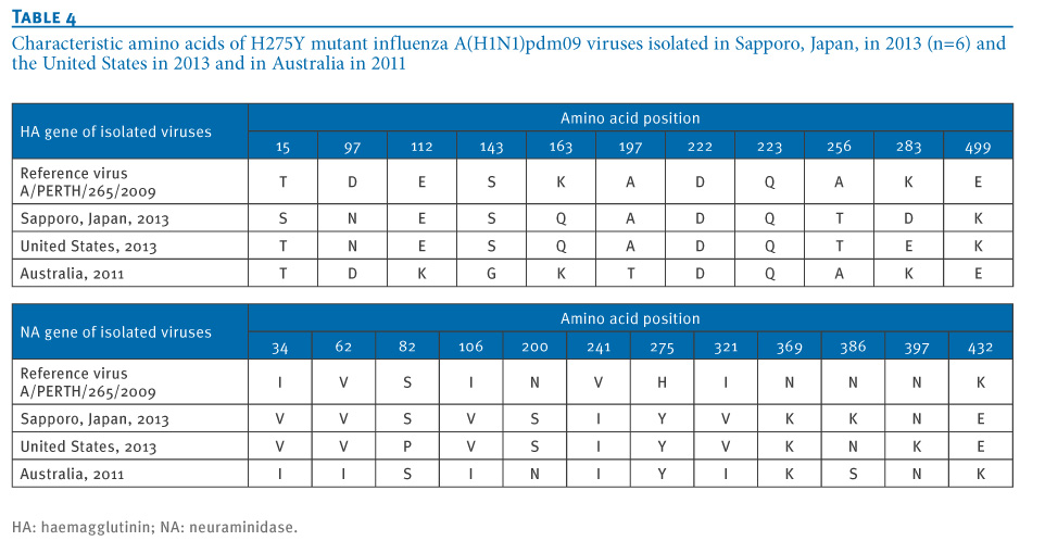
In the most recent 2013/14 Northern Hemisphere influenza season, a cluster of A(H1N1)pdm09 H275Y OR viruses that contained both the V241I and N369K PPMs plus an additional N386K NA mutation, was detected in Sapporo, Japan [53], during a period of the season where A(H1N1)pdm09 viruses contributed approximately 50% of the circulating influenza strains [54].

We’ve just come through an H1N1 dominated flu season in North America, and the incidence of H274Y has remained low, so other factors may be involved. The authors suggest:

Apart from NA PPMs, it may be that other properties, such as antigenic novelty, are also necessary for an OR virus to spread widely. In 2007–2008, the H275Y NA mutation became fixed in a new seasonal A(H1N1) antigenic variant (A/Brisbane/59/2007-like), suggesting that the antigenic novelty of the OR virus assisted its prolific spread.

In this vein, the authors warn:

A(H1N1)pdm09 viruses have now been circulating in humans for over four years, but are yet to undergo a significant antigenic change (as evidenced by the continued inclusion of A/California/7/2009 in the human seasonal influenza vaccine since 2009).

As the H1 component of the vaccine has been updated, on average, every 2.8 years (range 1 to 8 years), and the H3 component every 1.8 years (range 1 to 4 years) since 1980, it is reasonable to anticipate that A(H1N1)pdm09 viruses will undergo antigenic change in the near future.


The significance being that an antigenic change in the virus might be the spark needed to spread the resistant mutation, and at the same time would reduce the effectiveness of the current vaccine (and evade herd immunity), and therefore increase our need for effective antiviral medications.


Viruses and bacteria evolve and adapt very quickly. The sobering truth is pharmacological victories over them tend to be fleeting. New classes of drugs are going to be needed, along with prudent stewardship of the drugs currently in our arsenal.


The authors of this study wrap up by saying:


Here we demonstrate that contemporary A(H1N1)pdm09 viruses have acquired NA mutations which permit the acquisition of NA H275Y without compromising viral fitness. These mutations, which are now present in virtually all circulating A(H1N1)pdm09 viruses, enhance the surface expression and enzymatic activity of the A(H1N1)pdm09 H275Y NA protein in vitro and result in enhanced viral fitness in vivo.

Hence, the risk that H275Y A(H1N1)pdm09 viruses will spread globally, in a similar manner to OR seasonal A(H1N1) viruses in 2007–2008, now appears greater than at any time since the A(H1N1)pdm09 lineage emerged in 2009.

Thursday, March 06, 2014

CHP CDW Report On Antiviral Resistant Influenza In Hong Kong

image

Photo Credit – Wikipedia


# 8354


Last January, we looked at a Eurosurveillance journal reports (see Community Cluster Of Antiviral Resistant pH1N1 in Japan) that detailed a recent (between November and December 2013) cluster of resistant pH1N1 (with the H275Y mutation) in Sapporo, Japan.  Six genetically similar viruses were detected, although none of the patients had known contact with each other, which suggests a resistant strain may be starting to spread in that region.


The most common cause of  Neuraminidase Inhibitor (NAI) antiviral drugs (like oseltamivir aka Tamiflu ®) is the H275Y mutation - where a single amino acid substitution (histidine (H) to tyrosine (Y)) occurs at the neuraminidase position 275 (Note: some scientists use 'N2 numbering' (H274Y)).


Up until 2006 we only saw a smattering of oseltamivir resistant seasonal H1N1 cases, almost always attributed to `spontaneous mutations’  within a patient already receiving the drug.  While of concern to the patient being treated, it appeared to be poorly transmissible, and less than 1% of cases exhibited resistance.


But in 2008 the profile of antiviral resistant seasonal flu changed, and by the spring roughly 25% of European samples tested showed the H275Y mutation (see Increased Tamiflu Resistance In Seasonal Influenza).


By the end of 2008, nearly all of the samples tested in the United States were resistant to oseltamivir and the CDC was forced to issue major new guidance for the use of antivirals (see CIDRAP article With H1N1 resistance, CDC changes advice on flu drugs).


One benefit of the arrival of the H1N1 pandemic strain the following spring was that it effectively removed this resistant strain from circulation.  Despite some scattered clusters of resistance reported in Asia and Australia (see NEJM: Oseltamivir Resistant H1N1 in Australia), nearly 99% of the  pH1N1 viruses tested around the globe have remained sensitive to NA inhibiting drugs. 


The latest FluView report (week 8) indicated that of 3733 viruses tested this flu season in the United States, only 28 (0.8%) showed signs of NA Inhibitor resistance.

image


Today, Hong Kong’s CHP has a report in their Communicable Diseases Watch  (CDW) that indicates a slightly higher (but still < 2%) incidence rate of Oseltamivir resistance detected over the past several years, but reassuring finds that the overwhelming majority of viruses tested still remain sensitive to the drug, and reports no signs of community transmission.


Human Infections with Oseltamivir-resistant Influenza A(H1N1)pdm09 Virus in Hong Kong


Reported by Dr Henry YH Mou, Medical and Health Officer, Respiratory Disease Office, Surveillance and
Epidemiology Branch, CHP.


(EXCERPT)

From May 2009 to February 2014, more than 2,700 influenza A(H1N1)pdm09 viruses were tested for oseltamivir resistance in Hong Kong. Among them, a total of 46 reports (<2% of tested samples) of oseltamivir-resistant influenza A(H1N1)pdm09 virus were detected. The cases affected 26 males and 20 females with a male to female ratio of 1.3:1. Their ages ranged from 5 months to 85 years (median: 16 years). Most of them (87%) were known to acquire the infection locally. No epidemiological linkage was identified among the cases. The annual number of cases detected ranged from 1 to 17 during the period between 2009 and 2013. The monthly number of cases ranged from 0 to 6.

Fifteen cases (33%) had known exposure to a full course of oseltamivir before the collection of specimens. Among the 44 cases with information available, 26 cases (59%) enjoyed good past health. The remaining 18 cases had one or more underlying medical conditions such as hypertension, diabetes, chronic lung diseases, malignant conditions, etc. There was one fatal case affecting a 52 years old female who had multiple chronic medical conditions including hypertension, diabetes and depression. It was also noted that five cases were known to have conditions that resulted in immunosuppressed or immunocompromised state. Viral replication may persist in such patients for prolonged periods of time despite antiviral treatment and this can create a favourable environment for selection of drug-resistant strain.

Molecular tests showed the presence of nucleotide mutation resulting in H275Y amino acid substitution in the neuraminidase protein (N1) of all the oseltamivir-resistant viruses isolated. All were found to be sensitive to another neuraminidase inhibitor zanamivir.


So far, the vast majority of influenza A(H1N1)pdm09 viruses tested in Hong Kong remained sensitive to oseltamivir. Cases of oseltamivir-resistant viruses were sporadic and infrequently found and there is no evidence of onward transmission of oseltamivir-resistant influenza A(H1N1)pdm09 viruses in Hong Kong. Zanamivir remains a treatment option in patients with severe or deteriorating illness caused by oseltamivir-resistant virus. The CHP will continue to monitor the global and local situation of oseltamivir-resistant influenza viruses and remain vigilant for any further changes in influenza viruses that may have public health significance.

(Continue . . .)

While surveillance for antiviral resistance continues to be reassuring, scientists remember the remarkable speed by which seasonal influenza went from being almost 100% sensitive to being nearly 100% resistant.


So, as the report says, continued vigilance is required. 

Thursday, January 09, 2014

Eurosurveillance: Community Cluster Of Antiviral Resistant pH1N1 in Japan

image

Photo Credit – Wikipedia


# 8150


While it’s not easy to find anything good to say about a pandemic influenza virus, the 2009 pH1N1 strain had at least one saving grace; unlike the old seasonal H1N1 strain it replaced, it was overwhelming sensitive to Neuraminidase Inhibitor (NAI) antiviral drugs like oseltamivir (Tamiflu ®) and zanamivir (Relenza ®).


In 2006 we only saw a smattering of oseltamivir resistant seasonal H1N1 cases, almost always attributed to `spontaneous mutations’  within a patient receiving the drug.  While of concern to the patient being treated, it appeared to be poorly transmissible.


In the 2006-2007 flu season, laboratories found no resistant strains in Europe or Japan, and in less than 1% of samples from the United States.


Resistance in this viral strain was mostly caused by a mutation called H275Y, where a single amino acid substitution (histidine (H) to tyrosine (Y)) occurs at the neuraminidase position 275.


(Note: some scientists use 'N2 numbering' (H274Y) and some use 'N1 numbering' (H275Y))


The following year, during the 2007-2008 flu season, oseltamivir resistant viruses suddenly took flight, and by the spring of 2008 roughly 25% of European samples tested showed the H275Y mutation (see Increased Tamiflu Resistance In Seasonal Influenza).


By December of 2008 nearly all of the samples tested in the United States were resistant to oseltamivir and the CDC was forced to issue major new guidance for the use of antivirals (see CIDRAP article With H1N1 resistance, CDC changes advice on flu drugs).


The arrival of a the pandemic H1N1 in the spring of 2009 effectively removed this resistant strain from circulation, and in the five ensuing years, nearly 99% of the  pH1N1 viruses tested have remained sensitive to NA inhibiting drugs.  The latest FluView report (week 52) indicated that of 910 viruses tested, only 10 showed signs of NA Inhibitor resistance.


image

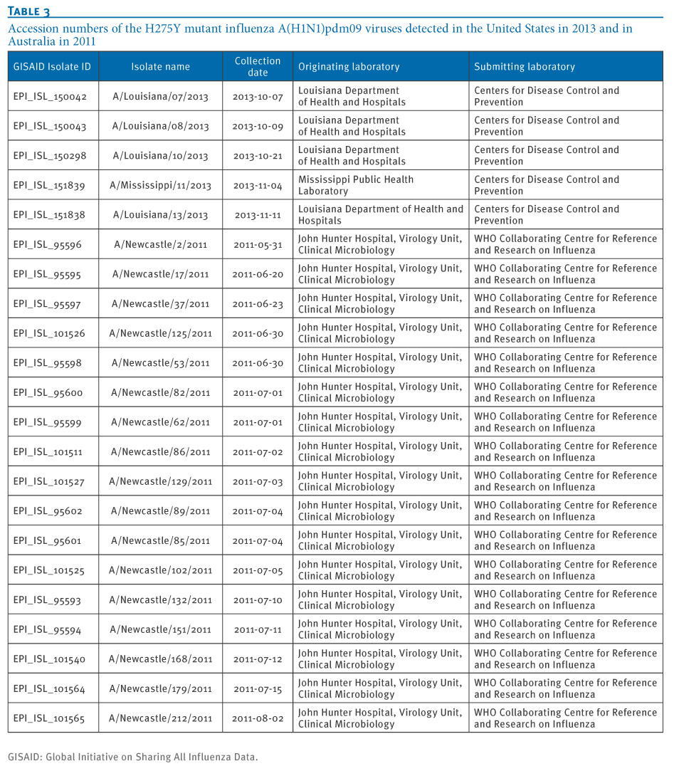
While 99% sensitivity is an excellent batting average in any league, 5 of the 10 resistant viruses were detected in Louisiana and Mississippi, suggesting a geographic clustering of cases.


Although we have seen some signs the pH1N1 might be figuring its way around our arsenal of antivirals, most often this occurs after a patient is placed on antivirals, occasionally resulting in a spontaneous mutation within the host. 


But we have seen some signs of community spread of resistant pH1N1 as well.


Readers with good memories will recall that in December of 2011, in NEJM: Oseltamivir Resistant H1N1 in Australia, we looked at a cluster of oseltamivir (Tamiflu ®) resistant H1N1 viruses in and around the Newcastle area of New South Wales.


The lead author of that NEJM correspondence was Aeron C. Hurt, Ph.D. from the World Health Organization (WHO) Collaborating Centre for Reference and Research on Influenza, North Melbourne, VIC, Australia.  


What Hurt and his colleagues found was evidence for the sustained community transmission of a resistant strain of the H1N1pdm09 virus.

After analyzing viral samples pulled from 182 patients seen in emergency departments, intensive care units, and doctor’s offices in New South Wales between May and August of 2011, they found 29 (16%) carried the H275Y resistance mutation.


In 2011, we also saw reports of a `mildly resistant’ version of pH1N1 – with a different mutation S247N (serine (S) to asparagine (N)) mutation at the neuraminidase position 247) – showing up with some frequency in  Australia, Brunei and Singapore (see Eurosurveillance: A `Mildly’ Resistant Strain of H1N1 Emerges).


But neither of these strains has gained much traction globally, and the World Health Organization still reports only about 2% of samples tested are resistant to our frontline antiviral drugs.


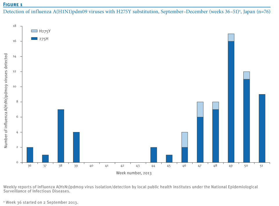
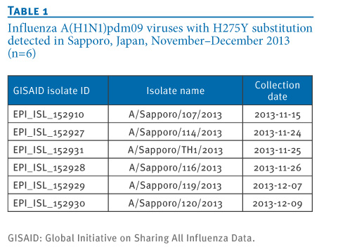
Today, we’ve a Rapid Communications  in the journal Eurosurveillance that details a recent community cluster of resistant pH1N1 (with the H275Y mutation) in Sapporo, Japan.  Six genetically similar viruses were detected, although none of the patients had known contact with each other, which suggests a resistant strain may be starting to spread in that region.


Rapid communications

A community cluster of influenza A(H1N1)pdm09 virus exhibiting cross-resistance to oseltamivir and peramivir in Japan, November to December 2013

E Takashita1, K Ejima1, R Itoh1, M Miura1, A Ohnishi2, H Nishimura3, T Odagiri1, M Tashiro ()1

  1. Influenza Virus Research Center, National Institute of Infectious Diseases, Tokyo, Japan
  2. Sapporo City Institute of Public Health, Hokkaido, Japan
  3. Virus Research Center, Sendai Medical Center, Miyagi, Japan

Citation style for this article: Takashita E, Ejima K, Itoh R, Miura M, Ohnishi A, Nishimura H, Odagiri T, Tashiro M. A community cluster of influenza A(H1N1)pdm09 virus exhibiting cross-resistance to oseltamivir and peramivir in Japan, November to December 2013. Euro Surveill. 2014;19(1):pii=20666. Available online: http://www.eurosurveillance.org/ViewArticle.aspx?ArticleId=20666
Date of submission: 30 December 2013


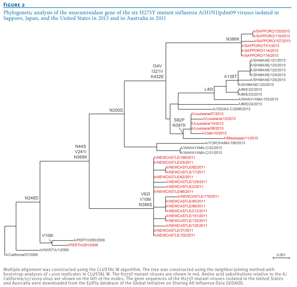
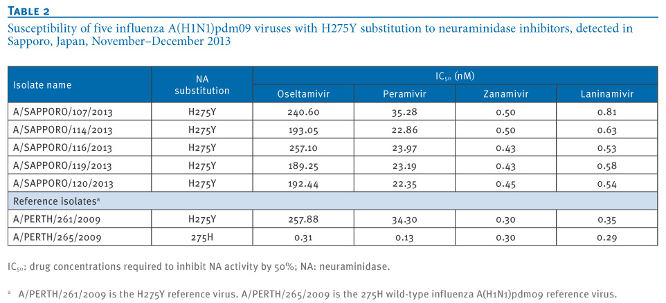
Six influenza A(H1N1)pdm09 viruses were detected in Sapporo, Japan, between November and December 2013. All six viruses possessed an H275Y substitution in the neuraminidase protein, which confers cross-resistance to oseltamivir and peramivir. No epidemiological link among the six cases could be identified; none of them had received neuraminidase inhibitors before specimen collection. The haemagglutinin and neuraminidase genes of the six viruses were closely related to one another, suggesting clonal spread of a single resistant virus.

(Continue . . .)

In their discussion, the authors write:

It has been shown that oseltamivir-resistant influenza A(H1N1) virus infection reduced the effectiveness of oseltamivir and this tendency was more apparent in children 0 to 6 years old [14-16]. Among patients from whom oseltamivir- and peramivir-resistant A(H1N1)pdm09 viruses have been detected in Japan, the percentage with no known exposure to NA inhibitors has increased significantly, from 16% during the pandemic period to 44% during the post-pandemic period [2]. These observations may suggest that human-to-human transmission with H275Y mutant viruses has increased gradually in the post-pandemic period. Consequently, surveillance of antiviral-resistant influenza viruses should be continued and strengthened, particularly for the choice of antiviral drugs.


Since 2009 we’ve seen sporadic cases of antiviral resistance show up in the new H1N1 virus, but only rarely have we seen clusters that suggest limited community spread.  So we are nowhere near the level of concern over antiviral resistance that we experienced in 2008.


That said,  we know that pharmacological victories over viruses and bacteria have always been fleeting at best. Pathogens – given enough time – have demonstrated a keen ability to evade each new generation of drugs we throw at them.


A reminder that in our ongoing battle against infectious diseases, that nature always bats last.

Tuesday, December 10, 2013

Nature Comms: H7N9 Gains Antiviral Resistance Without A `Fitness Penalty’

image


# 8060


If you follow influenza topics closely, you may know that when influenza viruses gain antiviral resistance they often (but  not always) take a performance hit when it comes to transmissibility.  Back in 2007, it was widely held  that the mutation that made the virus resistant to Tamiflu ® (H274Y), also reduced its biological fitness – suggesting that mutated versions of the viruses were unlikely to spread widely.


That notion was turned on its head when, a year later, the (old) seasonal H1N1 virus acquired resistance to oseltamivir, `broke with convention’  and spread like wildfire around the globe.


Fortunately, that highly resistant strain is no more, having been supplanted by the 2009 H1N1 pandemic virus. And resistant strains of the other two `human’ flu strains (H1N109 and H3N2) have failed to make much of an impact. 


But earlier this year we saw signs that at least some of the isolates collected from patients infected with the  H7N9 virus in China were resistant to antiviral medications (see EID Journal: R292K Substitution & Antiviral Resistance & mBio: Antiviral Resistance In H7N9).  Most of these mutations appear to have developed after the patient was placed on antiviral meds.


Unlike the H1N1 and H3N2 resistant strains – which picks up resistance from the H274Y (Histidine  to Tyrosine at neuraminidase position 275) – H7N9 resistant strains have the R292K Substitution (Arginine to Lysine at position 292 in the NA) – also known as Arg292Lys


Since we’ve no well documented human-to-human transmissions of this virus to look at, we really haven’t known if these resistant H7N9 strains – like their resistant H3N2 and 2009 H1N1 cousins – take a performance hit. 


Today, in a report that appears in Nature Communications, we get  evidence that suggests that resistant H7N9 viruses remain biologically `fit’ , competitive, and transmissible.


First a link to the Abstract (the full open access report is available at this link), then I’ll be back with a bit more.


Influenza A(H7N9) virus gains neuraminidase inhibitor resistance without loss of in vivo virulence or transmissibility

Rong Hai, Mirco Schmolke, Victor H. Leyva-Grado, Rajagowthamee R. Thangavel, Irina Margine, Eric L. Jaffe, Florian Krammer, Alicia Solórzano, Adolfo García-Sastre, Peter Palese & Nicole M. Bouvier

Without baseline human immunity to the emergent avian influenza A(H7N9) virus, neuraminidase inhibitors are vital for controlling viral replication in severe infections. An amino acid change in the viral neuraminidase associated with drug resistance, NA-R292K (N2 numbering), has been found in some H7N9 clinical isolates. Here we assess the impact of the NA-R292K substitution on antiviral sensitivity and viral replication, pathogenicity and transmissibility of H7N9 viruses. Our data indicate that an H7N9 isolate encoding the NA-R292K substitution is highly resistant to oseltamivir and peramivir and partially resistant to zanamivir.

Furthermore, H7N9 reassortants with and without the resistance mutation demonstrate comparable viral replication in primary human respiratory cells, virulence in mice and transmissibility in guinea pigs. Thus, in stark contrast to oseltamivir-resistant seasonal influenza A(H3N2) viruses, H7N9 virus replication and pathogenicity in these models are not substantially altered by the acquisition of high-level oseltamivir resistance due to the NA-R292K mutation.

(Continue . . . )


We’ve seen conflicting views on this virus’s pandemic potential (see TSRI: H7N9 Virus Still Binds Preferentially to Avian Receptors vs. Lancet: Tropism Of H7N9 In the Human Respiratory Tract vs.Nature: H7N9 Pathogenesis and Transmissibility In Ferrets & Mice ) over the summer and fall.


The good news is that  we’ve not seen any evidence that any of the H7N9 viruses to date are capable of efficiently transmitting among humans.


That could change, of course. But no one really knows how many adaptations are needed for this virus to become a pandemic contender.  Since human immunity to the H7N9 virus is likely to be minimal, and this virus is capable of producing significant morbidity and mortality, today’s findings are clearly important and a bit worrisome.


But none of this increases the odds that  the H7N9 virus will spark a pandemic.


It does suggest, however, that our pharmacological options for treating it could be limited, should that ever happen. 

Thursday, August 01, 2013

EID Journal: R292K Substitution & Antiviral Resistance

image


# 7534


With slim prospects of having an H7N9 influenza vaccine in the near term (see JAMA: Challenges Of Producing An Effective & Timely H7N9 Vaccine) society will have basically two tools to combat H7N9 should it erupt into a pandemic:


  • NPIs (Non Pharmaceutical Interventions like social distancing, school closures, hand hygiene & masks)
  • neuraminidase (NA) inhibiting antiviral drugs  (NAIs) like oseltamivir (Tamiflu ®) and Zanamivir (Relenza ®).


While most of the H7N9 cases reported in China have proven susceptible to these two antiviral drugs, a small but disturbing number of cases have developed resistance while receiving treatment.


Last May, in The Lancet: Antiviral Resistance In Two H7N9 Patients, we looked at a study that found that a mutation R292K (Arginine to Lysine at position 292 in the NA) – also known as Arg292Lys – already known to confer antiviral resistance to seasonal flu (see Resistant influenza A viruses in children treated with oseltamivir: descriptive study), appeared in two patients after several days of oseltamivir therapy.


In the middle of July, in mBio: Antiviral Resistance In H7N9, we saw a  study that warned that the standard laboratory tests for antiviral susceptibility can miss `mixed population’ infections (comprised of both resistant and susceptible strains), and that antiviral treatment could suppress the susceptible strains while allowing resistant strains to flourish.


And this particular mutation (R292K) can provide resistance not only to oseltamivir, but to zanamivir and peramivir as well.


While the number of resistant cases identified has remained relatively small, as we saw with the old (pre-2009) seasonal H1N1 virus – resistance can sometimes develop quickly and spread globally (see 2008 CIDRAP NEWS  article With H1N1 resistance, CDC changes advice on flu drugs).


Which beings us to an EID Journal study, published this week, that found that (as did the mBio study above) that conventional NI assays may not always reliably detect the R292K substitution.


They believe that the `fitness’ of resistant strains to replicate and spread is less than that of the NAI susceptible `wild type’ H7N9, and in the absence of antiviral exposure they would be unlikely to compete effectively against them. 


But when you inhibit the replication of susceptible viruses with antivirals, you create an environment in which resistant strains can flourish. 


Not clear at this time is how much of a `fitness’ hit these resistant viruses take. That is, whether they are likely to be a dead-end infection in a host, or can spread efficiently to others


Also unknown is just how much this R292K mutation actually reduces the effectiveness of antivirals in a clinical setting.  Nor what other mutations might exacerbate, or mitigate, this effect.


Follow the link below to read:


Volume 19, Number 9—September 2013

Dispatch

R292K Substitution and Drug Susceptibility of Influenza A(H7N9) Viruses

Katrina Sleeman1, Zhu Guo1, John Barnes, Michael Shaw, James Stevens, and Larisa V. GubarevaComments to Author

Author affiliations: Centers for Disease Control and Prevention, Atlanta, Georgia, USA

Abstract

Neuraminidase inhibitors are the only licensed antiviral medications available to treat avian influenza A(H7N9) virus infections in humans. According to a neuraminidase inhibition assay, an R292K substitution reduced antiviral efficacy of inhibitors, especially oseltamivir, and decreased viral fitness in cell culture. Monitoring emergence of R292K-carrying viruses using a pH-modified neuraminidase inhibition assay should be considered.


The recent emergence of an avian influenza A(H7N9) virus causing human infections in China (1,2) is of global concern. Most patients infected during this outbreak have experienced severe disease and required hospitalization; the mortality rate is 21% (3). Although epidemiologic investigations have revealed no evidence of sustained human-to-human transmission (4), suspected limited human-to-human transmission has been reported (3).


As with any emergent influenza virus, it is critical to assess the susceptibility of the influenza A(H7N9) outbreak virus to antiviral drugs, which are the first line of defense before an effective vaccine becomes available. Two classes of antiviral drugs are approved for management of influenza A infections, neuraminidase (NA) inhibitors (NAIs) and matrix 2 protein (M2) blockers (adamantanes). The outbreak viruses carry the established adamantane resistance marker, an S31N substitution in the M2 protein (2), leaving NAIs as the only licensed treatment option.


Among the 4 NAIs, oseltamivir and zanamivir are approved in many countries; peramivir has been approved in Japan, South Korea, and China; and laninamivir is approved only in Japan. In contrast to those for adamantanes, genetic markers of resistance to NAIs are often subtype specific and drug specific (5). Therefore, monitoring drug susceptibility of the influenza A(H7N9) viruses requires testing in phenotypic assays using all available NAIs.

<SNIP>
Conclusions

R292 is a highly conserved amino acid across all NA subtypes, and together with 2 other highly conserved residues (R118 and R371), it forms an arginine triad in the enzyme active site (5). R292K is a rare substitution and to date has only been reported in viruses collected from patients treated with oseltamivir (2,5). In addition to A/Shanghai/1/2013 isolate, there is evidence of additional influenza A(H7N9) isolates with the R292K substitution (11).


In this study, propagation of A/Shanghai/1/2013 (E1) isolate in eggs and in MDCK-SIAT1 cells resulted in reversion to wild-type (23% Arg in E1 to 100% in E1/S3), confirming results of previous studies with N2 subtype viruses (12). Therefore, fitness of the A/Shanghai/1/2013 R292K virus is probably compromised when replication occurs in the absence of an NAI. However, propagation of the E1 isolate in the presence of oseltamivir (100 nmol/L) resulted in enrichment of the R292K population (from 77% to 100%), demonstrating a growth advantage over the wild-type.


Replication of the E1 isolate in the presence of any NAI in cell culture might lead to enrichment with R292K, because even a small growth advantage would reduce the proportion of the wild type. The efficacy of NAIs in clinical management of influenza (H7N9) infection remains unknown and may be compromised to a certain extent when R292K is present.


Animal model studies are needed to aid in the understanding of clinical relevance of R292K. Reduction of NA activity caused by R292K may detrimentally affect transmission of the virus, as indicated by an R292K influenza A(H3N2) virus that showed reduced infectivity in mice (13,14) and ferrets (12,13,15) and was not transmitted among ferrets (12,15). The data reported here demonstrate the continued importance of monitoring drug susceptibility in emergent influenza viruses and highlight the challenges involved in laboratory assessment of NAI drug susceptibility testing.