Showing posts sorted by relevance for query Baskin. Sort by date Show all posts

Tuesday, August 24, 2010

Study: What Makes Avian Flu So Deadly


# 4830


Avian flu, particularly H5N1, has our attention because unlike regular influenza, it has a very high mortality rate.  Among those we know to have been infected, roughly 60% have died.


A rate well over 100 times higher than with regular flu.


Roughly 18 months ago we got a look at a study that compared the pathogenesis (disease progression) of non-human primates (macaques) infected with the H5N1 virus, seasonal flu, and with two altered viruses carrying genetic material from the 1918 Spanish Flu.


The study, entitled Early and sustained innate immune response defines pathology and death in nonhuman primates infected by highly pathogenic influenza virus by Carole Baskin et. al.  appeared PNAS (The Proceedings of the National Academy of Science).


At the time, I wrote a 3-part essay which attempted to put this complex study into layman’s terms. 


For those interested in learning more about the human immune system, cytokine production, and the pathogenesis of influenza the Baskin study and these three blogs may be of value:


Dissecting the Influenza Pathogenesis Study Pt. 1 (link fixed)

Dissecting the Influenza Pathogenesis Study Pt. 2
Dissecting the Influenza Pathogenesis Study Pt. 3


While the Baskin study is fascinating, and gives us considerable insight into what avian flu does to primates, it really doesn’t explain how or why.


To help us with those questions, researchers at Baylor College of Medicine and The University of Texas at Austin have studied the molecular structure of the avian flu virus, and have found 4 tiny amino acids dangling from the tip of a protein called NS1 that they believe at least partially explains this virulence.


Their research appears in the latest issue of the Journal of Virology.  You can access the abstract at the link below.


J. Virol. doi:10.1128/JVI.01278-10


The ESEV PDZ Binding-Motif of the Avian Influenza A Virus NS1 Protein Protects Infected Cells from Apoptosis through Directly Targeting Scribble


Hongbing Liu, Lisa Golebiewski, Eugene C. Dow, Robert M. Krug, Ronald T. Javier, and Andrew P. Rice


Admittedly, to non-scientists, this title is more than a little imposing.  


Apoptosis is programmed cellular death, while Scribble is a protein the body’s immune system uses to promote the early death (apoptosis) of virally infected cells. 


In this way, the immune system can help limit viral replication while it develops defenses (antibodies, cytokines, etc.) against the invader.


In the case of H5N1, the virus’s PDZ binding-motif works to  deactivates the host’s apoptosis defense mechanism, giving the virus a decided advantage.


Fortunately, for us mere mortals without a virology degree, we have a less technical background piece from the BCM news site which is well worth following the link and reading in its entirety. 


Avian flu virus protein turns off cell defense

HOUSTON -- (August 23, 2010) -- As the avian influenza A virus seeks to infect its bird hosts, it brings a special weapon to the fray – four tiny amino acids that hang off the end of a viral protein called NS1, said researchers from Baylor College of Medicine (www.bcm.edu) and The University of Texas at Austin in a report that appears in the current Journal of Virology.


This collection of molecules known as the PDZ binding-motif help make the avian virus – known to experts at H5N1 – particularly virulent and a threat to human populations everywhere.

(Continue . . . )

Saturday, February 14, 2009

Dissecting the Influenza Pathogenesis Study Pt. 1


# 2793


Last night we got our first look at a study that compared the pathogenesis (disease progression) of non-human primates (macaques) infected with the H5N1 virus, seasonal flu, and with two altered viruses carrying genetic material from the 1918 Spanish Flu.

The study, entitled Early and sustained innate immune response defines pathology and death in nonhuman primates infected by highly pathogenic influenza virus by Carole Baskin et. al.  appears in the latest issue of PNAS (The Proceedings of the National Academy of Science).

While there is much here to consider (and we'll get to that), the upshot is that:

The avian virus was found to significantly outpace not only run-of-the-mill influenza but even the highly virulent 1918 ressortants, in terms of its relentless pathogenicity.


I'm not going to try to critique this research, as I'm obviously unqualified to do so.   Someone with more letters after their name than EMT-II (Ret.) will have to take that on.


But what I'd like to do is explain, in layman's language, what the authors are reporting, and some of the implications of what they've learned.


And here I run into a bit of a dilemma.


Just about every facet of this study requires a bit of `background', to make it understandable.  At least if we're going to do more than give it a cursory look.


Even the title, which refers to the `innate immune response' requires a bit of explanation.   And the deeper you plunge into this paper, the more `detours' - areas that require a good deal of explanation -  you find.  


I have to ask myself. 


How many of my readers understand the difference between Type I and Type II Pneumocytes?  (I hope the number is low, since I had to look it up)


I suddenly realized that, in order to make all of this understandable to the widest number of readers, I'd have to write a multi-part post.   Not really a problem, of course.  After nearly 2800 blogs, what's a multi-part post?


So, while I fully expect we can get `there' from `here', I'll warn my readers in advance it may require a serpentine route.   As to how many installments it will take to get `there' - well, we'll know when we've arrived.

Given the long, and awkward title of the research paper, once I link to it in at the top of each post, I'm simply going to refer to it as The Baskin Study. 


So, without further ado,  I present Part I of my series Dissecting the Influenza Pathogenesis Study.


MEET THE PLAYERS


To perform a systematic comparison of several highly pathogenic influenza viruses, we inoculated cynomolgus macaques (8 animals per group) with either influenza A/Vietnam/1203/2004 (referred to as H5N1), 1918HA/NA:A/Texas/36/91 (1918HANA), 1918HA/NA/NS:A/Texas/36/91 (1918HANANS), or the parental H1N1 A/Texas/36/91 (Texas) virus. - The Baskin Study


The hodgepodge of letters and numbers above identify 4 very different influenza viruses, which researchers used to infect macaques in order to study their different disease processes.

To understand what these numbers and letters mean, we need to digress briefly. 


Influenza viruses come in 3 broad types; A, B, and C.  

  • Influenza `C' viruses are relatively rare, generally produce milder symptoms in humans, and are not considered to have epidemic or pandemic potential. 

  • `B' viruses, which are far more common, may produce serious illness and spark regional epidemics, but aren't viewed as `pandemic strains'.

  • It is only the `A' influenza strain - which is characterized by two surface glycoproteins known as hemagglutinin (HA) and neuraminidase (NA) - that mutates rapidly and can cause an influenza pandemic.


Influenza `A' viruses are identified by their HA and NA proteins.  There are 16 known HA subtypes and 9 known NA subtypes.  While many combinations are possible, currently only a few are in circulation.


Among humans, the H1N1, H1N2, and H3N2 viruses currently circulate globally.    H5s, H7s, and H9s are commonly found in birds.


The H1N1 viruses today are all descendants of the 1918 Spanish Flu.   The H3N2 viruses are descendants of the 1968 pandemic. 


The H1N2 virus, which was first detected in 2002, appears to be a hybrid, consisting of the hemagglutinin (HA) from the H1N1 virus and the neuraminidase (NA) from the H3N2 virus.


Influenza `A'  viruses are actually complex entities comprised of 10 genes on 8 separate RNA molecules (called: PB2, PB1, PA, HA, NP, NA, M, and NS)


image


This study compared 4 influenza viruses.

  • One wholly avian virus (H5N1), a clade 1 H5N1 virus collected from Vietnam in 2004.

  • One seasonal influenza virus (Texas) H1N1 A/Texas/36/91, which circulated widely in the mid and early 1990's.

  • One genetically modified seasonal H1N1 virus (1918HANA) with the HA and NA from the 1918 virus spliced to it.

  • And one genetically modified seasonal H1N1 virus (1918HANANS) with the HA, NA, and NS1 from the 1918 virus spliced to it.


The (HA) and neuraminidase (NA) surface proteins are known conveyors of virulence, and while the resultant reassortant viruses might not have been an exact match to the 1918 pandemic virus, they do represent a reasonable facsimile.
 
The NS1, grafted onto the 2nd 1918-like virus, is known to inhibit the host interferon response.   
 
Interferon is a natural protein produced by the cells of the immune system when they sense a viral or bacterial invader.  Interferons belong to the larger class of glycoproteins known as cytokines. 
 
 
The stage is now set. 
 
Thirty-four macaques were used in this study, four groups of 8 - with 2 `mock infected' to serve as a reference for host response without viral challenge.  
 
Each group of 8 were macaques were infected with a different strain of the virus via tracheal, nasal, conjunctival, and tonsillar routes.
 
On days 1, 2, 4, and 7 postinfection (PI), 2 animals per group were euthanized.  One macaque died from excessive pulmonary damage between day 6 and 7.
 
On day 7, the two `mock infected' animals were euthanized.  
 
Necropsies were performed on each animal and tissue and serum samples were subjected to numerous tests. 
 
In the next installment Dissecting the Influenza Pathogenesis Study Pt. 2 , we'll look at some of the results.


Friday, September 17, 2010

Study: Receptor Binding Changes With H1N1 D222G Mutation


# 4909


Although it’s nestled behind a pay wall at the Journal of Virology, the abstract from a study published ahead of print this week in the Journal of Virology gives us a tantalizing glimpse at research conducted on the D222G mutation that has been found in some isolates of the pandemic H1N1 (pdmH1N1) virus.


While we’ve discussed the D222G mutation before, this is an obscure enough subject as to make a review helpful.  I’ll keep it simple (essential so that I can follow, as well), so real scientists may wish to skim or skip ahead.


The `Norway’ or D222G (D225G in influenza H3 Numbering) mutation first announced by Norwegian Scientists last November has sparked repeated speculation that it might be associated with increased virulence.


This mutation had actually been detected months earlier, and in many other countries, but Norway was the first country to announce a possible link between that mutation and greater virulence.


This mutation involves a single amino acid change in the HA1 gene at position 222 from aspartic acid (D) to glycine (G).


The World Health Organization’s take on this mutation has been pretty consistent.  It is worth following, and studying, but there is no evidence (as yet) that it poses a substantial public health hazard.


In January, in a blog entitled WER Review: D222G Mutation In H1N1, I quoted the latest WHO report that stated:


`Based on currently available virological, epidemiological and clinical information, the D222G substitution does not appear to pose a major public health issue.’


This view is not universally held, however. There are some who have maintained that that the WHO is underestimating the impact of this mutation.


In March of this year, researchers from the Norwegian Institute of Public Health in Oslo reported that they found the mutation in 11 of 61 severe illness cases that they analyzed, but that it was not found in any of the 205 mild cases they looked at  (see CIDRAP Report On The H1N1 Mutation Debate).


The WHO WER Review reported that the overall prevalence of D222G was <1.8% (52 detections among >2755 HA sequences) in contrast to a rate of 7.1% in fatal cases. The WHO paper also reported on the occurrence of  two other mutations at this amino acid position, D222E and D222N, although their significance is unclear.


While this may sound like fairly damning evidence, it should be noted that mild cases have been detected with this D222G mutation in other studies, and most of the severe and fatal cases of pandemic H1N1 that have been examined did not have this mutation.


Which brings us to today’s study, which features an impressive pedigree and some very familiar names including Ab Osterhaus and  Ron Fouchier of the Erasmus Medical Center in Rotterdam.


This study was also supported by researchers from the Mt. Sinai School of Medicine in New York, the NIH, the University of Cambridge, the University of Maryland . . . among others.


First the abstract (hat tip Tetano on FluTrackers) slightly reformatted for readability, then a little discussion.


Virulence-associated substitution D222G in hemagglutinin of 2009 pandemic influenza A(H1N1) virus affects receptor binding.

Chutinimitkul S, Herfst S, Steel J, Lowen AC, Ye J, van Riel D, Schrauwen EJ, Bestebroer TM, Koel B, Burke DF, Sutherland-Cash KH, Whittleston CS, Russell CA, Wales DJ, Smith DJ, Jonges M, Meijer A, Koopmans M, Rimmelzwaan GF, Kuiken T, Osterhaus AD, Garcia-Sastre A, Perez DR, Fouchier RA.

Abstract

The clinical impact of the 2009 pandemic influenza A(H1N1) virus (pdmH1N1) has been relatively low. However, amino acid substitution D222G in the hemagglutinin of pdmH1N1 has been associated with cases of severe disease and fatalities.


Here, D222G was introduced in a prototype pdmH1N1 by reverse genetics, and the effect on virus receptor binding, replication, antigenic properties, and pathogenesis and transmission in animal models was investigated.


pdmH1N1 with D222G caused ocular disease in mice without further indications of enhanced virulence in mice and ferrets. pdmH1N1 with D222G retained transmissibility via aerosols or respiratory droplets in ferrets and guinea pigs.


The virus displayed changes in attachment to human respiratory tissues in vitro, in particular increased binding to macrophages and type II pneumocytes in the alveoli and to tracheal and bronchial submucosal glands.


Virus attachment studies further indicated that pdmH1N1 with D222G acquired dual receptor specificity for complex α2,3- and α2,6-linked sialic acids. Molecular dynamics modeling of the hemagglutinin structure provided an explanation for the retention of α2,6 binding.


Altered receptor specificity of the virus with D222G thus affected interaction with cells of the human lower respiratory tract, possibly explaining the observed association with enhanced disease in humans.


Testing here was done on mice, ferrets, guinea pigs, and on human cells in vitro, and each demonstrated (sometimes small) pathogenic differences between the D222G-engineered and regular pdmH1N1 virus.


In mice and ferrets, the D222G virus showed no increase in virulence with the exception of `ocular disease’ in mice (I’m guessing conjunctivitis, but without access to the full article, I can’t be certain).


Given the low incidence of the D222G mutation in the wild (less than 1.8%), it has been suggested that this mutation might render the virus less contagious, but ferret and guinea pig studies showed it retained transmissibility via aerosols and respiratory droplets.


The increased binding to type II pneumocytes in the alveoli (in vitro) is a particularly interesting finding, given that this was also observed in the Baskin Study of H5N1 vs human H1N1 viruses.


Seasonal H1N1 viruses, when they invade the lungs, are more likely to attack type I pneumocytes which handle the gas exchange (02 and C02) between the lungs and the blood stream.  


Type II pneumocytes are responsible for the production of surfactant with antimicrobial, immunomodulatory, and anti-inflammatory properties, and are the lung’s primary mechanism for repairing damaged cells.


Damaging them can significantly degrade the lung’s ability to recover from injury.


Which brings us to the last major finding, that D222G acquired dual receptor specificity for complex α2,3- and α2,6-linked sialic acids.


Familiar territory to regular readers of this blog, but at the risk of repeating myself:


A virus’s ability to bind to specific cells is controlled by its RBD or Receptor Binding Domain; an area of its genetic code that allows it to attach to, and infect, specific types of host cells.


RBD

(Very Simplified Illustration of RBDs)


Like a key into a padlock, the RBD must `fit’ in order to open the cell to infection.


Avian adapted influenza viruses bind preferentially to Alpha 2,3 receptor cells, which are commonly found in the digestive tract of birds.


Human adapted viruses have an affinity for the alpha 2,6 receptor cell, which populate the upper airway and lungs.


Humans have some avian-like alpha 2,3 receptor cells, particularly deep in the lungs. 


This has been suggested as the reason that when humans contract H5N1, it is usually a deep lung infection.  It has also been postulated that H5N1’s deeper lung infections may reduce human-to-human transmission, as sneezing is a less common symptom.   


This duel receptor affinity with the D222G mutation may help explain why some patients that contract it also develop more serious lung infections.


The operative word here being `may’. 


The bottom line here is that so far, whatever pathogenic differences this mutation may spark, it has had a relatively small effect on the overall mortality and morbidity of this virus. 


That could change, of course, if this mutation were to become more common, or if complementary concurrent changes to the genetic structure of the virus were to further enhance its virulence.


All in all, a fascinating piece of research, and one that advances our knowledge of this mutation considerably.  No, it doesn’t answer the `big question’, of whether this mutation will end up becoming a significant public health threat.


But scientific knowledge is gained incrementally. 


So stay tuned.


For more on the Baskin Study (Early and sustained innate immune response defines pathology and death in nonhuman primates infected by highly pathogenic influenza virus by Carole Baskin et. al.  that appeared PNAS), which looked at the comparative pathogenesis of seasonal H1N1, a 1918-like H1N1, and the H5N1 virus, you may enjoy my 3-part series available at the following links:


Dissecting the Influenza Pathogenesis Study Pt. 1

Dissecting the Influenza Pathogenesis Study Pt. 2

Dissecting the Influenza Pathogenesis Study Pt. 3

Thursday, September 03, 2009

Pathology Of Fatal H1N1 Lung Infections


# 3695


Despite all of the assurances that the novel H1N1 virus is `similar in severity to seasonal flu’, there are obviously differences in the way the novel virus behaves.


We’ve heard a great deal about the `age-shift’ to younger victims – something we often see with pandemic viruses – but so far we’ve heard very little about the pathogenesis of the virus; how the disease progresses inside the body.


Today Helen Branswell of the Canadian Press brings us a fascinating interview with the CDC’s chief pathologist regarding post-mortem examinations of the lungs of H1N1 fatalities.


I’ll just get out of the way and let Ms. Branswell take it from here.


Lung damage in fatal swine flu cases more bird flu than seasonal flu: expert

By Helen Branswell Medical Reporter (CP) 

TORONTO — The lungs of people who have died from swine flu look more like those of the victims of H5N1 avian influenza than those of people who succumb to regular flu, the chief of infectious diseases pathology at the U.S. Centers for Disease Control says.


Study of about 70 fatal H1N1 cases so far also reveals there may be more incidences of co-infections with bacteria than was earlier thought, Dr. Sherif Zaki told The Canadian Press in an interview.


The damage to lung tissue is consistent with that inflicted by ARDS or acute respiratory distress symptom, Zaki says, referring to an often-fatal, difficult-to-treat syndrome that can have a number of causes. The U.S. National Heart, Lung and Blood Institute estimates about 30 per cent of people who develop ARDS die.


"In terms of the disease, yes, it (H1N1) is remarkably different than seasonal flu," Zaki says. "The pathology looks very similar to H5(N1)."

(Continue . . . )


Up until now we’ve heard little about bacterial co-infections in fatal H1N1 cases.  Dr. Zaki makes the point that pneumococcal vaccines might be of value in younger people, to prevent some of these infections.


Although I don’t offer medical advice in this blog, I have suggested on many occasions that people should consult with their primary care provider about the advisability of taking the Pneumococcal polysaccharide vaccine (PPV) – even if you aren’t sure you fall into a recommended category.


A couple of blogs on the subject include:


Referral: Effect Measure On Pneumococcal Vaccines
CDC Issues Pneumococcal Vaccine Recommendations


Given the comparisons being made between the lung damage wrought by the novel H1N1 virus and H5N1 Bird flu, it might be an appropriate time to take another look at the PNAS paper entitled Early and sustained innate immune response defines pathology and death in nonhuman primates infected by highly pathogenic influenza virus by Carole Baskin et. al., which analyses the pathogenesis of the H5N1 virus compared to seasonal flu, and 1918-like viruses.


With much trepidation I wrote a 3-part-layman’s guide to this paper last January.  It was an ambitious undertaking, but to my delight the study’s author Carole Baskin, read it and commented positively on it. 


Dissecting the Influenza Pathogenesis Study Pt. 1
Dissecting the Influenza Pathogenesis Study Pt. 2
Dissecting the Influenza Pathogenesis Study Pt. 3


I’m not sure that we can assume that the same damage observed at the cellular level with H5N1 is occurring with H1N1, but upon gross examination the two appear to be producing similar pathologies.


In any event, these three essays are a good review of the immune system, and our defense against viruses – novel and otherwise.

Monday, February 16, 2009

Dissecting the Influenza Pathogenesis Study Pt. 3


# 2799


This is third and last part of my extended look at the recent PNAS paper entitled Early and sustained innate immune response defines pathology and death in nonhuman primates infected by highly pathogenic influenza virus by Carole Baskin et. al., which analyses the pathogenesis of the H5N1 virus compared to seasonal flu, and 1918-like viruses.


  • In  Part I, which appeared on Saturday, we examined the 4 different viruses used in this study, and the methods used by the research team.


  • In Part II, which appeared on Sunday, we looked at the innate immune system and began looking at the results of this study.


This series is not intended to be a scientific review, but instead a layman's tour of the paper.


Today, we'll finish looking at the findings, and talk about about what it might mean.  In Part II I listed the 5 major findings, and we discussed the first 3.  


Today we'll cover points 4 & 5.


4.   The H5N1 infection also resulted in prolonged margination of circulating T lymphocytes and notable apoptosis of activated dendritic cells in the lungs and draining lymph nodes early during infection.


The loss of dendritic cells (named after, but not to be confused with the projections from neurons) through premature apoptosis (programmed cell death) in the lungs and draining lymph nodes is viewed by these researchers as a key finding.


image


Dendritic cells are a special type of immune cell, most commonly found in tissues that are exposed to the outside environment (skin, lungs, digestive tract), that boosts immune responses by showing antigens on its surface to other cells of the immune system.  


In the simplest language, dendritic cells help `teach' the adaptive immune system how to recognize a virus, and are key in the creation of pathogen-specific antibodies.  


A loss of dendritic cells at the site of infection (lungs) could negatively impact the body's ability to generate post-infection immunity.


In other words, catching and surviving the virus may not guarantee future immunity to the virus.


5.   While both 1918 reassortant viruses also were highly pathogenic, the H5N1 virus was exceptional for the extent of tissue damage, cytokinemia, and interference with immune regulatory mechanisms, which may help explain the extreme virulence of HPAI viruses in humans.


The primates infected with the H5N1 virus began to experience rapid, serious, and potentially permanent tissue damage in the lungs.  


Unlike the seasonal, and 1918-like viruses which attacked type I pneumocytes, the H5N1 virus seems to target type II pneumocytes.


Type II pneumocytes are not only more numerous in the lungs, they are responsible for producing surfactant with antimicrobial, immunomodulatory, and anti-inflammatory properties.  


A loss of type II pneumocytes severely degrades the lungs ability to fight off an infection, and to repair damaged tissue.


The most widely published graphic from this study has been this dramatic representation of the relative generation of 45 key cytokines in the host animals.


Red, as you might imagine, indicates a greater production of cytokines.  Green signals a reduction.


 image


Seasonal influenza (H1N1-Texas) and the two 1918-like ressortants all generated increased cytokine production - particularly in the first 4 days of infection.   


By day 7, however, the production of cytokines was fading.


In terms of quantity, the 1918-like viruses produced considerably higher numbers of cytokines than the seasonal virus.


But the expression of cytokines by hosts infected with the H5N1 virus elevates very early in the infection, and remains elevated across the board for the entire 7 day observation period. 


Both in terms of quantity, and duration, the H5N1 virus elicits an extreme immune response in the host animals. 


The authors refer to this it as  early, sustained, and relentless.   


Which pretty well describes it.


The authors of this study summarize these findings this way:


In conclusion, H5N1 virulence is a multipronged mechanism that causes severe lung pathology with potentially permanent tissue damage within 24 h PI, accompanied by excessive and sustained type I IFN, inflammation, and innate immune induction.


          *        *        *        *         *         *        *


In November of 2006 I wrote a blog called  Not Your Father’s Influenza , which outlined in very broad terms, some of the differences between seasonal influenza and the (thus far) highly lethal H5N1 virus.


After reading WHO (World Health Organization) report entitled Influenza Research At the Human And Animal Interface, I wrote:


While H5N1 is indeed an influenza virus, calling it the `flu’ is like calling a Hurricane a patch of bad weather.


It doesn’t even begin to describe it.


Obviously, with a CFR (case fatality ratio) of 60% among identified human infections, we've known for some time that the H5N1 virus isn't any ordinary flu. 


Today, courtesy of this research by Carole Baskin et. al., we now have a much better understanding of  Why the virus is so deadly.


And it turns out, there isn't just one thing, but rather a host of reasons.


From a scientific standpoint, all of this is fascinating.  We are not only seeing a unique pathogenesis, we are learning more and more about how the immune system works.


But the question I'm sure everyone is asking is: 


If the H5N1 virus ever goes pandemic, will it retain this unprecedented lethality?


We know that the H5N1 virus must undergo some (unknown) number of genetic changes in order to become easily transmissible.   The hope is, these changes will also reduce its virulence.


It is a popularly held theory that an outbreak from a virus that is too deadly will burn itself out quickly.  To spread effectively, you need mobile, yet infectious, vectors. 


A disease that sickens, incapacitates, and kills too quickly loses too many opportunities to spread.  Many researchers point to the localized, but short-lived outbreaks of Ebola in central Africa as prime examples.


But not everyone is convinced that this virulence for transmissibility tradeoff is inevitable.  


There are simply no guarantees.


Of course, there are no guarantees that the H5N1 virus will ever become a pandemic strain.  


The next pandemic could well come from an H2, or an H7, or even an H9 virus, or perhaps from a reassortment of the H5 virus with another strain.


But whatever the cause, this research has provided us with a much more detailed look at the pathogenesis of influenza viruses in non-human primates.  


And that will hopefully help doctors work on new avenues of treatment.

Tuesday, January 04, 2011

A Little Background On H11 Avian Influenzas


# 5204


Overnight news of an avian H11N1 virus, detected in an Eurasian Spoonbill in India, crossed the wires.  While not exactly startling news, it is interesting.


First a little background (feel free to skip ahead if this is familiar territory), then more on this finding. 


Fair warning: I’ll have to keep this simple, so I can understand it.  Real experts may wish to avert their eyes . . .


Influenza A viruses have 8 gene segments (PB2, PB1, PA, HA, NP, NA, M1, M2, NS1, NS2) and are broadly categorized by their HA (hemagglutinin) and NA (neuraminidase) genes.


Scientists have identified 15 16 different serotypes of Hemagglutinin (HA) and 9 serotypes of Neuraminidase (NA), which could theoretically yield 144 different HA/NA combinations.


Just over 100 have actually been isolated in nature.


But the flu universe is actually far more diverse than that might lead to you believe, since influenza’s 8 gene segments are largely interchangeable parts.


If you were to take two different influenza A viruses (say H3N2 and H5N1) and reshuffle them using reverse genetics, you can come up with 254 possible reassortments.


reshuffle


In fact, this has been done in the laboratory, as I detailed nearly a year ago in PNAS: H3N2 And H5N1 Reassortment.


Admittedly, many of these combinations might never occur naturally, but they do illustrate the potential that influenza has for generating reassortments.


And none of this begins to  consider the thousands of potential single amino acid substitutions that can further `customize’ a viral strain.


This mixing and matching of influenza viruses goes on constantly in nature, and while most of these combinations don’t turn out to be `biologically fit’, and are therefore evolutionary dead ends – more than a few prove competitive and capable of spreading.


Which brings us to today’s report in the Times of India.


H11N1 strain isolated in one Eurasian Spoonbill: Report

Neha Madaan, TNN, Jan 4, 2011, 06.57am IST


While H11 avian viruses have been detected in aquatic birds in other parts of the world, this is the first time it has been found in India.  And this particular H11N1 is – according to this report – a new reassortment, distinct from other H11N1’s detected elsewhere around the world.


Proof once again that nature’s laboratory is open, and operating, 24/7.


H11 viruses have been known to infect humans, although how often this happens is completely unknown.   Like many avian viruses, H11 is thought to produce only mild symptoms, and is therefore likely to go undetected. 


A fascinating bit of research, published in 2006 in the CDC’s EID Journal , looked for avian influenza antibodies in duck hunters and wildlife professionals.


Avian Influenza among Waterfowl Hunters and Wildlife Professionals

James S. Gill,* Comments to Author Richard Webby,† Mary J.R. Gilchrist,* and Gregory C. Gray‡
*University of Iowa Hygienic Laboratory, Iowa City, Iowa, USA; †St Jude Children's Research Hospital, Memphis, Tennessee, USA; and ‡University of Iowa College of Public Health, Iowa City, Iowa, USA

ABSTRACT

We report serologic evidence of avian influenza infection in 1 duck hunter and 2 wildlife professionals with extensive histories of wild waterfowl and game bird exposure. Two laboratory methods showed evidence of past infection with influenza A/H11N9, a less common virus strain in wild ducks, in these 3 persons.


While antibodies to the relatively rare H11N9 virus were detected in these 3 individuals, antibodies for the far more common H4 and H6 avian viruses (known to circulate in American ducks) were not.


Which leads one to ask, why not?


One theory is that humans are less likely to develop long-lasting antibodies when exposed to avian-type influenza viruses.  


In 1991, Beare and Webster published a study where 40 volunteers were inoculated with three avian influenza strains (H4N8, H6N1, and H10N7).  


While 11 subjects had mild clinical symptoms, none developed a detectable antibody response.


Replication of avian influenza viruses in humans.

Beare AS, Webster RG.

Clinical Research Centre, Harvard Hospital, Salisbury, Wiltshire, U.K.

Abstract

Volunteers inoculated with avian influenza viruses belonging to subtypes currently circulating in humans (H1N1 and H3N2) were largely refractory to infection. However 11 out of 40 volunteers inoculated with the avian subtypes, H4N8, H6N1, and H10N7, shed virus and had mild clinical symptoms: they did not produce a detectable antibody response.


This was presumably because virus multiplication was limited and insufficient to stimulate a detectable primary immune response. Avian influenza viruses comprise hemagglutinin (HA) subtypes 1-14 and it is possible that HA genes not so far seen in humans could enter the human influenza virus gene pool through reassortment between avian and circulating human viruses.


This is a theme revisited in a more recent study, the PNAS paper entitled Early and sustained innate immune response defines pathology and death in nonhuman primates infected by highly pathogenic influenza virus by Carole Baskin et. al., which analyses the pathogenesis of the H5N1 virus compared to seasonal flu, and 1918-like viruses.


In that study, the authors found:


The H5N1 infection also resulted in prolonged margination of circulating T lymphocytes and notable apoptosis of activated dendritic cells in the lungs and draining lymph nodes early during infection.


In the simplest language, dendritic cells help `teach' the adaptive immune system how to recognize a virus, and are key in the creation of pathogen-specific antibodies.


Theoretically, a loss of dendritic cells at the site of infection (lungs) could negatively impact the body's ability to generate post-infection immunity. 


Even when clinical symptoms are severe.


You can read more about the Baskin Study in my 3-part series (Part 1, Part 2, Part 3).


Other possibilities as to why H11 viruses were detected in these subjects, but not the more common avian strains, include:


  • H11 viruses may be more able to infect humans
  • H11 viruses may induce a relatively strong immune response
  • Available serologic tests are more sensitive to H11 than to other strains


The authors of the 2006 Iowa study conclude by saying:


The relative lack of antibody response in our study population, who had substantial exposures to waterfowl with influenza A infections, and in inoculated volunteers from Beare and Webster (12) suggests that avian influenza infections in humans exposed to wild waterfowl may occur more commonly than we are able to detect with current methods.


Although the sample size of our study was relatively small, our results suggest that handling wild waterfowl, especially ducks, is a risk factor for direct transmission of avian influenza virus to humans.


Every year billions of people around the world experience flu-like symptoms that can stem from any of hundreds of different viruses.


Of those, only the tiniest fraction are tested to see what pathogen caused the illness.


In recent months we’ve seen the emergence of a small number of trH3N2 swine influenza infections.   Like avian influenzas, swine-origin influenzas probably jump to humans more often than we realize. 


Luckily, these viruses generally produce mild symptoms, and are not well enough adapted to humans to replicate, or spread, efficiently.  Every once in a great while, though, a biologically `fit’ virus makes the jump.


Which is why increased surveillance in humans and in livestock - around the world - is desperately needed. Early detection of an emerging virus might allow for containment.


But even if that isn’t possible, the sooner we know about a new virus, the sooner we can begin to produce a vaccine.

Wednesday, December 23, 2009

Cytokine Storm Warnings


# 4188


Many scientists subscribe to the theory that novel viruses (like the 1918 pandemic, H5N1, and now novel H1N1) can unleash what is known as a `cytokine storm’ in some patients with a robust immune system.


Last February, in part 2 of my 3-part look at the Baskin Influenza Pathogenesis study  I wrote this description of how we think a cytokine storm comes about.


Our innate immune system throws just about everything but the kitchen sink at an unrecognized infection.


In response our bodies spike a fever while natural killer cells and phagocytes rush to fight the infection.  Our bodies produce protein and cytokine rich fluids at the site of the infection and cells throughout our body release inflammatory mediators.


Generally all of these things are good things, as they help fight the invading pathogen, although they are what make us so miserable when we have an infection.


Unfortunately, it is possible to have too much of a good thing.


On very rare occasions the body's innate immune system can overreact, go into overdrive, and overwhelm and damage the body's own organs - sometimes resulting in death.


This process is commonly called a `Cytokine Storm', although it is actually a poorly understood phenomenon, and not without controversy.


Cytokines, broadly speaking, are a category of signaling molecules that are used extensively in cellular communication.


They are often released by immune cells that have encountered a pathogen, and are designed to alert and activate other immune cells to join in the fight against the invading pathogen.


This  `Cytokine Storm' has been described as a positive feedback loop, where immune cells - encountering a pathogen - secrete  signaling cytokines which call more immune cells to the site of infection - which in turn secrete more cytokines - which call even more immune cells to join in the fight . . .


This cascade of immune cells rushing to the infection can, in rare instances, literally kill the patient. Their lungs can fill with fluid (which makes a terrific medium for a bacterial co-infection), and cells in the lungs (Type 1 & Type II Pneumocytes) can sustain severe damage.


Today, we’ve a press release in advance of the publication of a study to appear next month in American Thoracic Society's American Journal of Respiratory and Critical Care Medicine, that looks at the autopsy results of 21 pandemic flu victims.


Researchers found three separate types of severe lung damage, and point to signs of a cytokine storm response in some of these cases.


This from the American Thoracic Society.


Contact: Keely Savoie
ksavoie@thoracic.org
212-311-58620
American Thoracic Society


Researchers find new patterns in H1N1 deaths

Brazilian researchers have performed the first-ever autopsy study to examine the precise causes of death in victims of the H1N1 swine flu.


"The lack of information on the pathophysiology of this novel disease is a limitation that prevents better clinical management and hinders the development of a therapeutic strategy," said lead author, Thais Mauad, M.D., Ph.D., associate professor of the Department of Pathology at São Paulo University, in Brazil.


The results of their study will be published in the January 1 issue of the American Thoracic Society's American Journal of Respiratory and Critical Care Medicine.


The researchers examined 21 patients who had died in São Paulo with confirmed H1N1 infection in July and August, 2009. Most were between the ages of 30 and 59. They found that three-quarters (76 percent) of the patients had underlying medical conditions such as heart disease or cancer, but there was no clear complicating medical condition in the remaining quarter. All presented a progressive and rapidly fatal form of the disease.


While previous data has shown that most patients with a non-fatal infection have fever, cough and achiness (myalgia), Dr. Mauad noted that "most patients with a fatal form of the disease presented with difficulty breathing (dyspnea), with fever and myalgia being less frequently present."


All patients died of severe acute lung injury, but there were three distinct patterns of the damage to their lungs, indicating that the infection killed in distinct ways. "All patients have a picture of acute lung injury," said Dr. Mauad. "In some patients this is the predominant pattern; in others, acute lung injury is associated with necrotizing bronchiolitis (NB); and in others there is a hemorrhagic pattern."


"Patients with NB are more likely to have a bacterial co-infection. Patients with heart disease and cancer are more likely to have a hemorrhagic condition in their lungs. It is important to bear in mind that patients with underlying medical conditions must be adequately monitored, since they are at greater risk of developing a severe H1N1 infection," said Dr. Mauad. In these patients, H1N1 infection may present as a potential fatal disease, requiring early and prompt intensive care management, including protective ventilation strategies and adequate hemodynamic management. "We found that 38 percent of these patients had a bacterial infection (bronchopneumonia). This has important consequences because these patients need to receive antibiotic therapy, in addition to antiviral therapy."


The researchers also found evidence of an influenza-associated "cytokine storm," an aberrant immune response in the lungs of certain individuals, which was almost certainly involved in the pathogenesis in these fatal cases of the H1N1 infection. "[This] suggests that an overly vigorous host inflammatory response triggered by the viral infection may spill over to and damage lung tissue, thereby causing acute lung injury and fatal respiratory failure," noted John Heffner, M.D., past president of the ATS.

(Continue . . . )


More pieces of the puzzle that will, in time, hopefully give us a better understanding how this novel virus, and perhaps other pandemic strains, affect their hosts.


For a better understanding of the pathogenesis of influenza, the innate immune system, and the role of cytokines I would invite you to read my 3-part look at the Baskin study.

Dissecting the Influenza Pathogenesis Study Pt. 1

Dissecting the Influenza Pathogenesis Study Pt. 2
Dissecting the Influenza Pathogenesis Study Pt. 3

Thursday, December 13, 2012

BMC: Exploring The `Age Shift’ Of Pandemic Mortality


image

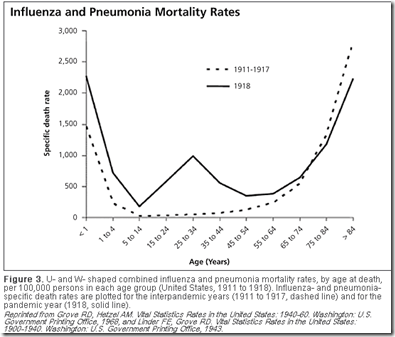
The infamous `W shaped curve’ of the 1918 pandemic clearly shows that the death rates among those in their teens, 20s, and 30s was much higher than was normally seen in previous influenza years. Those over the age of 65, however, saw a reduction in mortality during the pandemic.


# 6778


Seasonal influenza can strike people of any age, but exacts its greatest toll on the elderly – those over the age of 65 – whose weaker immune systems (and comorbid conditions) often render them less able to fight off the infection.


Exact numbers remain elusive, since influenza is only rarely cited as the primary cause of death. If a cause of death (beyond`natural causes’) is given, comorbidities like COPD, heart disease, asthma are far more likely to listed on a death certificate.


Still, estimates are that 90% of seasonal flu mortality occurs in those over the age of 65 (cite CDC Pink book).


In 2010, (see Study: Years Of Life Lost Due To 2009 Pandemic), researchers estimated the median age of death due to seasonal influenza-related illness in the United States to be 76.


In contrast, pandemic influenza strains, at least during the first few years after their introduction, often produce a dramatic `age shift’ downward in mortality. 


The CDC’s estimate of average and median age of death due to the 2009 Pandemic virus reads:


Based on two CDC investigations of confirmed 2009 H1N1-related deaths that occurred during the spring and fall of 2009, the average age of people in the U.S. who died from 2009 H1N1 from April to July of 2009 was 40. The median age of death for this time period was 43. From September to October of 2009, the average age of people in the U.S. who died from 2009 H1N1 was 41, and the median age was 45.


Admittedly, younger fatalities are more likely to be investigated, and documented, than those that occur among the elderly, but still . . . this is a significant shift.


And it corresponds closely to the results of the Years Of Life Lost Study mentioned above, which found the mean age of death from the novel H1N1 virus to be half that of seasonal flu, or 37.4 years.


In terms of years of life lost (YLL), the average pandemic flu death has a many fold greater impact than the average seasonal flu fatality.   


This same pattern was repeated (to greater and lesser degrees) during the 1918, 1957, and 1968 pandemics  . . .  along with the 1977 return of the H1N1 virus after an absence of 20 years.


All of which has led to a good deal of speculation.


What drives this age shift?  Why were apparently healthy, younger flu victims, with robust immune systems more likely to die from pandemic flu?


Although not universally accepted, one popular theory has centered around the production of a `cytokine storm’, which is believed to be the product of a robust immune system typically found in younger, healthier individuals.


Cytokines are a category of signaling molecules that are used extensively in cellular communication. They are often released by immune cells that have encountered a pathogen, and are designed to alert and activate other immune cells to join in the fight against the invading pathogen.


This cascade of immune cells rushing to the site of infection, that if it races out of control, can literally kill the patient.


The patient’s lungs can fill with fluid (which makes a terrific medium for a bacterial co-infection), and cells in the lungs (Type 1 & Type II Pneumocytes) can sustain severe damage.


You can find more on this theory in these earlier posts:


Study: Calming The Cytokine Storm
Cytokine Storm Warnings

The Baskin Influenza Pathogenesis Study

Pt. 1               Pt. 2            Pt. 3


Another theory has held that older populations are more likely to have been exposed to a similar influenza strain in the past and are more likely to carry some level of immunity to the emerging pandemic strain.


This was clearly the case in 1977, when the H1N1 virus – supplanted by the H2N2 virus in 1957 – made an unexpected comeback.  Those born after the virus last circulated in the mid 1950s – were the hardest hit age group.


Again with the 2009 H1N1 pandemic virus, those born before the early 1950s appeared to have higher levels of immunity, resulting in fewer severe outcomes among older individuals.


All of which serves as prelude to a research article, published yesterday in BMC Medicine, that looks at the age shift during pandemic outbreaks.


The age distribution of mortality due to influenza: pandemic and peri-pandemic

Tom Reichert, Gerardo Chowell and Jonathan A McCullers

Background

Pandemic influenza is said to 'shift mortality' to younger age groups; but also to spare a subpopulation of the elderly population. Does one of these effects dominate? Might this have important ramifications?

Methods

We estimated age-specific excess mortality rates for all-years for which data were available in the 20th century for Australia, Canada, France, Japan, the UK, and the USA for people older than 44 years of age. We modeled variation with age, and standardized estimates to allow direct comparison across age groups and countries. Attack rate data for four pandemics were assembled.

Results

For nearly all seasons, an exponential model characterized mortality data extremely well; For seasons of emergence and a variable number of seasons following, however, a subpopulation above a threshold age invariably enjoyed reduced mortality. 'Immune escape', a stepwise increase in mortality among the oldest elderly, was observed a number of seasons after both the A(H2N2) and A(H3N2) pandemics. The number of seasons from emergence to escape varied by country. For the latter pandemic, mortality rates in four countries increased for younger age groups but only in the season following that of emergence. Adaptation to both emergent viruses was apparent as a progressive decrease in mortality rates, which, with two exceptions, was seen only in younger age groups. Pandemic attack rate variation with age was estimated to be similar across four pandemics with very different mortality impact.

Conclusions

In all influenza pandemics of the 20th century, emergent viruses resembled those that had circulated previously within the lifespan of then-living people. Such individuals were relatively immune to the emergent strain, but this immunity waned with mutation of the emergent virus. An immune subpopulation complicates and may invalidate vaccine trials. Pandemic influenza does not 'shift' mortality to younger age groups; rather, the mortality level is reset by the virulence of the emerging virus and is moderated by immunity of past experience. In this study, we found that after immune escape, older age groups showed no further mortality reduction, despite their being the principal target of conventional influenza vaccines. Vaccines incorporating variants of pandemic viruses seem to provide little benefit to those previously immune. If attack rates truly are similar across pandemics, it must be the case that immunity to the pandemic virus does not prevent infection, but only mitigates the consequences.

The complete article is available as a provisional PDF.


The entire article is worthy of your attention, and the authors delve into a good many areas, including future pandemic mitigation planning, and vaccine strategies. 


But essentially the authors propose that all recent influenza pandemics (over the past century) have involved `recycled’ flu strains to which some portion of the population had previously been exposed to.


They conclude:


Pandemics do not ‘shift’ mortality to younger ages

From this study, it is evident that pandemics do not ‘shift’ mortality to younger ages. Rather, the
entire mortality level is simply reset to the virulence level of the emergent virus. This reset is accompanied by immunoprotection in older age groups, which is determined by their level of previous experience with viruses similar to that emerging. 


In other words, were older populations not carrying some vestiges of immunity from previously flu encounters, these researchers suggest they would suffer the same levels (or higher) of mortality and morbidity as do younger populations.


The authors also point out that initial levels of immunity to emerging (or more properly, re-emerging) influenza viruses in older populations tends to wane in subsequent seasons, leading to what they call Immune Escape: `a stepwise increase in mortality among the oldest elderly’.


Does this blow the whole cytokine storm theory out of the water?


Not necessarily, although it does call into question just how much of an impact it has on the perceived `age shift’ in pandemic flu cases.


These two theories need not be mutually exclusive, however, and so I wouldn’t rule out the possibility that both may play a part in driving pandemic mortality demographics.

Tuesday, August 19, 2025

Cell: Immunopathogenesis of Lethal H5N1 Avian influenza Virus Clade 2.3.4.4b Infection in Macaques

 
Credit Cell Immunity

#18,843

In 2009 we took a deep (3 blog) dive into a landmark PNAS study - Early and sustained innate immune response defines pathology and death in nonhuman primates infected by highly pathogenic influenza virus by Carole Baskin et. al. - which compared the pathogenesis of an older (2004) H5N1 virus, seasonal flu, and with two altered viruses carrying genetic material from the 1918 Spanish Flu in macaques. 

The key finding from this study was that severe morbidity and mortality in macaques infected with H5N1 was linked to a  severe `Cytokine storm'; a strong, early, and sustained innate immune response, marked by elevated pro-inflammatory cytokines and severe lung damage (notably, apoptosis of dendritic cells). 
 
As we've discussed previously, the HPAI H5N1 viruses circulating today are a far cry from the clades and variants that circulated in the early 2000's. They've evolved into new clades and subclades, and have reassorted into numerous genotypes (and subtypes).
The Cambodian clade 2.3.2.1e H5N1 virus appears to produce a nearly 50% CFR (case fatality rate), but the U.S. clade 2.3.4.4b `bovine' B3.13 genotype has a reputation for producing relatively mild symptoms in humans (primarily conjunctivitis, mild fever, cough, and mild flu-like symptoms).
This B3.13 genotype encompasses a wide range of variants, including at least one isolate (huTX37-H5N1) which carried the PB2-E276K mutation (glutamic acid → lysine at position 627), which is associated with increased mammalian adaption and pathogenicity.   
While there are undoubtedly other amino acid changes (e.g. HA-T143A, PB2 D701N, PB2 Q591K, HA Q226L, etc.) that could increase the pathogenicity of HPAI H5N1, the E627K mutation is always at or near the top of the list.

All of which brings us to a new study, published yesterday in Cell Immunity, which revisits the pathogenicity of the new bovine B3.13 genotype (with E627K) in two types of macaques (Cynomolgus and Rhesus). 

This lengthy, and at times technical, report finds that the `mild' bovine B3.13 genotype - when aided and abetted by the PB2-E627K mutation - can be every bit as deadly in macaques as the older H5N1 viruses from Vietnam the mid 2000s.  

It also finds - much like we saw in 2009 - that ` . . . acute inflammation, cytokine storm, and immune dysregulation are hallmarks of H5N1 disease pathogenesis in both cynomolgus and rhesus macaques.'

For now, E627K remains rarely reported in the B3.13 genotype of H5N1, but it has turned up in other genotypes, including the D1.1 genotype variant which caused severe infections in both Canada and the United States. 

Due to its length I've only posted the abstract, and a few excerpts.  Follow the link to read the full report in its entirety.  I'll have a brief postscript after the break. 

Immunopathogenesis of lethal H5N1 avian influenza virus clade 2.3.4.4b infection in macaques
Hanne Andersen1 ∙ Malika Aid2 ∙ Jonathan J. Stone3 ∙ … ∙ Mark G. Lewis1 ∙ Amanda J. Martinot3 ∙ Dan H. Barouch2,6,7 dbarouch@bidmc.harvard.edu
Published online August 18, 2025
DOI: 10.1016/j.immuni.2025.07.020


Highlights
  • Cynomolgus macaques developed lethal H5N1 influenza pneumonia
  • Rhesus macaques demonstrated dose-dependent H5N1 influenza mortality
  • Surviving animals were protected against high-dose re-challenge
  • H5N1 influenza was characterized by severe inflammation and immune exhaustion
Summary

The H5N1 avian influenza virus clade 2.3.4.4b outbreak represents a major pandemic threat for humans, with some reported cases of severe and fatal respiratory illness. A key unanswered question is the pathogenesis of severe H5N1 disease following respiratory infection.
In this study, we explored mechanisms of pathogenesis of severe H5N1 disease in cynomolgus and rhesus macaques following infection with the H5N1 isolate A/Texas/37/2024 (huTX37-H5N1). Cynomolgus macaques developed severe pneumonia that was lethal in 100% of macaques by 7 days post-infection. By contrast, rhesus macaques demonstrated dose-dependent mortality, and surviving animals showed protective immunity against high-dose re-challenge.
A multi-omics analysis demonstrated that H5N1 infection was characterized by robust induction of proinflammatory cytokines, innate immune cells, complement, coagulation, apoptosis, and immune exhaustion pathways.
Taken together, our data indicate inflammation and immune dysregulation as key mechanisms of H5N1 pathogenesis in nonhuman primates.

        (SNIP) 

Discussion

In this study, we observed that huTX37-H5N1 infection of cynomolgus and rhesus macaques by the respiratory route resulted in severe and rapidly progressive consolidative necrotizing pneumonia across a broad range of challenge doses. Infection was lethal in 100% (24 of 24) of cynomolgus macaques and in 33% (3 of 9) of rhesus macaques in the titration studies.
Lung consolidation was also more extensive and severe in cynomolgus macaques than in rhesus macaques. Transcriptomic profiling showed enrichment of proinflammatory and immune exhaustion pathways following challenge as well as reduction of NK, T, and B cell activation pathways and increased expression of T cell exhaustion markers, particularly in cynomolgus macaques.
These findings suggest that acute inflammation, cytokine storm, and immune dysregulation may be key contributors to the pathogenesis of H5N1 severe disease.
(SNIP)
In summary, our data demonstrate severe clinical disease with H5N1 clade 2.3.4.4b in cynomolgus and rhesus macaques when inoculated by the respiratory route, particularly in cynomolgus macaques. Although most human cases have been mild, several cases of severe and critical respiratory disease in humans have been reported, suggesting the importance of understanding the pathogenesis of severe H5N1 disease.
Our findings define key features of pathogenesis and immunity to H5N1 clade 2.3.4.4b infection in cynomolgus and rhesus macaques. Future studies could define the role of T cell responses and T cell exhaustion for H5N1 disease pathogenesis. Moreover, these models of H5N1 infection in nonhuman primates should prove useful in evaluating candidate vaccines and therapeutics.

Limitations of the study

This study is limited to the evaluation of one strain of H5N1 influenza in rhesus and cynomolgus macaques, and it is possible that other H5N1 strains may have different characteristics. Moreover, the pathogenicity of H5N1 influenza in macaques may differ from that in humans, potentially because of higher inoculation doses and different challenge routes. In addition, this study involves an H5N1 isolate with the human adaptation mutation PB2-E627K as a model for severe disease. This mutation is currently rare in bovine H5N1 isolates, and thus it is possible that future human infections will have other adaptation mutations.

        (Continue . . . )

Interestingly, while cynomolgus and rhesus macaques are closely related primate species - and are both used in biomedical research - there are some key differences which may  help explain the higher mortality in cynomolgus macaques over rhesus. 
  • Cynomolgus macaques tend to be slightly smaller than rhesus macaques, and are generally easier to handle (less aggressive).
While neither species likely serve as ideal analogs for human exposure to H5N1, they do provide us with a sobering range of non-human primate responses to infection with this specific H5N1 B3.13 isolate. 

But most of all, this study reaffirms that our currently mild `bovine' genotype may only lack one or two crucial fixed mutations to make it a genuine public health concern. 

Assuming that some other clade/genotype doesn't get there first. 

Sunday, December 22, 2013

Details & Background On the H5N2 Poultry Outbreak In Hebei Province

image

Photo Credit CRIENGLISH


# 8099


When it comes to variety, avian influenzas come in more flavors than you’ll find in a Baskin-Robbins store. Which explains  why in the past couple of months our attentions have been divided between H5N1 in Cambodia and Indonesia, H7N9 in Mainland China, an upstart H10N8 (again in China), and now H5N2 in Hebei Province, China.


While we worry most about H5N1 and H7N9 – primarily because both have been shown to produce severe illness and even death in humans – all H5 and H7 varieties are of concern, since these subtypes have a history of morphing from low-pathogenic strains to highly pathogenic strains if not controlled.  


Which is why all H5 and H7 outbreaks are reportable to the OIE, and quarantine and immediate culling are the preferred course of action.


Yesterday China reported an outbreak of H5N2 at a poultry farm in Baoding City, Hebei Province.  Reports today indicate that more than 120,000 birds have been culled, and that 11 people who were in direct contact with the poultry have been placed in isolation (and probably given Tamiflu ®), but none have shown signs of infection.


First a report from Sina  News, then I’ll be back with more on the history of H5N2


Hebei people infected with H5N2 bird flu yet (Alt Translation Hebei infection of H5N2 avian flu nobody has yet been)

CNA (2013-12-22 18:18)

(CNA Taipei 22 (Xinhua)) Hebei Baoding Nanshiqu officials said today, raising the local chicken farm H5N2 bird flu has been effectively controlled occurred, 11 were in direct contact with the relevant chickens were isolated observation, it was not found infected.

China News Service reported, Baoding Nanshiqu Agriculture Secretary Zhang Guang pointed out that, in addition to the above measures, culling chickens participating Nanshiqu work were monitored twice daily, were not unusual. Meanwhile, the official has the region's comprehensive agricultural and sideline products and fragmented market of live poultry trade to clean up and banned.

Zhang also said that the official and farm stocks of eggs into the marketing ruin handling , disposal of tracing the source of the product sold. Meanwhile, the live poultry within 3-5 km range chicken farm near the outbreak, have been carried out culling.

H5N2 outbreak was discovered south downtown Baoding Zhu Village tiger phoenix chicken farm was built in 1992, is a self-employed business , keeping more than 120,000 laying hens only, no chickens. 4000 chickens on the 17th because the epidemic of death was November 27 to buy from overseas. 1021222


It should be noted that we’ve seen both HPAI (Highly pathogenic) and LPAI (Low Pathogenic) H5N2 outbreaks in the past, notably in Taiwan, Canada, the United States, Korea, Japan, and Mexico.


The isolation (and usually, antiviral treatment) of those who have been in direct contact with infected birds is standard procedure, and should not be viewed as being particularly alarming.  While not normally thought of as a serious threat to public health (see WHO: Human Risk From H5N2 Is Low), we have seen some limited evidence of H5N2’s ability to infect humans in recent years.


Eighteen months ago, in Taiwan: Three Poultry Workers Show H5N2 Antibodies, we looked at a report that three poultry workers and officials working in animal quarantine have tested positive for antibodies for the H5N2, but all remained healthy and asymptomatic (note: refer to article for other possible causes of seropositivity)..  


Some earlier H5N2 studies include:


J Epidemiol. 2008;18(4):160-6. Epub 2008 Jul 7.

Human H5N2 avian influenza infection in Japan and the factors associated with high H5N2-neutralizing antibody titer.

Ogata T, Yamazaki Y, Okabe N, Nakamura Y, Tashiro M, Nagata N, Itamura S, Yasui Y, Nakashima K, Doi M, Izumi Y, Fujieda T, Yamato S, Kawada Y.

Arch Virol. 2009;154(3):421-7. Epub 2009 Feb 3.

Serological survey of avian H5N2-subtype influenza virus infections in human populations.Yamazaki Y, Doy M, Okabe N, Yasui Y, Nakashima K, Fujieda T, Yamato S, Kawata Y, Ogata T.


While these reports are suggestive of prior H5N2 human infection – particularly among poultry workers – we haven’t seen any good evidence that it has produced significant or serious human illness. 


The caveat being, that up until the emergence of H7N9 in March of this year, H7 avian flu viruses had been generally regarded as posing a minor threat to human health, and until this past week, we’d never seen a human infection with the H10N8 virus.


With influenza viruses, the only constant is change.  What may be relatively benign today, may become virulent or increase its host range tomorrow.


Which makes the rapid and intense response to bird flu outbreaks  – such as we are seeing  with H5N2 in China right now – the prudent course of action.