Showing posts with label The Lancet. Show all posts

Wednesday, April 01, 2015

CIDRAP On The Lancet EV-D68 Analysis

image

CDC EV-D68 Fact Sheet


# 9890


In August of last year, a seldom seen (at least, in North America) non-polio enterovirus D-68 (EV-D68) appeared in America’s Midwest and quickly spread across the nation, causing a wide spectrum of respiratory illness, predominantly in young children and adolescents (see Kansas City Outbreak Identified As HEV 68).


At roughly the same time, a coincident rise in cases of neurological illness with AFP (acute flaccid paralysis) or limb weakness – often associated with a recent respiratory illness – was reported across the country.


A month later the CDC  issued a HAN: Acute Neurologic Illness with Focal Limb Weakness of Unknown Etiology in Children, alerting doctors around the country to be aware of this trend, and providing information on reporting cases. While primarily associated with respiratory symptoms, EV-D68 had previously been linked to neurological presentations, including a small cluster of cases the previous winter (see Acute Flaccid Paralysis Cases In California).


Although a causal link wasn’t established, due to the timing and the fact that other enteroviruses have been linked to neurological illnesses, there was a high degree of suspicion that the two conditions were linked.


Enteroviruses encompass a large family of small RNA viruses that include the three Polioviruses, along with myriad non-polio serotypes of Human Rhinovirus, Coxsackievirus, echovirus, and human, porcine, and simian enteroviruses.  We’ve looked at EV-71 and the Coxsackieviruses on numerous occasions in regards to AFP (Acute Flaccid Paralysis) and HFMD (see here,  here & here).


According to the CDC  Non-Polio Enteroviruses (NPEVs) cause 10 to 15 million – mostly mild and often asymptomatic – infections in the United States each year, primarily among infants, children, and teenagers.


Fever, runny nose, sneezing, coughing, a skin rash or mouth blisters, and body and muscle aches are the most commonly reported symptoms.


First isolated in 1962, but only rarely reported in North America since, in 2011 – in MMWR: Clusters Of HEV68 Respiratory Infections 2008-2010 – we looked at a recent increase in EV-68 associated clusters reported in Asia, Europe, and the United States during 2008--2010.


We continued to watch the progress of this most recent outbreak over the winter (see CIDRAP: Likely That Polio-like Illness & EV-D68 Are Linked  & Eurosurveillance: Acute Flaccid Paralysis Following EV-D68 Infection – France), and the most recent CDC NCIRD update on last year’s outbreak was posted in early March.


Update

From August 2, 2014 to March 2, 2015, CDC has verified reports of 115 children in 34 states who developed acute flaccid myelitis that meets CDC’s case definition. CDC continues to collaborate with partners nationally to investigate reported cases, risk factors, and possible causes of this condition.

  • The median age of the children was about 8 years.
  • Almost all of them were hospitalized; some were put on breathing machines.
  • Most patients had fever and/or respiratory illness before onset of neurologic symptoms.
  • About two thirds of the children who have been observed (median 19 days) after their illness reported some improvement in symptoms, while about one third showed no improvement. Only two of the children have fully recovered.


          This week a genetic analysis of this emerging enterovirus was published in The Lancet -  A novel outbreak enterovirus D68 strain associated with acute flaccid myelitis cases in the USA (2012–14): a retrospective cohort study – that not only strengthened the link between the EV-D68 infection and paralytic complications, but also identified the virus as being part of a new clade (B1) with polio-like changes that emerged about 5 years ago.


          Since Robert Roos of CIDRAP News  has already done a terrific job covering this study, I’ll simply refer you to last night’s story for the rest of the details.  Follow the link to read:


          Genetic study boosts evidence for EV-D68 in polio-like cases

          Robert Roos | News Editor | CIDRAP News

          |

          Mar 31, 2015

          A careful study of children at two Colorado and California hospitals who had polio-like illnesses strengthens the evidence that cases were related to enterovirus-D68, which caused a widespread outbreak of severe respiratory illnesses in US children last year, according to a report published today in The Lancet Infectious Diseases.

          The virus was found in 12 of 25 patients who had the polio-like condition, called acute flaccid myelitis (AFM), and the proportion probably would have been higher if respiratory samples had been collected earlier, the researchers said. In addition, a thorough genomic search for other viruses in the neural fluid of 14 of the patients found no signs of any other virus that could have caused the patients' limb weakness and related problems.

          "These findings strengthen the putative association between enterovirus D68 and acute flaccid myelitis and the contention that acute flaccid myelitis is a rare yet severe clinical manifestation of enterovirus D68 infection in susceptible hosts," the authors wrote.

          (Continue . . . )

          Saturday, January 31, 2015

          CIDRAP News On The Lancet Oseltamivir (Tamiflu ®) Meta-Analysis

          image

          Photo Credit – Wikipedia


          # 9552


          I had plans this morning to write an overview of meta-analysis of Tamiflu effectiveness published January 29th in The Lancet, but I’m happy to note that last night Robert Roos of CIDRAP News has beaten me to it. Since I’m unlikely to improve upon his reportage, I’ll direct you to his excellent review – which you’ll want to read in its entirety - after which I’ll return with a little bit more.


          Meta-analysis supports oseltamivir use in adults, notes side effects

          Robert Roos | News Editor | CIDRAP News

          Jan 30, 2015

          A comprehensive new meta-analysis on the controversial topic of oseltamivir's effectiveness found that the drug reduces the duration of influenza symptoms and the risk of hospitalization in adults and adolescents, while increasing the risk of nausea and vomiting.

          A US-British team, with Arnold S. Monto, MD, of the University of Michigan as senior author, included in the analysis all randomized controlled trials sponsored by Roche, the drug's manufacturer, as well as other relevant trials. The study, reported yesterday in The Lancet, was funded by Roche, but the researchers worked independently.

          (Continue.  . . )


          Over the past few years we’ve seen the demonization of influenza antivirals in the media (see Daily Mail: Ministers blew £650MILLION on useless anti-flu drugs), warnings of potential aberrant psychiatric behavior (see 2007 New Worries On Tamiflu), and repeated Cochrane group analyses that have found insufficient evidence that the drug reduces influenza complications.


          Add in some serious foot-dragging by manufacturer Roche in releasing all of their testing data, and Tamiflu has become an easy drug for the public, and some doctors, to distrust.


          Despite all of this `baggage’  the CDC, ECDC, UK’s PHE, and other public health agencies have steadfastly supported the early use of oseltamivir in the treatment of severe flu (see this week’s CDC Antiviral Letter to Providers and ECDC Influenza Season Risk Assessment).


          The reason?

          Even without the `gold standard’ Randomized controlled trials (RCTs) that the Cochrane group relies on for their analyses, we’ve seen numerous observational studies that lend support to the use of antivirals in severe influenza.


          A few I’ve written about in the past include:


          Their main finding was antiviral therapy - principally oseltamivir - initiated within 48 hours of onset, reduced the likelihood of severe outcomes, namely admission to a critical care unit or death, by 49 to 65%.


          Added to this, we now have this new meta-analysis of the data from all published and unpublished clinical trials from 1997-2001, involving more than 4,300 patients. Patients with influenza (not just an ILI), who received the drug within 36 hours of onset of symptoms saw a reduction in the duration of their illness of 21% and a significant reduction in the risk of developing pneumonia or requiring hospitalization.


          While nausea (9.9% vs 6.2% in controls) and vomiting (8.0% vs 3.3%)  were common side effects, no serious adverse reactions were reported, with no increase in psychiatric or neurological symptoms.


          For uncomplicated influenza in a healthy individual (essentially what the Cochrane studies looked at), antivirals probably offer limited benefits.


          But for severe influenza, or for people at risk of complications . . .


          The preponderance of evidence shows that taking antivirals early can limit the severity and duration of symptoms – and for patients at risk of complications – that  could help keep them out of the hospital . . .  or worse.

          Thursday, November 20, 2014

          Lancet: Mackay & Arden On Ebola In Semen Of Convalescent Men

          image

          Credit CDC PHIL


          # 9348


          Last August, Dr. Ian Mackay writing on his VDU Blog, posted Ebola virus in semen is the real deal...., which he followed up 10 days later with  Ebola: Blood, sweat and tears, where Ian described the (somewhat limited) research to date on the level of EBOV detection in other body fluids – like tears, sweat and saliva.


          Given how easily Ebola has spread these past few months in Africa – and all apparently via infected body fluids – the more we know about the risks, the better.


          This week we’ve seen India isolate a man with Ebola-infected semen, more than six weeks after he was pronounced cured (Sept 30th) and released from a Liberian hospital. While this may provide additional data on the duration of viral shedding via semen, as a public health measure, many consider his isolation both excessive and unnecessary.


          In a case of exceedingly good timing, yesterday Ian Mackay  and Dr. Katherine Arden published a review of the potential for semen to carry the EBOV in The Lancet.


          Ebola virus in the semen of convalescent men

          Ian M Mackay,Katherine E Arden
          The Lancet Infectious Diseases - 19 November 2014
          DOI: 10.1016/S1473-3099(14)71033-3

          Semen is one of the body fluids from which Ebola virus disease can be contracted, presumably by delivery of the infectious virus to a mucosal surface. WHO notes that Ebola viruses can be transmitted via the semen of a man who is convalescing, for up to 7 weeks after recovery from an Ebola virus infection.1 The European Centre for Disease Prevention and Control2 and Public Health Agency of Canada3 both report the 7 week figure, and the latter also refers to a period of 61 days. The United States Army Medical Research Institute of Infectious Diseases4 suggests a 3 month period, recommending that survivors avoid sexual contact.

          (Continue . . . )

          Tuesday, September 02, 2014

          The Lancet: Series On Emerging Respiratory Tract Infections

          image

          Pneumonia Etiology Unknown – CDC PHIL


          # 9029


          This is mostly a head’s up, since I’ve not had time to read any of these papers. But as they appear to be both interesting and on point for this blog, so I decided to point my readers in their direction.


          Among the authors you’ll find such familiar names as  Ziad Memish, Christian Drosten, Alimuddin Zumla, and Jaffar Al-Tawfiq.


          This series is provided for free by The lancet, although (free) registration is required for full access.


          Emerging respiratory tract infections

          Published September 2, 2014

          Executive summary

          Emerging and re-emerging respiratory tract infections and the growing problem of antimicrobial resistance challenge diagnostic, treatment, prevention, and control strategies. Respiratory tract infections with epidemic and pandemic potential have plagued people since the dawn of human history. Comprehensive assessments of the global burden of disease show that respiratory tract infections are some of the commonest causes of morbidity and mortality worldwide, imposing a huge burden on health services. Several epidemics of new viral respiratory tract infections, most of which emerged from interactions between people and animals, have occurred in the past century.


          The Lancet Infectious Diseases Series on emerging respiratory tract infections contains five state-of-the-art papers developed and written by international groups of experts with experience in management of emerging respiratory tract infections. The Series covers the topics of surveillance, pandemic potential, transmission and the environment, diagnosis, and treatment. The focus is on the emerging viral pathogens (particularly influenza, adenovirus, and coronaviruses) that pose the greatest threat, although bacterial, mycobacterial, and fungal pathogens are included where appropriate

          Comment

          Emerging respiratory tract infections

          Zumla and others

          Full Text | PDF

          Series

          Surveillance for emerging respiratory viruses

          J A Al-Tawfiq and others


          Full Text | PDF

          Emerging infectious diseases and pandemic potential: status quo and reducing risk of global spread

          B McCloskey and others

          Full Text | PDF

          Emerging viral respiratory tract infections—environmental risk factors and transmission

          P Gautret and others

          Full Text | PDF

          Rapid point of care diagnostic tests for viral and bacterial respiratory tract infections—needs, advances, and future prospects

          A Zumla and others

          Full Text | PDF


          Emerging novel and antimicrobial-resistant respiratory tract infections: new drug development and therapeutic options

          A Zumla and others

          Full Text | PDF

          Wednesday, March 19, 2014

          The Lancet: Effectiveness Of NAI Antivirals In Reducing Mortality In Hospitalized H1N1pdm09 Cases

          image


          # 8387


          If you want to start a row on the Internet, simply say something positive about Roche’s neuraminidase inhibitor (NAI) antiviral Tamiflu ® (oseltamivir). 


          Between large government outlays for stockpiles of the drug, an inherent distrust of `Big Pharma’,  a lack of Randomized Controlled Trials (RCTs) to gauge the effectiveness of the drug, a Cochrane group analysis that found insufficient evidence to show whether the drug reduces influenza complications and transmission, and the apparent reluctance of Hoffmann-La Roche  to release unpublished trial data . . .


          Suffice to say its an easy drug to disparage - at least - until you need it (see Dec 2013’s Spot Shortages Of Tamiflu Reported In Some Regions). 


          With all of this negative press (which let’s face it, sells newspapers & drives web traffic), it would be easy to assume that Tamiflu is overrated, a waste of money, and not worth risking the (usually mild) side effects.


          Yet despite these critics, the CDC continues to recommend its use (as do many other public health agencies around the world), and as recently as last week, the CDC released a fresh set of antiviral guidelines for clinicians:


          Why, you might ask, would the CDC express confidence in a drug that been so disparaged online and in the media?


          Well, despite the critics, there are studies that show that Tamiflu can significantly reduce morbidity and mortality associated with influenza.


          Ideally what researchers want to see are a series of well mounted Randomized controlled trials (RCTs) – long considered the `gold standard’ for drug research. But these types of studies are expensive, and difficult to conduct ethically when trying to evaluate a potentially life saving drug.


          So what we are often left with are observational studies, which critics such as the Cochrane group often discard as being unreliable. With few studies that can meet their requirements, there is – in their opinion - insufficient evidence to show whether the drug reduces influenza complications and transmission.


          But observational studies are not without value, and often they are the only evidence available.  Over the years we’ve looked at a number of them,  including:


          Their main finding was antiviral therapy - principally oseltamivir - initiated within 48 hours of onset, reduced the likelihood of severe outcomes, namely admission to a critical care unit or death, by 49 to 65%.


          Today The Lancet presents a new observational study (funded by F Hoffmann-La Roche), conducted by researchers at The University of Nottingham – that reviewed more than 29,000 hospitalized H1N1pdm cases across 38 countries between 2009 and 2011, and found that the administration of NAI antivirals was associated with a 19% reduction in mortality compared to receiving no NAI treatment at all.


          And among those who received the NAI antiviral within the first 48 hours of admission, the mortality risk was halved. Pregnant women who received the drug swiftly saw a 54% reduction in mortality.


          While funding was received from Roche for the study, the researchers stated that `No data were requested from nor provided by pharmaceutical companies.’ .  First a link to the study (which contains much more detail), followed by a link to an accompanying commentary from Alicia M Fry of the CDC’s Influenza Division.


          Effectiveness of neuraminidase inhibitors in reducing mortality in patients admitted to hospital with influenza A H1N1pdm09 virus infection: a meta-analysis of individual participant data


          Jonathan S Nguyen-Van-Tam, et. al.

          EXCERPTS

          Findings

          We included data for 29 234 patients from 78 studies of patients admitted to hospital between Jan 2, 2009, and March 14, 2011. Compared with no treatment, neuraminidase inhibitor treatment (irrespective of timing) was associated with a reduction in mortality risk (adjusted odds ratio [OR] 0·81; 95% CI 0·70—0·93; p=0·0024). Compared with later treatment, early treatment (within 2 days of symptom onset) was associated with a reduction in mortality risk (adjusted OR 0·48; 95% CI 0·41—0·56; p<0·0001).

          Early treatment versus no treatment was also associated with a reduction in mortality (adjusted OR 0·50; 95% CI 0·37—0·67; p<0·0001). These associations with reduced mortality risk were less pronounced and not significant in children. There was an increase in the mortality hazard rate with each day's delay in initiation of treatment up to day 5 as compared with treatment initiated within 2 days of symptom onset (adjusted hazard ratio [HR 1·23] [95% CI 1·18—1·28]; p<0·0001 for the increasing HR with each day's delay).

          Interpretation

          We advocate early instigation of neuraminidase inhibitor treatment in adults admitted to hospital with suspected or proven influenza infection.

          (Continue Reading Full Text)

          The related commentary may be read at:


          Effectiveness of neuraminidase inhibitors for severe influenza

          Alicia M Fry aEmail Address

          The neuraminidase inhibitors (NAIs) were licensed for use for the treatment of uncomplicated influenza on the basis of results from phase 3 placebo-controlled, randomised clinical trials and are the only influenza-specific treatment option recommended for use. 1 However, the greatest potential clinical and public health benefits of NAI treatment are associated with more severe influenza-associated illness and outcomes, including admission to hospital and death. Thus, in the absence of placebo-cont ...

          (Continue . . . )

          Today’s study adds to the preponderance of evidence that oseltamivir (and other NAI antivirals) provide substantial benefits – particularly when administered early and in cases of severe influenza.  For on this topic you may wish to review these earlier blogs.

          CDC Research On Benefits Of Antivirals For Uncomplicated Influenza
          New Scientist: Don’t Stop Stockpiling Tamiflu
          The CDC On The Value Of Antivirals


          Wednesday, February 26, 2014

          The Lancet: H9N2’s Role In Evolution Of Novel Avian Influenzas

          image

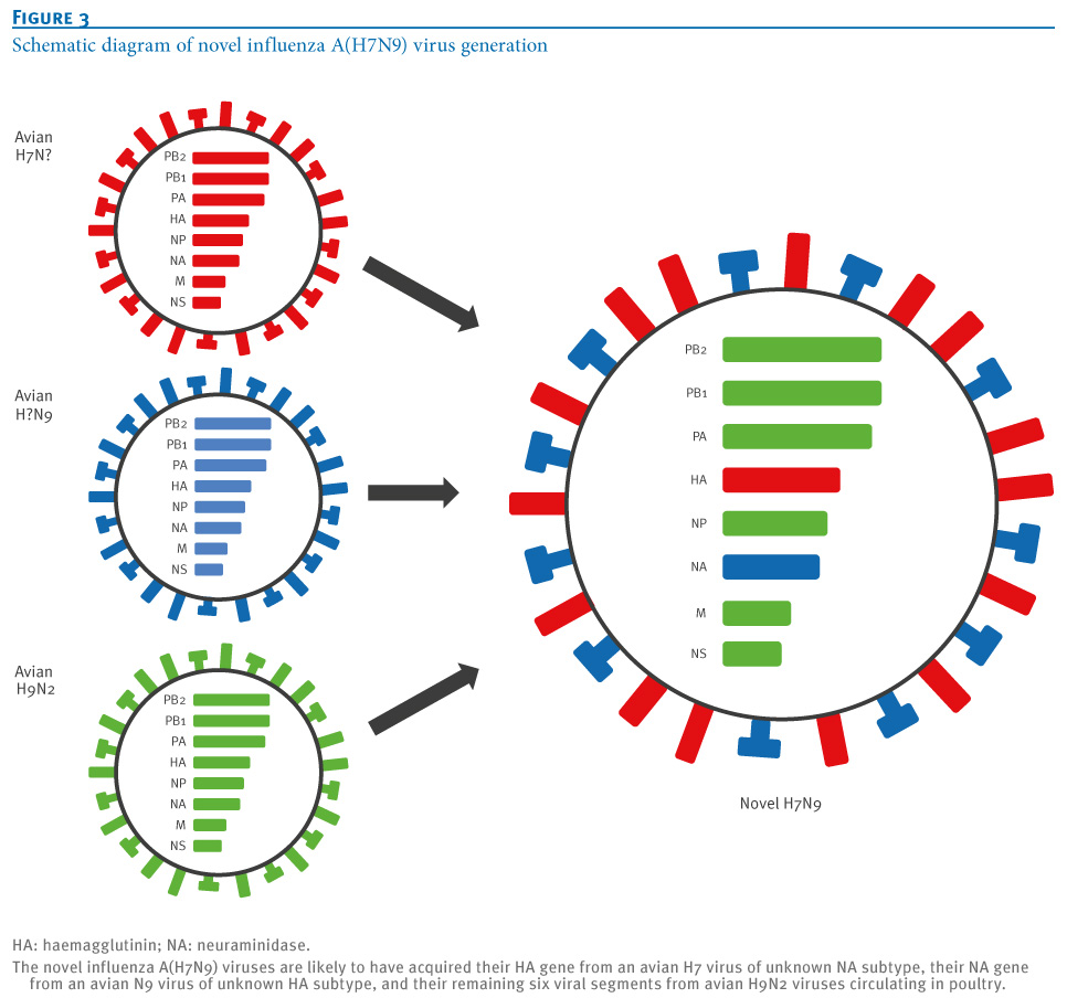
          Schematic Diagram of Novel A(H7N9) Generation- Credit Eurosurveillance


          # 8329


          The three novel avian flu strains that currently worry scientists the most – H5N1, H7N9, and the recently observed H10N8 – all share several important features,  which we’ve discussed previously.


          1. They all first appear to emanate from Mainland China
          2. They all appear to have come about through viral reassortment in poultry
          3. And most telling of all, while their HA and NA genes differ - they all carry the internal genes from the avian H9N2 virus


          The avian H9N2 virus – unlike the H5 and H7 avian viruses – is not considered a `reportable’ disease by the OIE since it is viewed as a relatively stable LPAI (Low Pathogenic Avian Influenza), not prone to evolving into a more dangerous HPAI form.  It is, however:


          1. Believed ubiquitous across much of Asia’s poultry population
          2. Has occasionally infected humans (see Hong Kong: Isolation & Treatment Of An H9N2 Patient)
          3. And is viewed as having at least some `pandemic potential’ (see H9N2: The Other Bird Flu Threat)


          As the diagram at the top of this blog shows, the H7N9 virus is a combination (reassortment) of several different avian flu viruses – with six of its eight genes contributed by the H9N2 virus ( shown in green).  An evolutionary pathway similar to that followed by the H5N1 virus in the mid-1990s, and the recently emerging H10N8 virus in China.

          Although categorized by their two surface proteins (HA & NA) Influenza A viruses have 8 gene segments (PB2, PB1, PA, HA, NP, NA, M1, M2, NS1, NS2).

          image

          Shift, or reassortment, happens when two different influenza viruses co-infect the same host swap genetic material.  New hybrid viruses may be the result of multiple reassortments, with gene contributions coming from several parental viruses.


          In the past, we’ve looked at the propensity of the H9N2 virus to reassort with other avian flu viruses (see PNAS: Reassortment Of H1N1 And H9N2 Avian viruses &  PNAS: Reassortment Potential Of Avian H9N2) which have shown the H9N2 capable of producing `biologically fit’ and highly pathogenic reassortant viruses.


          In 2010 (see Study: The Continuing Evolution Of Avian H9N2) we looked at computer modeling (in silica) that warned the H9N2 virus has been slowly evolving towards becoming a `more humanized’ virus.


          All of which serves as prelude to a brief report appearing today in The Lancet, where Chinese researchers warn of the threat posed by the H9N2 virus, and call for prompt and bold action to prevent the `next pandemic virus’ from emerging from China’s poultry industry. 


          The report is a short one, well worth reading,  and quite to the point. I’ve  only excerpted its conclusions (bolding mine).  Follow the link below to read it in its entirety.


          Poultry carrying H9N2 act as incubators for novel human avian influenza viruses

          Di Liu a, Weifeng Shi b, George F Gao a c 

          (EXCERPT)

          Although the contribution of H9N2 genes to infection in human beings needs to be determined, these genes probably enable H7N9 virus to survive and be transmitted within poultry, because dynamic reassortments of H7N9 with H9N2 genes have been observed,5 suggesting that H7N9 virus evolved in poultry to become a virus that infects human beings. Hence, reassortment between the prevalent poultry H9N2 viruses (providing genetic segments) and the influenza virus from wild birds could make the influenza evolve to adapt to domestic hosts. Poultry, especially in live markets, would have a pivotal role during the emergence of a novel influenza virus of avian origin.


          Several subtypes of avian influenza viruses in poultry are capable of infecting human beings, and the next avian influenza virus that could cause mass infections is not known. Therefore, slaughter of poultry carrying H9N2—the incubators for wild-bird-origin influenza viruses—would be an effective strategy to prevent human beings from becoming infected with avian influenza.


          We call for either a shutdown of live poultry markets or periodic thorough disinfections of these markets in China and any other regions with live poultry markets.


          The shutdown of live poultry markets has been a stated goal by Chinese authorities for many years due to the H5N1 threat, but thus far, only limited (and usually temporary) shutdowns have been orchestrated. Despite pretty good evidence that the shutdown of live markets last spring helped quell China’s H7N9 outbreak (see The Lancet: Poultry Market Closure Effect On H7N9 Transmission), there remains strong public pressure to keep them open.


          Even more problematic is their call to cull H9N2 infected chickens.  


          As most infected poultry are asymptomatic, it would require extensive (and expensive) surveillance and testing just to identify these birds. And of course, given the likely incidence of the virus in Asian poultry,  the economic losses would be substantial. 


          At least in China, the MOA (Ministry of Agriculture’s) policy has seemed to revolve around deflecting concerns over avian flu in their poultry supply, rather than addressing it in an organized and substantive manner (see  China’s MOA Disputes Poultry As Source Of H7N9 Infections). 


          As we saw last year in  EID Journal: Predicting Hotspots for Influenza Virus Reassortment, China ranks as one of the globe’s top breeding grounds for new flu strains. Which makes the control of these emerging viruses in all the more important. 


          For more on reassortment risks, you may wish to revisit:


          Eurosurveillance:The Evolving Threat From New, Reassorted H7N9 Viruses
          Lancet: Clinical & Epidemiological Characteristics Of A Fatal H10N8 Case
          Viral Reassortants: Rocking The Cradle Of Influenza

          Tuesday, December 17, 2013

          The Lancet: Identification Of MERS Virus In Camels

          image

          Photo Credit Wikipedia


          #8080


          Since early August, when the first evidence was presented (see Lancet: Camels Found With Antibodies To MERS-CoV-Like Virus), researchers have known that some camels on the Arabian peninsula had been exposed to a MERS-like virus, but the exact virus was undetermined. 


          It could have been the same MERS-CoV that has been infecting humans for the past two years, or it could have been a close relative.


          Subsequent studies have incremented our knowledge, but we’ve not had solid evidence linking the human virus to the camel antibodies.  That is, until now.


          On November 27th, Qatari officials announced that the MERS virus had been detected in Camels on a farm where two people had become infected (see Qatar Supreme Council of Health Statement On MERS-CoV In Camels), and that further investigations were underway by the local Health Ministry and RIVM laboratory and Erasmus Medical Centre in the Netherlands. 


          Last night The Lancet published a study, led by Marion Koopmans, DVM, PhD, head of virology at the Laboratory for Infectious Diseases at the RIVM in the Netherlands, that determined that the human viruses and the camel viruses were almost an identical match. So close, in fact, that they were unable to determine whether the humans or the camels were infected first.


          While pretty much confirming that both humans and camels were infected with the same virus, the report states: “We cannot conclude whether the people on the farm were infected by the camels or vice versa, or if a third source was responsible.”


          First, a link to the article’s abstract, then a link to coverage by Helen Branswell and by Robert Roos at CIDRAP.


          Middle East respiratory syndrome coronavirus in dromedary camels: an outbreak investigation

          Bart L Haagmans PhD a †, Said H S Al Dhahiry PhD b †, Chantal B E M Reusken PhD c †, V Stalin Raj PhD a †, Monica Galiano PhD d, Richard Myers PhD d, Gert-Jan Godeke BSc c, Marcel Jonges MSc c, Elmoubasher Farag MPH e, Ayman Diab MPH e, Hazem Ghobashy PhD e, Farhoud Alhajri BSc e, Mohamed Al-Thani ABCM e, Salih A Al-Marri ABFM e, Hamad E Al Romaihi ABCM e, Abdullatif Al Khal PhD e, Alison Bermingham PhD d, Prof Albert D M E Osterhaus PhD a, Dr Mohd M AlHajri ABCM e , Prof Marion P G Koopmans PhD a c

          Summary

          Findings

          We obtained samples from 14 camels on Oct 17, 2013. We detected MERS-CoV in nose swabs from three camels by three independent RT-PCRs and sequencing. The nucleotide sequence of an ORF1a fragment (940 nucleotides) and a 4·2 kb concatenated fragment were very similar to the MERS-CoV from two human cases on the same farm and a MERS-CoV isolate from Hafr-Al-Batin. Eight additional camel nose swabs were positive on one or more RT-PCRs, but could not be confirmed by sequencing. All camels had MERS-CoV spike-binding antibodies that correlated well with the presence of neutralising antibodies to MERS-CoV.

          Interpretation

          Our study provides virological confirmation of MERS-CoV in camels and suggests a recent outbreak affecting both human beings and camels. We cannot conclude whether the people on the farm were infected by the camels or vice versa, or if a third source was responsible.


          Helen Branswell brings us additional remarks by lead author Dr. Marion Koopmans, in her report entitled:


          Questions remain about MERS and camels

          By Helen Branswell The Canadian Press

          A new scientific paper confirms that camels on a farm in Qatar were sick with MERS earlier this fall.

          But the report does not make clear whether camels infected people on the farm in question, whether people infected camels or whether an unidentified third species infected both.

          In fact, the senior author of the study says the evidence as it exists cannot determine which way the virus spread.

          And virologist Marion Koopmans, of the Dutch National Institute of Public Health, says it is unlikely that the mystery will be cleared up in this case.

          (Continue . . . )


          And last stop, Robert Roos writing for CIDRAP NEWS files this report:


          Nearly identical MERS-CoV strains found in camels, humans

          MERS-CoV

          Robert Roos | News Editor | CIDRAP News

          Dec 16, 2013

          Researchers today reported that dromedary camels on a farm in Qatar were infected with a strain of Middle East respiratory syndrome coronavirus (MERS-CoV) nearly identical to that found in two people associated with the farm. The findings point to an outbreak that involved both camels and humans, but they don't answer the key question of whether camels infected humans or the other way around.

          Qatari health officials announced Nov 27 that the virus had been found in camels on the farm. Today's report in the Lancet Infectious Diseases spells out the science behind the announcement and says the findings mark the first definitive confirmation of the virus in camels.

          (Continue . . . )

          Saturday, November 23, 2013

          CDC Research On Benefits Of Antivirals For Uncomplicated Influenza

          image

          Photo Credit – Wikipedia


          # 8004


          In clinical medicine there are a lot of things that we think we know – based on observational studies – but for which rigorous randomized controlled trials (RCTs) have not been conducted. Sometimes it is impractical (or even unethical) to subject patients to an RCT, especially if it involves withholding a potentially lifesaving drug from a `control’ cohort.


          So we are often left to rely on less desirable, but still useful, observational studies to gauge the value of treatments or therapies.


          Over the past decade one of the drugs that has fallen into this pharmaceutical limbo is oseltamivir (Tamiflu ®), a product of Roche laboratories, which has been stockpiled by many nations to combat a flu pandemic.  Observational studies have shown that this drug can significantly reduce morbidity and  mortality in severe cases of flu, and modestly reduce the duration of symptoms in uncomplicated seasonal flu.


          But robust RCTs have not been conducted to quantify these benefits, and `RCT purists’ like this Cochrane group analysis – that do not consider `observational studies’ to be solid evidence - have found insufficient evidence to show whether the drug reduces influenza complications and transmission (see 2012 CIDRAP article Review renews questions about oseltamivir benefits).


          Add to this a prolonged reluctance on the part of Roche laboratories to release all of their clinical trial data, and a not totally undeserved reputation of `Big Pharma’ to massage test results, and the result has been a vociferous backlash against the government stockpiling of Tamiflu in some quarters (see Dr. Ben Goldacre Opinion Piece).


          While academics and activists tend to have a dim view of Roche and their antiviral drug, clinicians obviously see value in oseltamivir,  and continue to prescribe it.  The CDC continues to recommend its use – particularly for high-risk influenza patients - or for the treatment of novel flu (see 2012 blog The CDC Responds To The Cochrane Group’s Tamiflu Study). 


          With a new avian (H7N9) virus in the wings and H5N1 still simmering in Asia and the Middle East, the CDC recently reiterated their support for the use of oseltamivir in the treatment of severe, or novel, influenza infection (See H7N9: Updated CDC Guidance For Antiviral Treatment).


          Yesterday, the CDC published a news release on an RCT conducted in Bangladesh on the benefits of Oseltamivir in uncomplicated seasonal flu.  A category of illness where one would expect the least amount of benefit to taking an antiviral. A few excerpts, and a link to The Lancet Study, then I’ll return with a bit more.


          CDC Research Confirms Benefits of Flu Antiviral Drugs, Even Beyond 2 Days After Symptoms Start

          New research confirms benefits of the influenza antiviral medication oseltamivir in treating children with uncomplicated flu illness and shows that treatment can be beneficial even beyond the two-day window recommended as a cut-off for treatment in the drug’s package insert.

          A new study on influenza (flu) antiviral drugs by CDC authors was released today in The Lancet Infectious Diseases. This study is the first clinical trial to note a significant reduction in the duration of illness and virus shedding in children when influenza antiviral treatment was initiated more than 2 days after the onset of influenza (flu) symptoms. These findings confirm the benefits of using the antiviral drug oseltamivir to treat flu illness and suggest that some children will benefit when treatment is initiated beyond 2 days, which is the recommended cut-off for treatment in the current package insert.


          The patients in this double-blind, randomized, placebo-controlled study were mostly children (average age: 5 years) in an urban setting in Bangladesh with laboratory-confirmed influenza infection and no additional flu-related complications. Patients were treated with either oseltamivir (a type of flu antiviral drug known as a “neuraminidase inhibitor”) or a placebo (e.g., a shot of saline). Researchers observed when patients began oseltamivir treatment — either less than 48 hours or 48 hours or more after illness onset — and collected information about the duration of flu symptoms using standardized forms collected from daily household visits. In addition to documenting the duration of flu symptoms, researchers also measured viral shedding, which is virus detection at various times after the patients were enrolled. The detection of live virus in respiratory secretions is thought to be associated with how contagious a person is to others.


          Among all children receiving oseltamivir within 5 days of illness onset, researchers found that overall flu symptoms were reduced by one day compared with those treated with placebo (3 days versus 4 days). This finding is consistent with results from other flu antiviral studies that started treatment within 2 days of illness onset. The results also show that oseltamivir treatment reduced the amount of live virus that was isolated from respiratory specimens by 12% to 50% compared with placebo regardless of whether treatment was started before or after 2 days since illness onset. This finding is especially important because no other study has shown reduced viral shedding in similar proportions regardless of whether treatment is started less than or more than 48 hours after flu symptoms begin.

          (Continue . . . )

          The article is available in The Lancet Infectious Diseases: “Efficacy of oseltamivir treatment started within 5 days of symptom onset to reduce influenza illness duration and virus shedding in an urban setting in Bangladesh: a randomised placebo-controlled trialExternal Web Site Icon.”


          Of course, governments aren’t stockpiling Tamiflu for uncomplicated seasonal flu.  They have purchased millions of doses in anticipation of a severe pandemic.  And while RCTs on treating severe influenza with the drug are scant, we have seen some pretty compelling observational and anecdotal data.


          In 2010 an observational study appearing in JAMA  (see Study: Antivirals Saved Lives Of Pregnant Women) strongly suggested that Tamiflu was life saving for some patients with pandemic flu. And again in 2010, in BMJ: Efficacy of Oseltamivir In Mild H1N1, we saw a study which suggested that the administration of oseltamivir may have significantly reduced the incidence of pneumonia among otherwise healthy pandemic H1N1 patients.


          In December of 2012, in Study: The Benefits Of Antiviral Therapy During the 2009 Pandemic we looked at a meta-analysis of 90 observational studies that appeared in the Journal of Infectious Diseases that spanned nearly 35,000 patients, 85% of whom has laboratory confirmed H1N1.


          Their main finding was antiviral therapy - principally oseltamivir - initiated within 48 hours of onset, reduced the likelihood of severe outcomes, namely admission to a critical care unit or death, by 49 to 65%.


          And lastly, for those who question the value of Tamiflu in an avian flu pandemic, in Study: Antiviral Therapy For H5N1, we saw the largest study to date on outcomes of H5N1 patients who either received, or did not receive, antiviral treatment. The research appears in the IDSA’s  Journal of Infectious Diseases. The bottom line is essentially out of 308 cases studied, the overall survival rate was a dismal 43.5%.


          But . . . of those who received at least one dose of Tamiflu . . .  60% survived . . .  as opposed to only 24% who received no antivirals.

          While today’s study may not completely mollify the critics, the preponderance of evidence continues to show that antivirals – including Tamiflu – can have a substantial positive therapeutic effect on influenza, particularly in high risk patients.

          Thursday, November 14, 2013

          Taiwan CDC: Epidemiological Analysis Of Human H6N1 Infection

          image


          # 7975


          Early in June of this year (see Taiwan CDC Reports Human Infection With Avian H6N1) we learned of the first known human infection with the avian H6N1 strain of influenza.  The patient was a 20-year-old female, diagnosed in May of this year, no doubt  picked up because of the heightened surveillance for H7N9 in Taiwan. 

          Today, Taiwan’s CDC published an epidemiological analysis of that case in The Lancet Respiratory Medicine, which highlights the need to be prepared for known novel influenza threats (like H5N1, H7N9, H3N2v), but also for something emerging from out of left field (which is exactly what happened with the swine-origin H1N1 pandemic virus of 2009).

          First a link and a short except from the Abstract, followed by the press release from Taiwan’s CDC.


          Human infection with avian influenza A H6N1 virus: an epidemiological analysis


          Sung-Hsi Wei*, Ji-Rong Yang*, Ho-Sheng Wu*, Ming-Chuan Chang*, Jen-Shiou Lin, Chi-Yung Lin, Yu-Lun Liu, Yi-Chun Lo, Chin-Hui Yang, Jen-Hsiang Chuang, Min-Cheng Lin, Wen-Chen Chung, Chia-Hung Liao, Min-Shiuh Lee, Wan-Ting Huang, Pei-Jung Chen, Ming-Tsan Liu, Feng-Yee Chang


          Summary

          (Excerpts)

          Interpretation This is the first report of human infection with a wild avian influenza A H6N1 virus. A unique clade of H6N1 viruses with a G228S substitution of haemagglutinin have circulated persistently in poultry in Taiwan. These viruses continue to evolve and accumulate changes, increasing the potential risk of human-to-human transmission. Our report highlights the continuous need for preparedness for a pandemic of unpredictable and complex avian influenza

          (Continue . . . )

          Press Releases

          Article describing world’s first case of human infection with avian influenza A(H6N1) virus confirmed in Taiwan to be published in international medical journal ( 2013-11-14 )
          Article describing world’s first case of human infection with avian influenza A(H6N1) virus confirmed in Taiwan to be published in international medical journal


          The Taiwan Centers for Disease Control (Taiwan CDC) announced an article describing the world’s first case of human infection with avian influenza A (H6N1) virus confirmed in Taiwan in June, 2013 had been submitted to The Lancet Respiratory Medicine by scientists working at Taiwan CDC. At the time of writing, this article has been accepted and will be published in the journal on November 14, 2013.

          A genetic analysis of the avian influenza A (H6N1) virus isolated from this case shows that the virus is closest to that from poultry in Taiwan, suggesting that this virus originates from poultry in Taiwan. Notably, the virus had a G228S substitution in the haemagglutinin (HA) protein that might increase its ability to infect human cells. In addition, based on the sequences of the NA protein, the virus is susceptible to neuraminidase (NA) –inhibitors such as Oseltamivir and Relenza.

          Taiwan CDC further explained that the case’s condition improved after administering antivirals and the case fully recovered after appropriate medical treatment.  Among the 36 close contacts of the case, none has been found to be infected with avian influenza A (H6N1) virus. Moreover, Taiwan CDC conducted enhanced influenza surveillance in patients that visit the 3 hospitals and 7 clinics located within the 8-km radius of the case’s residence for three months. A cumulative total of 178 influenza-like illness (ILI) specimens have been tested through this surveillance and none has been tested positive for avian influenza A (H6N1) virus. On the other hand, thus far in 2013, the contracted virology laboratories in the nation have tested a cumulative total of 6,985 respiratory specimens collected from the community and none has been tested positive for avian influenza A (H6N1) virus. Based on the results of the epidemiological investigation, the case presenting mild pneumonia is a sporadic case and testing of the close contacts of the case has not yielded any evidence of human-to-human transmission of this virus in the community. Both the health and agricultural authorities will continue to reinforce avian influenza surveillance in humans, poultry and the environment.

          Taiwan CDC points out that avian influenza A (H6N1) virus is commonly found in poultry. No avian influenza A (H6N1) virus had been detected in humans till the world’s first case was confirmed in Taiwan.  To prevent avian-to-human transmission of avian influenza infections, Taiwan CDC advises poultry vendors to take relevant personal precautions and the general public to avoid unnecessary direct contact with poultry and birds or their droppings.  In addition, the public is also advised to practice good personal hygiene such as washing hands frequently and consuming only thoroughly cooked poultry and eggs to reduce the risk of infection.  If influenza-like illness symptoms or conjunctivitis develop, please put on a surgical mask, seek immediate medical attention and inform your doctor of your exposure history to birds/poultry, job contents and travel history. 

          (Continue . . .)

          Thursday, October 31, 2013

          The Lancet: Poultry Market Closure Effect On H7N9 Transmission

          Photo: ©FAO/Tariq Tinazay

          Credit FAO 


          # 7922


          We’ve a report in The Lancet this morning that quantifies the benefits of closing live poultry markets in order to curb the spread of the H7N9 virus.  This infection control practice  – while costly, and not particularly popular among many in China – was credited with dramatically reduced the number of human infections with the virus in a very short period of time last spring.


          Since that time, caveats about visiting live poultry markets have been included in nearly every public health statement issued by Hong Kong (and others) to travelers headed to mainland China. Last month Zeng Guang, chief epidemiologist at the China’s CDC, urged:


          . . . .  the public to take precautions such as to avoid staying in public places for long and to be aware of good personal hygiene, including frequent hand-washing. In addition, to prevent H7N9 infection, "people shouldn't go to live poultry markets", he said. - Chinese CDC: Be Alert For H7N9


          While poultry markets were closed in April and May in H7N9 affected regions, most have since re-opened, sparking fears that another round of human H7N9 cases may emerge this winter.


          Effect of closure of live poultry markets on poultry-to-person transmission of avian influenza A H7N9 virus: an ecological study

          Hongjie Yu MD a †, Joseph T Wu PhD e †, Dr Benjamin J Cowling PhD e , Qiaohong Liao MD a, Vicky J Fang MPhil e, Sheng Zhou MD a, Peng Wu PhD e, Hang Zhou MD a, Eric H Y Lau PhD e, Danhuai Guo PhD f, Michael Y Ni MPH e, Zhibin Peng MD a, Luzhao Feng MD a, Hui Jiang MD a, Huiming Luo MD b, Qun Li MD c, Zijian Feng MD c, Yu Wang PhD d, Dr Weizhong Yang MD d , Prof Gabriel M Leung MD e

          Findings

          85 human cases of avian influenza A H7N9 virus infection were reported in Shanghai, Hangzhou, Huzhou, and Nanjing by June 7, 2013, of which 60 were included in our main analysis. Closure of LPMs reduced the mean daily number of infections by 99% (95% credibility interval 93—100%) in Shanghai, by 99% (92—100%) in Hangzhou, by 97% (68—100%) in Huzhou, and by 97% (81—100%) in Nanjing. Because LPMs were the predominant source of exposure to avian influenza A H7N9 virus for confirmed cases in these cities, we estimated that the mean incubation period was 3·3 days (1·4—5·7).

          Interpretation

          LPM closures were effective in the control of human risk of avian influenza A H7N9 virus infection in the spring of 2013. In the short term, LPM closure should be rapidly implemented in areas where the virus is identified in live poultry or people. In the long term, evidence-based discussions and deliberations about the role of market rest days and central slaughtering of all live poultry should be renewed.


          We’ve seen in China, Indonesia, and in other countries attempts to close or strictly regulate live bird markets in the past – only to be met with tremendous public resistance (see 2009 blog China Announces Plan To Shut Down Live Poultry Markets In Many Cities).  Their ambitious plan, announced 4 years ago to `shut live poultry markets in all large and medium-sized cities throughout China’, obviously never happened.


          Purchasing live market birds is deeply ingrained in the culture, as it reassures the buyer that the bird is both fresh and healthy.   


          When dealing with a highly pathogenic (in poultry) virus like H5N1 - where birds often sicken and die - vendors and the public can usually see the effect of the virus.  Not so with H7N9, as birds can carry it and remain healthy, while in humans it can produce severe illness.


          Convincing vendors, and the public, that their perfectly healthy looking duck or chicken is a threat to human health can be a tough sell. 


          Add to that the economic impact of closing live markets – the often conflicting goals and mandate of  China’s Ministry of Agriculture - and prospects for a quick and permanent solution to the sale of live market birds appears low.

          Wednesday, May 29, 2013

          Lancet: Clinical Findings On 2 French MERS-CoV Cases


          image


          # 7337


          From The Lancet today, our first good look at the details of an apparent nosocomial transmission of the novel coronavirus at a hospital in France. First a link to the report, then a link to a Helen Branswell article on the prolonged incubation period observed in this case.


          Clinical features and viral diagnosis of two cases of infection with Middle East Respiratory Syndrome coronavirus: a report of nosocomial transmission


          Benoit Guery, Julien Poissy, Loubna el Mansouf, Caroline Séjourné, Nicolas Ettahar, Xavier Lemaire, Fanny Vuotto, Anne Goffard, Sylvie Behillil, Vincent Enouf, Valérie Caro, Alexandra Mailles, Didier Che, Jean-Claude Manuguerra, Daniel Mathieu, Arnaud Fontanet, Sylvie van der Werf, and the MERS-CoV study group*


          Summary


          Background Human infection with a novel coronavirus named Middle East Respiratory Syndrome coronavirus (MERS-CoV) was first identified in Saudi Arabia and the Middle East in September, 2012, with 44 laboratory-confirmed cases as of May 23, 2013. We report detailed clinical and virological data for two related cases of MERS-CoV disease, after nosocomial transmission of the virus from one patient to another in a French hospital.

          Findings

          (Excerpt)

          The two patients shared the same room for 3 days. The incubation period was estimated at 9–12 days for the second case. No secondary transmission was documented in hospital staff despite the absence of specific protective measures before the diagnosis of MERS-CoV was suspected. Patient 1 died on May 28, due to refractory multiple organ failure.

          Interpretation Patients with respiratory symptoms returning from the Middle East or exposed to a confirmed case should be isolated and investigated for MERS-CoV with lower respiratory tract sample analysis and an assumed incubation period of 12 days. Immunosuppression should also be taken into account as a risk factor


          The article (which is of most interest to clinicians) provides a detailed review of the clinical findings on both patients, including lab results, radiographs and CT scans, and genetic analysis of specimens. 


          Helen Branswell, who can always be counted upon to cut through the clutter, looks at one of the key findings – that the incubation period for the second patient may have been as long as 12 days.


          MERS incubation period may be longer than suspected, study of French cases hints

          By: Helen Branswell, The Canadian Press

          Wednesday, May. 29, 2013 at 11:10 AMTORONTO - A new study suggests the incubation period for the new MERS coronavirus may be longer than has been believed up until now.


          French doctors report that a man who caught the virus from an infected patient he shared a hospital room may have gone 12 days before developing symptoms.


          They suggest people who have symptoms of the disease and have travelled to the Middle East or have been in contact with a known MERS patient within the past 12 days should be isolated and investigated as a possible case.

          (Continue . . .)

          Tuesday, May 28, 2013

          The Lancet: Antiviral Resistance In Two H7N9 Patients

          image


          # 7328


          On Sunday, in The Taiwan H7N9 Patient & Antiviral Resistance, we looked at Taiwan’s only H7N9 patient, who spend more than a month in the hospital, and who developed resistance to oseltamivir while undergoing treatment.


          Today, the Lancet carries a report of two more cases (out of 14 studied) – this time in Shanghai – who developed resistance to our first line antivirals during their course of treatment for the H7N9 virus.


          The authors found that a mutation R292K (Arginine to Lysine at position 292 in the NA) – also known as Arg292Lys – already known to confer antiviral resistance to seasonal flu (see Resistant influenza A viruses in children treated with oseltamivir: descriptive study), appeared in two patients after several days of oseltamivir therapy.


          The article, which is available as a PDF file, is called:


          Association between adverse clinical outcome in human disease caused by novel influenza A H7N9 virus and sustained viral shedding and emergence of antiviral resistance


          Yunwen Hu, Shuihua Lu, Zhigang Song, Wei Wang, Pei Hao, Jianhua Li, Xiaonan Zhang, Hui-Ling Yen, Bisheng Shi, Tao Li, Wencai Guan, Lei Xu, Yi Liu, Sen Wang, Xiaoling Zhang, Di Tian, Zhaoqin Zhu, Jing He, Kai Huang, Huijie Chen, Lulu Zheng, Xuan Li, Jie Ping, Bin Kang, Xiuhong Xi, Lijun Zha,Yixue Li, Zhiyong Zhang, Malik Peiris, Zhenghong Yuan


          Interpretation: Reduction of viral load following antiviral treatment correlated with improved outcome. Emergence of  NA Arg292Lys mutation in two patients who also received corticosteroid treatment led to treatment failure and a poor  clinical outcome.


          The emergence of antiviral resistance in A/H7N9 viruses, especially in patients receiving corticosteroid therapy, is concerning, needs to be closely monitored, and considered in pandemic preparedness planning.


          While most of the H7N9 patients we’ve had information about appear to have benefited from oseltamivir treatment, we have seen a worrying number of failures.


          Helen Branswell has more on this in her article:


          Study warns drug resistance develops easily with new H7N9 bird flu

          By: Helen Branswell, The Canadian Press

          Tuesday, May. 28, 2013 at 8:01 AM |

          TORONTO - A new study warns that resistance to the main flu drug Tamiflu seems to develop easily in infections with the new H7N9 bird flu.


          The authors say early treatment with Tamiflu or other drugs in that class is still the best approach for treating these cases when they arise.

          (Continue . . . )


          Of note, both patients received relatively low-dose corticosteroid treatment (40mg/day) during part of their hospitalization, and researchers question whether this might have contributed to their development of resistance.


          Higher dose steroid treatment (250-500+ mg/day) has been tried for both H5N1 and SARS in the past. While patients often showed showed short-term improvement, long-term survival rates were less than encouraging. 


          The good news here is we are getting patient treatment and outcome data on these H7N9 infections from China with remarkable speed, and in far greater detail, than we ever did for H5N1 or SARS.


          Which means that if this virus does manage to spread beyond the Chinese mainland, doctors around the world will have a much better idea of what they will be dealing with.