Showing posts with label EID. Show all posts

Tuesday, September 10, 2013

Webcast: CDC Director On International Disease Threats

image

Credit National Press Club


# 7746


CDC Director Dr. Thomas R. Frieden will appear today at the National Press Club in Washington D.C. to give a timely talk about the major health issues facing the world today – in particular focusing on emerging pathogens that can rapidly spread globally – in  a talk called `The Cough Heard Round The World’.

While no one is making any predictions for this fall and winter, the emergence of a new avian influenza (H7N9) in China, along with a steady flow of MERS-CoV reports from the Middle East, has public health officials on increased alert.


These are not the only `pandemic contenders’, of course.  H5N1 continues to circulate, we’ve been watching several variant swine influenzas over the past couple of years, and there’s always the possibility of having something come out of left field; Virus X . . . the one that isn’t on our radar, yet.


And there are other pathogenic travelers, that while unlikely to produce large epidemics, can spread to new regions and cause serious public health concerns.  Like dengue, Malaria, Chikungunya, Cholera, SFTS, CCHF, Lassa Fever, Ebola . . . .   the list is long, and growing.


Dr. Frieden’s talk, and a Q&A afterward, will be webcast live today.  Details are available from the following press release and from the National Press Club. This event will be webcast starting at 12:50 pm, September 10, 2013 —  live video at this link.


CDC Director to speak on the state of nation’s health security at the National Press Club

“The Cough Heard ‘Round the World”

What

Centers for Disease Control and Prevention (CDC) Director Dr. Tom Frieden  will speak on the state of our nation’s health security at the National Press Club’s Luncheon on Tuesday, September 10, 2013.  Dr. Frieden’s talk, The Cough Heard ‘Round the World, will identify imminent dangers to our health security – superbugs, disasters, and leading causes of death – and the strategies the nation’s health protection agency can use to thwart them.

A Q&A session will follow his remarks. Questions may be submitted for Dr. Frieden by emailing the National Press Club president (president@press.org with the subject line FRIEDEN) before 10:00 a.m. (ET) on September 10th.

When

Tuesday, September 10, 2013
1:00 p.m. (ET)

Where

National Press ClubExternal Web Site Icon
529 14th Street NW
Washington, DC 20045

Dr. Frieden’s remarks will also be broadcast live at 1:00 p.m. (ET) on the National Press Club’s website (http://press.org/External Web Site Icon). For more information about the event, click hereExternal Web Site Icon


Monday, January 28, 2013

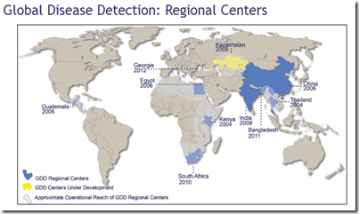
Bangladesh: Updating Nipah & The CDC Director’s Visit

image

Credit – The CDC’s GDDER


# 6888


I wrote at length yesterday on the Nipah virus, and the current outbreak in Bangladesh (see Bangladesh: Nipah Returns, so I’ll not bother repeating those points this morning. 


What is new this morning is a report from Xinhua News indicating that three more Nipah cases (two fatal) have been reported in Bangladesh, and the arrival on Saturday of U.S. CDC Director Thomas Frieden in Dhaka to discuss their plans to assist that nation in strengthening disease surveillance.


First the report from Xinhua on the latest Nipah cases.


Virus Nipah strikes back in Bangladesh, claims 8 more lives within one week

2013-01-28 10:00:41 GMT2013-01-28 18:00:41(Beijing Time) by Naim-Ul-Karim


DHAKA, Jan. 28 (Xinhua) -- Bangladesh on Monday reported eight more deaths from infection with the deadly Nipah virus within one week as the bat-borne disease has struck early and hard.


The country's Institute of Epidemiology, Disease Control & Research (IEDCR) confirmed two more deaths Sunday from the Nipah infection, tallying the toll at eight from Jan. 22.


"Outbreak of the deadly Nipah virus has claimed eight lives since January 22," Mahmudur Raman, head of the IEDCR, told Xinhua Monday.


"As of today, there are 11 cases from eight Bangladesh districts including Dhaka and eight of them died," he said, adding "Three Nipah-infected patients remain critically ill."

(Continue . . .)


The Institute of Epidemiology, Disease Control and Research (IEDCR) in Bangladesh has far more to deal with than just the Nipah virus. The 150+ million inhabitants of Bangladesh are also subject to H5N1 bird flu, Dengue, Anthrax, and Chikungunya, along with many other diseases.


The United States has been providing ongoing technical, financial, and logistical support to Bangladesh for several years to assist them in upgrading their disease detection and surveillance systems.  Hence the visit this past weekend by the CDC’s Director.


The news site BDNews24 has details of Director Frieden’s 2-day visit.  Follow the link to read:


Bangladesh to be 'Global Disease Detection site'

Senior Correspondent,  bdnews24.com

Published: 2013-01-27 14:56:52.0 Updated: 2013-01-27 18:24:00.0

The US Centres for Disease Control and Prevention (CDC) is strengthening its ties with Bangladesh as part of its initiative to protect the global community from the urgent public health threats.

(Continue . . . )


Improving disease surveillance, detection, and prevention in those regions where emerging diseases are most apt to occur is not only the humanitarian thing to do, it also helps to keep the world from being blind-sided by emerging infectious disease threats.


We’ve far too many `blind spots’ around the world where a novel virus could emerge, and take hold in the population, before it could be identified and (hopefully) contained.


To counter that threat, the CDC has developed a Global Disease Detection and Response division (GDDER).


image


Global Health - Global Disease Detection and Emergency Response

The Division of Global Disease Detection and Emergency Response (GDDER) protects Americans and the global community from urgent public health threats and provides public health relief for humanitarian emergencies. Activities include:

  • Detecting and containing emerging health threats
  • Deploying CDC experts 24 hours a day, 7 days a week at host country requests for technical assistance and other support
  • Building capacity by providing technical assistance in support of International Health Regulations (IHR)
  • Promoting policies for public health and bio-security
  • Improving the health of populations affected by complex humanitarian emergencies.


According to the GDDER website, nations are selected by the following criteria:


  • Public health significance: The country has a high population density or history of infectious diseases or expected potential for emerging diseases;
  • Country commitment: The country supports and values partnership with CDC and will actively engage in collaborative activities and identify new partners;
  • Established CDC presence: The country has an established, effective working relationship with CDC and supports CDC staff in-country;
  • Established regional reach: The country has the infrastructure and regional stature to serve as regional resource, or is already acting as a regional leader in other arenas;
  • International partner presence: The country has other U.S. Government agencies and international partners operating in-country.

The GDDER is a program that will hopefully not only help alert us of an emerging infectious disease threat, but that may also provide a chance to stop it in its tracks before it can spread.

While there are no guarantees of success, this is a public health advantage that previous generations could only have dreamed of.

Sunday, January 06, 2013

EID Journal: XDR-TB/HIV Treatment Outcomes

image

(From the 2011 TB Progress Report)


# 6828


Despite great advances made against tuberculosis since the introduction of antibiotics in the 1940s, in recent years we’ve seen the rise of new drug resistant strains of this killer disease; MDR-TB (Multi-drug Resistant Tuberculosis) and XDR-TB (Extensively Drug Resistant Tuberculosis).


Although the numbers have decreased in recent years, in 2010 1.4 million deaths were attributed TB, and it remains one of the three greatest causes of death of women (ages 14-44) in the world.


In 2009, the NIH had this to say about the global spread of the disease, including the fact that about 1 in 5 active cases of TB are also co-infected with HIV.


Today, one-third of the world’s population is thought to be infected with Mycobacterium tuberculosis (Mtb), the microbe that causes TB.


An estimated 13.7 million people have the active form of the disease. In 2007, approximately 9.27 million people developed TB, of whom 1.37 million were HIV positive, and 1.75 million died, including 456,000 individuals co-infected with HIV.


And in 2010, the World Health Organization announced:


Drug-resistant tuberculosis now at record levels

18 MARCH 2010 | GENEVA | WASHINGTON DC -- In some areas of the world, one in four people with tuberculosis (TB) becomes ill with a form of the disease that can no longer be treated with standard drugs regimens, a World Health Organization (WHO) report says.


Also from the World Health Organization:


Tuberculosis and HIV


The risk of developing tuberculosis (TB) is estimated to be between 20-37 times greater in people living with HIV than among those without HIV infection. In 2010, there were 8.8 million new cases of TB, of which 1.1 million were among people living with HIV.

In response to demands from countries, WHO recommends 12 TB/HIV collaborative activities, including the Three I's for HIV/TB. The WHO HIV/AIDS and TB Departments and their partners, including community groups, work collaboratively on joint HIV/TB advocacy, policy development and implementation in countries.

The Three I’s

  • Intensified TB case finding
  • Isoniazid preventive therapy
  • Infection control for TB.


While the incidence of co-infection with XDR-TB and HIV is rising, little is known about the effectiveness of treatment of these patients.


We’ve some new research, appearing in the CDC’s EID Journal, indicating that among a small cohort of patients followed in South Africa over 2 years – disappointingly - only 22% were cured or successfully completed treatment.


A few excerpts ( reparagraphed for readability), but follow the link to read the entire study:


Treatment Outcomes for Extensively Drug-Resistant Tuberculosis and HIV Co-infection

Max R. O’Donnell , Nesri Padayatchi, Charlotte Kvasnovsky, Lise Werner, Iqbal Master, and C. Robert Horsburgh

Abstract

High mortality rates have been reported for patients co-infected with extensively drug-resistant tuberculosis (XDR-TB) and HIV, but treatment outcomes have not been reported. We report treatment outcomes for adult XDR TB patients in KwaZulu-Natal Province, South Africa. Initial data were obtained retrospectively, and outcomes were obtained prospectively during 24 months of treatment.


A total of 114 XDR TB patients were treated (median 6 drugs, range 3–9 drugs); 82 (73%) were HIV positive and 50 (61%) were receiving antiretroviral therapy. After receiving treatment for 24 months, 48 (42%) of 114 patients died, 25 (22%) were cured or successfully completed treatment, 19 (17%) withdrew from the study, and 22 (19%) showed treatment failure.


A higher number of deaths occurred among HIV-positive patients not receiving antiretroviral therapy and among patients who did not show sputum culture conversion. Culture conversion was a major predictor of survival but was poorly predictive (51%) of successful treatment outcome.


Discussion

The main findings of our study were a high mortality rate (42%) and a low rate of successful treatment outcomes (22%) for XDR TB patients after completion of 24 months of treatment in a setting with a high incidence of HIV.


All deaths in this cohort occurred in the first 12 months after start of treatment. Predictors of deaths in this cohort included TB-specific (TB culture conversion) and HIV-specific (ART use) factors. Consistent with findings in other studies of treatment of drug-resistant TB/HIV, HIV was not independently associated with death (12,13,20).


Although HIV was not independently associated with death, use of ART among HIV-infected patients was associated with improved survival.


Sex appeared to modify the association between death and HIV because female sex was associated with higher survival rates among HIV-negative XDR TB patients but with higher death rates in women co-infected with HIV than in men co-infected with HIV.


However, this finding was not significant in all strata. TB culture conversion was a useful predictor of survival and treatment outcome. However, it was not sufficiently sensitive in this cohort to be a surrogate for successful TB treatment outcome, given the number of patients who ultimately showed treatment failure (n = 7), defaulted (n = 7), or died (n = 4) after TB culture conversion.


The authors conclude by saying:


Although not addressed by our study, improvements in treatment outcomes for patients co-infected with MDR TB and HIV will require changes in HIV- and TB-related factors. For HIV, these include more rapid HIV testing for early initiation of ART, appropriate monitoring of CD4 T-cell counts, HIV virus load testing, appropriate opportunistic infection prophylaxis, and improvement in ART adherence.


Although not addressed by our study, we recommend that for TB these improvements include widespread implementation of rapid diagnostics, particularly for smear-negative disease; early drug susceptibility testing for first-line and second line agents; improvement in adherence for second-line TB drugs; development of more effective anti-TB drugs and regimens; and guidance of drug selection by timely and ongoing drug susceptibility testing.


While often overshadowed by other issues, the HIV AIDS epidemic (exacerbated by TB) in South Africa (and much of Sub-Saharan Africa) continues to devastate the populace.


The UNAIDS website lists the following grim statistics for South Africa, a country that has seen more than a 10 year-drop in life expectancy since HIV began to spread in the 1980s:


HIV AND AIDS ESTIMATES (2011)

Number of people living with HIV
5,600,000 [5,300,000 - 5,900,000]
 
Adults aged 15 to 49 prevalence rate
17.30% [16.60% - 18.10%]
 
Adults aged 15 and up living with HIV
5,100,000 [4,900,000 - 5,400,000]
 
Women aged 15 and up living with HIV
2,900,000 [2,700,000 - 3,000,000]
 
Children aged 0 to 14 living with HIV
460,000 [410,000 - 520,000]
 
Deaths due to AIDS
270,000 [240,000 - 300,000]
 
Orphans due to AIDS aged 0 to 17
2,100,000 [2,000,000 - 2,300,000]

Wednesday, October 24, 2012

Asymptomatic Pigs: Revisited

image

Credit Wikipedia


# 6660


A couple of months ago in EID Journal: Flu In Healthy-Looking Pigs we looked at a report indicating that it isn’t always possible to identify pigs carrying an influenza virus based simply on their appearance.


A dispatch from the EID Journal reported that nearly 1 in 5 healthy-looking pigs they tested at the Minnesota State Fair during the 2009 H1N1 influenza pandemic were actually infected with a flu virus.


This came out during the midst of several swine variant flu outbreaks this summer, involving several hundred people across 10 states. Viruses apparently contracted from pigs on display at local fairs (see MMWR: H3N2v Related Hospitalizations In Ohio – Summer 2012).


While fair officials were turning away pigs with overt signs of infection, this report suggested that perhaps those measures might not be sufficient.


Today, a pair of studies from Ohio State University -  one that finds a surprisingly large percentage of flu infected swine to be asymptomatic - and another that establishes just how closely linked the human and swine variant strains of influenza this summer really were.


In both cases, the lead author is Andrew Bowman, a  Ph.D. candidate in veterinary preventive medicine at Ohio State.


First, from the EID Journal, research that found more than 80% of the pigs that tested positive for influenza at the Ohio State fair between 2009 and 2011 showed no signs of illness.


Subclinical Influenza Virus A Infections in Pigs Exhibited at Agricultural Fairs, Ohio, USA, 2009–2011

Andrew S. BowmanComments to Author , Jacqueline M. Nolting, Sarah W. Nelson, and Richard D. Slemons
Author affiliations: The Ohio State University, Columbus, Ohio, USA
Abstract

Agricultural fairs are associated with bidirectional, interspecies transmission of influenza virus A between humans and pigs. We examined pigs exhibited at agricultural fairs in Ohio during 2009–2011 for signs of influenza-like illness and collected nasal swab specimens from a representative subset of these animals.


Influenza virus A was recovered from pigs at 12/53 (22.6%) fairs during the 3-year sampling period. Pigs at 10/12 (83.3%) fairs from which influenza virus A was recovered did not show signs of influenza-like illness. Hemagglutinin, neuraminidase, and matrix gene combinations of the isolates were consistent with influenza virus A concurrently circulating among swine herds in the United States.


Subclinical influenza virus A infections in pigs at agricultural fairs may pose a risk to human health and create challenges for passive surveillance programs for influenza virus A in swine herds.

(Continue . . . )


Ohio State University has published a lengthy press release that discusses both papers.


Studies: Pigs Look Healthy But Test Positive for Flu at Fairs; Flu Transmission Seen Between Pigs and Humans

COLUMBUS, Ohio – More than 80 percent of pigs that tested positive for influenza A virus at Ohio county fairs between 2009 and 2011 showed no signs of illness, according to a new study.


Ohio State University researchers tested 20 pigs each at 53 fair events over those three summers and found at least one flu-positive pig at 12 fairs – almost a quarter of fairs tested.


The influenza strains identified in pigs in this study include H1N2 and H3N2 viruses – strains that have been circulating in pigs since 1998. In 2011, all of the H3N2 and H1N2 isolates found in pigs at the fairs contained a gene from the 2009 pandemic strain of H1N1, which is similar to the H3N2v strain causing human illness this year.


Though this finding alone is no cause for panic, it does show how quickly influenza viruses can change, said Andrew Bowman, lead author of the study and a Ph.D. candidate in veterinary preventive medicine at Ohio State.


In a second study led by Bowman, researchers compared the genomic sequences of influenza A viruses recovered in July 2012 from pigs and people. The analysis, showing a greater than 99 percent genetic similarity among the viruses, confirms that pigs and humans were infected with the same virus, indicating interspecies transmission.

(Continue . . .)


This second study appears in the journal Emerging Microbes & Infections.


Although the timing of the illnesses (and initial sub-typing) in humans and swine at the Ohio State Fair last July strongly suggested interspecies transmission, a >99% genomic match pretty much erases all doubt.


Molecular evidence for interspecies transmission of H3N2pM/H3N2v influenza A viruses at an Ohio agricultural fair, July 2012

Andrew S Bowman1, Srinand Sreevatsan2, Mary L Killian3, Shannon L Page4, Sarah W Nelson1, Jacqueline M Nolting1, Carol Cardona2 and Richard D Slemons1


Evidence accumulating in 2011–2012 indicates that there is significant intra- and inter-species transmission of influenza A viruses at agricultural fairs, which has renewed interest in this unique human/swine interface.


Six human cases of influenza A (H3N2) variant (H3N2v) virus infections were epidemiologically linked to swine exposure at fairs in the United States in 2011. In 2012, the number of H3N2v cases in the Midwest had exceeded 300 from early July to September, 2012.


Prospective influenza A virus surveillance among pigs at Ohio fairs resulted in the detection of H3N2pM (H3N2 influenza A viruses containing the matrix (M) gene from the influenza A (H1N1) pdm09 virus). These H3N2pM viruses were temporally and spatially linked to several human H3N2v cases.


Complete genomic analyses of these H3N2pM isolates demonstrated >99% nucleotide similarity to the H3N2v isolates recovered from human cases. Actions to mitigate the bidirectional interspecies transmission of influenza A virus between people and animals at agricultural fairs may be warranted.

(Continue . . .)


While the vast majority of the swine-to-human flu transmissions this year have involved the H3N2v virus, as you can see from the following table, small number of H1N1v and H1N2v infections have been reported as well. 


image


Given the limits of surveillance, testing, and reporting – we really don’t know what the normal `background rate’ of these types of novel flu infections are in humans.


The smattering of reports since 2005 indicate they may be fairly rare, but are certainly not unheard of.


The majority of human swine variant flu infections this summer were relatively mild (out of 300+ infections, 16 were hospitalized, and 1 died), but it is always of concern anytime a novel influenza virus jumps from another species to humans.


Each time a novel flu jumps from a pig to a human it gives the virus another opportunity to adapt to human physiology. The CDC’s most recent assessment on the H3N2v virus reads:


It's possible that sporadic infections and even localized outbreaks among people with this virus will continue to occur.

While there is no evidence at this time that sustained human-to-human transmission is occurring, all influenza viruses have the capacity to change and it's possible that this virus may become widespread.


So far, the severity of illnesses associated with this virus in people has been similar to the severity of illnesses associated with seasonal flu virus infections. Limited serologic studies indicate that adults may have some pre-existing immunity to this virus while children do not.


CDC is closely monitoring human infections with all novel influenza viruses, including H3N2v viruses, and will provide more information as it becomes available.


With further evidence of asymptomatic influenza infection in pigs, fair officials and public health agencies will need to decide on the best policies to limit future exchanges (in either direction) of flu viruses between people and pigs.

Monday, October 08, 2012

Diary From The HMNZ Tahiti During The 1918 Pandemic

image

Troop Ship Tahiti in Wellington Harbor, circa 1918-19  Unknown Photographer


# 6617


For years historians, epidemiologists, and virologists have been attempting to peel back the cobwebs of time in order to analyze the deadliest pandemic in human history; the 1918 Spanish Flu Pandemic.


John Barry’s The Great Influenza: The Epic Story of the Greatest Plague in History, has probably done more to reawaken memories of that awful time than any other source, but many gaps in our knowledge remain.


Jeffrey  K. Taubenberger and David Morens - both researchers at NIAID – have added considerably to our understanding of the H1N1 virus and the events surrounding its emergence. Taubenberger was the first to sequence the the genome of the 1918 Spanish Flu virus while David Morens is a prominent medical historian.


See Morens & Taubenberger on Influenza’s History for a fascinating look back at influenza through the ages. Highly recommended.


Spanish Flu broke out in the spring and summer of 1918, while WWI was still underway. It so devastated troops on both sides of the conflict that historians believed it helped to hasten the end of the war.


Soldiers and sailors – living in cramped and often unhygienic quarters – bore the early brunt of the pandemic, while troop trains and ships helped to spread it around the globe.


While there are many horrific accounts from the pandemic – including some small villages in Alaska entirely wiped out – some of the best documented events occurred onboard troop ships. 


One of the most famous was the HMNZ Troop Carrier Tahiti, which during August-September of 1918 carried 1217 troops and crew (almost double what the ship was rated to carry) from New Zealand to Plymouth, England with provisioning stops at Cape Town and Sierra Leone. 


Since fever was reported in Sierra Leone, no crew or passengers reportedly went ashore , but locals came aboard to coal the ship.  Within a few days of leaving port, half the men on the ship were sick, and in a matter of days, more than 80 would perish.


The University of Otago has an well done 1-page synopsis of the investigation, which you can access here.


image


The detailed report, on which this exhibit is based, appeared in the CDC’s EID Journal in December of 2010.

Historical Review

Mortality Risk Factors for Pandemic Influenza on New Zealand Troop Ship, 1918

Jennifer A. SummersComments to Author , Nick Wilson, Michael G. Baker, and G. Dennis Shanks


Adding another dimension to this story, Jennifer A. Summers returns to the October, 2012 edition of the EID Journal with excerpts from a recently uncovered diary, kept by one of the troops aboard that ship.


Pandemic Influenza Outbreak on Troop Ship—Diary of a Soldier in 1918

Jennifer A. Summers
Abstract

A newly identified diary from a soldier in 1918 describes aspects of a troop ship outbreak of pandemic influenza. This diary is the only known document that describes this outbreak and provides information not officially documented concerning possible risk factors such as overcrowding and the suboptimal outbreak response by military leaders. It also presents an independent personal perspective of this overwhelming experience.

(Continue . . . )


The diary entries make fascinating reading, as does the commentary provided by the author. Well worth following the link and to read in its entirety.


For more on the history and impact of the 1918 Spanish flu, you may wish to pay a visit to Flu.gov’s Pandemic history page, with offerings such as:


Monday, October 01, 2012

EID Journal: Challenges To Defining TDR-TB


image

(From the 2011 TB Progress Report)


# 6600


In January of this year reports began to emerge out of India regarding what was being called Totally Drug Resistant (TDR) tuberculosis (see Crof’s report India: New TB strain is "totally drug-resistant" (updated)).


While not an officially recognized term, TDR-TB was portrayed by the media as a frightening escalation of the existing classifications of Multi-drug resistant (MDR-TB) and Extensively-drug resistant (XDR-TB).


Resistant forms of Tuberculosis have come about primarily as the result of incomplete, irregular, or inappropriate treatment and management of infected patients.


A few days later (January 14th), the World Health Organization updated their Drug-resistant tuberculosis FAQ where they take exception to the term TDR-TB.


Why are these terms not yet recognised by WHO?

Terms such as “totally drug resistant” have not been clearly defined for tuberculosis. While the concept of “total drug resistance” is easily understood in general terms, in practice, in vitro drug susceptibility testing (DST) is technically challenging and limitations on the use of results remain: conventional DST for the drugs that define MDR and XDR-TB has been thoroughly studied and consensus reached on appropriate methods, critical drug concentrations that define resistance, and reliability and reproducibility of testing

Around the same time we saw an ECDC Comment On Drug Resistant TB In India. One of the points being made in the comments section (excerpted below) is that the term TDR-TB is as yet not well defined, and may be misleading.


Total drug resistant TB is a relative notion and depends on the local drugs available and tested on. This term/expression should either be avoided or should be defined worldwide. The World Health Organization (WHO) has internationally-endorsed treatment recommendations for the treatment of drug-susceptible, MDR-TB and XDR-TB.


In March, as part of my World TB Day Roundup, the World Health Organization released this statement on the supposed TDR-TB, cautioning:


More evidence and better diagnostics needed before redefining severe forms of drug-resistant TB says WHO

Note for the media

23 March 2012 | Geneva - Reports of tuberculosis (TB) cases with severe patterns of drug resistance are increasing, said experts who attended a WHO meeting in Geneva on 21-22 March. Participants stressed that the emergence of drug resistance should be a wake-up call for Ministries of Health. The group urged the global TB community to make greater efforts to prevent drug resistance and scale up provision of appropriate care and management to avoid a scenario where TB becomes incurable.

Insufficient evidence

The meeting concluded that there is currently insufficient evidence to adopt new case definitions for drug-resistant TB. Drug susceptibility testing (DST), which is key to defining new levels of drug resistance, lacks accuracy for several of the drugs that are used to treat multi drug-resistant (MDR) and extensively drug resistant (XDR)-TB. Secondly, there is insufficient correlation of DST results with clinical response to treatment for several drugs currently used to treat XDR-TB. Thirdly, new drugs are currently undergoing clinical trials, and could prove effective against drug resistant strains. The meeting urged diagnostics companies and TB laboratories to develop better diagnostic tests and also agreed that WHO and technical partners should develop more detailed guidance on XDR-TB treatment.

(Continue . . . )


All of which brings us to a new article appearing ahead of print in the CDC’s EID Journal that looks at the:


Challenges and Controversies in Defining Totally Drug-Resistant Tuberculosis

Peter Cegielski , Paul Nunn, Ekaterina V. Kurbatova, Karin Weyer, Tracy L. Dalton, Douglas F. Wares, Michael F. Iademarco, Kenneth G. Castro, and Mario Raviglione
Abstract

In March 2012, in response to reports of tuberculosis (TB) resistant to all anti-TB drugs, the World Health Organization convened an expert consultation that identified issues to be resolved before defining a new category of highly drug-resistant TB.


Proposed definitions are ambiguous, and extensive drug resistance is encompassed by the already defined extensively drug-resistant (XDR) TB. There is no evidence that proposed totally resistant TB differs from strains encompassed by XDR TB.


Susceptibility tests for several drugs are poorly reproducible. Few laboratories can test all drugs, and there is no consensus list of all anti-TB drugs. Many drugs are used off-label for highly drug resistant TB, and new drugs formulated to combat resistant strains would render the proposed category obsolete. Labeling TB strains as totally drug resistant might lead providers to think infected patients are untreatable. These challenges must be addressed before defining a new category for highly drug-resistant TB.

(Continue . . . )


Given the complexities of evaluating for total drug resistance, attaching the label TDR-TB to these strains may well be both premature and inaccurate.


But the sobering fact remains, if you are infected with one of these XDR-TB strains - and no drug is available to you that can treat it – for you, it may as well be totally resistant.


Of course, this disparity of available medical treatment exists for a great many diseases around the world, and is not just limited to TB.


The authors of the EID study conclude with this warning:


As countries increase treatment of MDR and XDR TB, it is inevitable that resistance to second-line drugs will increase. New drugs and better diagnostic tools are needed urgently for patients with highly drug-resistant TB.


For more on the global spread of TB, you may wish to revisit:


WHO-ECDC: Joint Report On Tuberculosis In Europe
Resistant TB: The Limits Of Surveillance & Reporting
WHO: Blood Tests To Detect Active TB Unreliable

Tuesday, September 25, 2012

WHO: Coronavirus Not SARS


image

Coronavirus – Credit CDC PHIL


image


# 6578


We are just three days into the emerging coronavirus story (see here, here, and here) , and the short message above, tweeted this morning by the World Health Organization, deserves repeating.


Despite coming from the same family of viruses as SARS (as do many other, far less pathogenic viruses), this new coronavirus is not SARS - and until more testing can be completed - we don’t know how much of a danger it actually presents.


The WHO continued to tweet:


image

image

image


Kidney failure, while not unheard of in SARS, was not a common presenting symptom back in 2002-2003.  And SARS transmitted readily from human to human. Thus far, we haven’t seen signs of that sort of transmission with this new virus.


None of which is to say this virus couldn’t prove to be a bigger public health threat down the road, only that it is premature to think of this virus as the next global health crisis.


The media has been quick to refer to this virus as SARS-like, but it remains to be seen just how much the two viruses really have in common.


The discovery of new - even deadly - viruses that can afflict humans or other mammals is not uncommon. And most of the time, after an initial flurry of breathless news reports, the threat is found to be less than of pandemic proportion.


To provide a little perspective, a few viral discoveries in recent years that sparked initial hyperbolic headlines, but have (so far, anyway) failed to present a major public health threat:


  • We’ve watched a number of triple-reassortant swine flu (Variant) viruses (H1N1v, H1N2v, H3N2v) make tentative jumps into human hosts (see An Increasingly Complex Flu Field), and so far they’ve failed to spread efficiently, or to produce substantial levels of morbidity.
  • In August of this year (see New Phlebovirus Discovered In Missouri) the CDC announced the detection of a novel tick-borne virus in America’s heartland.  Despite being detected in 2009, only two cases have been reported.
  • In March of this year, we learned of a new H17 flu subtype, carried by bats in an unusual host: bats (see A New Flu Comes Up To Bat).
  • In November of 2011 we saw a major die-off of seals in New England, that was eventually traced to a new mammalian adapted influenza virus mBio: A Mammalian Adapted H3N8 In Seals.  
  • In October of 2008  doctors in Zambia and South Africa ran across a mysterious, previously unclassified virus that caused hemorrhagic symptoms in its victims similar to Ebola (see Lujo Virus: Newly Identified Arenavirus) While highly contagious, and fatal in 4 of the 5 identified victims, it has not reappeared since 2008.


And if you want to go back a few more years, you can add Nipah, Hendra, H5N1, Ebola, Marburg . . . .


The truth is, scientists – with better tools available today – are indentifying `new’ viruses all of the time. A few well distributed viruses that until recently, were unknown, include:


  • The human metapneumovirus (HMPV) was identified in Dutch children with bronchiolitis about a decade ago.  Since then, it has been found to be ubiquitous around the world, and responsible for a significant percentage of childhood respiratory infections . . . yet until 2001, no one knew it existed.
  • Human Bocavirus-infection (HBoV) wasn’t identified until 2005, when it was detected in 48 (9.1%) of 527 children with gastroenteritis in Spain (cite).  It has since been found around the globe using PCR testing.


And the list grows longer every year.


While most will prove to be less than devastating in impact, we need only look at HIV, the 1918 H1N1 pandemic, and the pandemic viruses of 1957 and 1968 to realize that novel viruses can sometimes emerge and cause incredible morbidity and death.


According to well respected anthropologist and researcher George Armelagos of Emory University, we are entering the Third Epidemiological Transition.

This third transition began in the late 1970s or early 1980s, and is hallmarked by newly emerging infectious diseases, re-emerging diseases carried over from the 2nd transition (which began with the industrial revolution, and added chronic, non-infectious, degenerative diseases), and a rise in antimicrobial resistant pathogens.


When you combine those factors with an increasingly mobile global population of about 7 billion people, and huge increases in the number of animals being raised for food consumption (often in environments conducive to the spread of diseases), and you have a recipe for explosive growth in diseases.


Hence the need for continual surveillance, which will help us spot – and if we are lucky, even contain – the next pandemic before it can spread widely.

Thursday, August 16, 2012

EID Journal: Flu In Healthy-Looking Pigs


image

Credit Wikipedia


# 6497


Over the past month we’ve been watching a small but growing number of human infections with a novel H3N2v influenza virus - most of which are associated with direct contact with pigs being displayed at county and state fairs.


Efficient and sustained human-to-human transmission has not been established by the CDC, and so their recommendations at this time revolve around preventing disease transmission from pigs to humans.


Earlier this week the CDC issued advice to Fair organizers, that included:


Animal Health Recommendations:
  • Monitor animals daily for signs of illness, including discharge from nose and/or eyes, lethargy (sleepiness), no appetite, fever, or sometimes coughing. Ensure that a veterinarian, such as the fair vet or state vet, is notified of any ill animals.
  • Ill pigs, animals suspected or known to be infected with influenza viruses, and animals from herds with a recent history of respiratory disease should not be exhibited. They should be immediately isolated or sent home.


Yesterday, a dispatch appearing in the CDC’s EID Journal illustrated just how difficult identifying and separating influenza-infected pigs from the rest can be. 


It’s called:


Volume 18, Number 9—September 2012
Dispatch

Influenza A(H1N1)pdm09 Virus among Healthy Show Pigs, United States

Article Contents

Gregory C. Gray , Jeffrey B. Bender, Carolyn B. Bridges, Russell F. Daly, Whitney S. Krueger, Michael J. Male, Gary L. Heil, John A. Friary, Robin B. Derby, and Nancy J. Cox

Abstract

Within 5 months after the earliest detection of human influenza A(H1N1)pdm09 virus, we found molecular and culture evidence of the virus in healthy US show pigs. The mixing of humans and pigs at swine shows possibly could further the geographic and cross-species spread of influenza A viruses.


Asymptomatic carriage of viruses is not uncommon in humans, of course.  A few examples we’ve looked at in the past include:


  • During the 2009 pandemic I wrote They Walk Among Us, that looked at the difficulties of identifying those who might be infectious based on symptoms such as fever.
  • Earlier this year, in The Very Common Cold, we looked at a study of rhinovirus among college students tested over an 8 week period – that found asymptomatic infections led symptomatic infections by a factor of 4 to 1.
  • And last year, in EID Journal: Pre-Symptomatic Influenza Transmission, we saw evidence of presymptomatic spread of the H1N1 virus in three clusters in Japan, which also suggests that asymptomatic carriers ought to be able to spread the virus as well.


The idea that pigs might carry influenza viruses asymptomatically, therefore, is hardly surprising. But the amount of available scientific research has been limited.


Yesterday’s study found that nearly 1 in 5 healthy-looking pigs they tested were actually infected with a flu virus.


Last night Lisa Schnirring and Robert Roos of CIDRAP NEWS  wrote extensively on this study, and so at this point, I’ll simply invite you to read their excellent report.


Study finds flu in healthy-looking pigs at state fairs

Lisa Schnirring and Robert Roos * Staff Writers

Aug 15, 2012 (CIDRAP News) – Testing of a sampling of pigs shown at the Minnesota State Fair during the 2009 H1N1 influenza pandemic revealed that 19% of them were infected with flu viruses, even though they looked healthy, according to a new study.


The findings highlight the challenges of preventing pigs and humans from passing flu viruses back and forth at fairs and swine shows, especially this summer when several states are tracking human illnesses from a novel H3N2 virus that has been detected in both pigs and people.

(Continue . . . )


During the 2009 H1N1 pandemic Japan, India, and China were among the nations that attempted to identify, interdict, and isolate those who might be carrying the H1N1 virus when they entered their country.


While their efforts may have slowed the introduction of the virus, they certainly didn’t stop it.  Earlier blogs on these attempts include:


Japan: Quarantine At Ports Ineffective Against Pandemic Flu
Experts: Extreme Measures Won’t Stop The Flu


Their failure was likely due to the large number of presymptomatic, and asymptomatic carriers of the virus that arrived without showing signs of illness.


And the same is likely true with pigs on display at county fairs this fall.


Removing symptomatic pigs certainly makes sense - and will certainly reduce the risks of spreading the virus - but it is unlikely to totally eliminate it.


Which means that additional human cases of H3N2v are likely, even with the enhanced biosecurity measures in place at county fairs.


Before anyone gets freaked out over the risks of going to the county fair, so far this summer there have been only about 200 confirmed cases of this H3N2v flu, involving 5 brief hospitalizations and no deaths. 


During roughly the same time period, there have been nearly 700 West Nile Infections, involving hundreds of hospitalizations, and 26 deaths.


So if you are looking to lower your risks of illness when you go to the county fair this fall, it only makes sense to carry and use a hand sanitizer, and avoid eating or drinking around animal displays. 


But to avoid serious illness, the better advice is to wear an insect repellant whenever you go outside, as your risks of contracting viral illness are greater right now from infected mosquitoes, than they are from sick pigs.


image

Tuesday, August 07, 2012

EID Journal: Persistence Of H5N1 In Soil


image

Photo Credit – FAO


# 6478


The notion that the H5N1 `bird flu’ virus can persist in the environment – for hours or days (or perhaps even weeks) - is hardly new, yet very little is really known about how, and where, the virus resides outside of a living host.


As H5N1 is primarily a gastrointestinal malady in birds it is believed that the virus is commonly spread in the wild via shared feces-contaminated pond and lake waters (see Bogor: H5N1 Detected In Retention Pond).


But there appear to be other routes of transmission as well.


In June of 2010, we saw a study (see Birds Of A Feather . . . .) in PLoS One, suggesting that waterfowl may be spreading avian flu viruses because their preening oils bind the virus to their feathers.


Another study conducted by researchers at the  National Institute of Animal Health, Tsukuba, Ibaraki, Japan was reported in the August 2010 issue of Applied and Environmental Microbiology.


They determined that the H5N1 virus may persist on the dropped feathers from infected ducks and may therefore spread to the environment. 


Applied and Environmental Microbiology, August 2010, p. 5496-5499, Vol. 76, No. 16
0099-2240/10/$12.00+0     doi:10.1128/AEM.00563-10

Persistence of Avian Influenza Virus (H5N1) in Feathers Detached from Bodies of Infected Domestic Ducks
Yu Yamamoto, Kikuyasu Nakamura, Manabu Yamada, and Masaji Mase


The surprising part of this study is how long these feathers retained some degree of viral contamination at various temperatures.


At 4°C (39F) the virus was detectable for 160 days, while at the higher temperature 20°C (68F), the virus was detected for 15 days.


We saw another study from 2010, which appeared in Environmental Science and Technology, titled:


Environmental Persistence of a Highly Pathogenic Avian Influenza (H5N1) Virus

Joseph P. Wood, Young W. Choi, Daniel J. Chappie, James V. Rogers, and Jonatha

n Z. Kaye

DOI: 10.1021/es1016153

Copyright © 2010 American Chemical Society


Researchers conducted tests on four inanimate materials (glass, wood, galvanized metal, and top soil) to determine how long – and under what environmental conditions – the virus could survive.


They adjusted factors such as  temperature, relative humidity, and simulated sunlight and checked the samples over a period of 13 days. The virus was most persistent at lower temperatures, and on surfaces such as glass and steel.


Their conclusion?: under the right conditions, the virus could be expected to persist beyond 13 days.


Earlier this year we looked at a study published in the journal Influenza and Other Respiratory Viruses that examined environmental samples taken in Cambodia between April 2007 and February 2010 during several bird flu outbreaks (see Environment: a potential source of animal and human infection with influenza A (H5N1) virus  Gutiérrez, Buchy et al.)

Out of 246 samples taken around farms with outbreaks, 19% of dust, mud and soil samples showed contamination from the H5N1 virus.


Admittedly, just because RT-PRC testing was able to detect a virus in a sample doesn’t necessarily mean that the virus is viable.  But it does give us an idea of the environment spread of the virus.


All of which brings us to a letter, again from  Ramona A. Gutiérrez and Philippe Buchy of the Institut Pasteur in Cambodia, that appears in September’s edition of the CDC’s EID journal.


Volume 18, Number 9—September 2012
Letter

Contaminated Soil and Transmission of Influenza Virus (H5N1)

To the Editor: Highly pathogenic avian influenza (HPAI) virus (H5N1) has been responsible for 603 confirmed human cases worldwide, including 356 that resulted in death, and for >7,000 epizootic outbreaks (1,2). Direct contact between hosts is the main mechanism of transmission for avian influenza viruses, but the possible role of the environment as a source of HPAI virus (H5N1) infection has been rarely studied, particularly in the context of countries where the virus is enzootic or epizootic (37). To determine if contaminated soil contributes to the transmission cycle of HPAI virus (H5N1), we used experimental and simulated field conditions to assess possible transmission in chickens.

(Continue . . . )


Essentially, these researchers took 3 types of soil, described as:


(1) sandy topsoil collected from around rice fields in Phnom Penh Province, Cambodia;

2) building sand purchased from a local building company; and

3) soil-based compost purchased from a local tree nursery

. . . and inoculated samples with low to high doses of the H5N1 virus. They then introduced these soil samples to the bottom of cages where chickens were housed, and then tested the chickens for infection over the next several days.

You can read the entire letter for more details on their methods and materials. The results are summarized in the chart below:

image


Essentially, sandy topsoil collected from rice fields proved to be a poor environment for transmitting the H5N1 virus, while soil-based compost proved highly effective. 


The authors believe that the highly acidic nature of the sandy topsoil may work to inactivate viral particles.


Since one of the methods used to control and contain an avian flu outbreak is environmental decontamination, knowing which types of soil are unlikely to harbor and transmit the virus can save time, and reduce the use of harsh (and often scarce) chemicals in the environment.


Interestingly, the authors also found evidence to suggest that exposure to moderately contaminated soil may help poultry to develop a protective immune response to the virus.


H5N1 is not the only viral contender to spark the next pandemic, but due to its apparent high lethality, it is the one we tend to concentrate on the most.  


Fortunately the virus remains adapted primarily to avian physiology – not human - and must mutate further if it is to become an imminent public health threat. 


But with 20+ clades of the virus now circulating, and numerous opportunities to expose and infect other hosts (human, swine, mammal, and avian), the concern is this virus may one day succeed.
Wednesday, July 18, 2012

EID: Environmental NDM-1 Detected In Vietnam

image

Inoculated MacConkey agar culture plate cultivated colonial growth of Gram-negative, small rod-shaped and facultatively anaerobic Klebsiella pneumoniae bacteria. – CDC PHIL.


# 6440


In a study reminiscent of one we saw published in April of last year (see Lancet Study: NDM-1 In New Delhi Water Supply), the CDC’s EID Journal has a letter by R. Isozumi et al. that appears (ahead of print) in their August issue, that reveals the detection of the NDM-1 gene in a river in Hanoi.


blaNDM-1–positive Klebsiella pneumoniae from Environment, Vietnam

Rie Isozumi , Kumiko Yoshimatsu, Tetsu Yamashiro, Futoshi Hasebe, Binh Minh Nguyen, Tuan Cuong Ngo, Shumpei P. Yasuda, Takaaki Koma, Kenta Shimizu, and Jiro Arikawa


By way of explanation, blaNDM-1 is the gene responsible for creating the NDM-1 (New Delhi metallo-β-lactamase) enzyme that can make many types of bacteria resistant to a wide spectrum of antibiotics.


Of particular concern, this enzyme is carried by a plasmid – a snippet of portable DNA  - that can be transferred to other types of bacteria (see Study: Adaptation Of Plasmids To New Bacterial Species).


Over the past few years we have seen a worrisome expansion of β-lactamase enzymes in bacteria, and they are slowly eroding the value of much of our antibiotic arsenal.


Those that inhibit the antimicrobial actions of the (formerly resistant) Carbapenem class of antibiotics – called carbapenemases – are of particular concern. Carbapenems are often used as the drug of last resort for treating difficult bacterial infections, including Escherichia coli (E. coli) and Klebsiella pneumoniae.


So when the gene responsible for the NDM-1 enzyme begins to show up in the environment, doctors and researchers take notice.


The author’s of today’s EID Journal report state their reasons for concern:


The possible appearance of bacteria harboring blaNDM-1 in Vietnam is of concern because cultural and economic links between Vietnam and India are strongly established, including extensive person-to-person exchanges that could enable easy exchange of pathogens. In addition, Vietnam faces a serious problem of antimicrobial drug resistance because drugs are freely available and used in an indiscriminate fashion. Thus, once blaNDM-1–positive bacteria colonize persons in Vietnam, they would be able to spread easily and pose a serious public health threat.


To look for environmental blaNDM-1, researchers examined water samples taken from 20 locations within 10 km of Hanoi, Vietnam. Samples were collected from rivers, lakes, and standing water in the streets.


The authors report finding the NDM-1 enzyme producing gene in two locations – 3km apart – in the Kim Nguu River, which flows through the city.  They write:


We harvested several species of bacteria from the 2 seepage samples positive for blaNDM-1: Acinetobacter baumannii, Klebsiella pneumoniae, Pseudomonas aeruginosa, P. fluorescens/putida, and P. luteola


They also report finding 2 other βeta-lactamases (blaTEM-1 and blaCTX-M-3) that were highly resistant to another class of antibiotics called aminoglycosides (which include neomycin, streptomycin & tobramycin)

The authors conclude by saying:

Wide-scale surveillance of environmental and clinical samples in Vietnam and establishment of a strategy to prevent further spread of blaNDM-1 are urgently needed.

It’s been nearly 2 years since The Lancet published a study (see NDM-1: A New Acronym To Memorize)  by Walsh, Toleman, Livermore, et al. that awakened the world to the  emergence and growing prevalence of the NDM-1 enzyme.


Since that time, we’ve seen a slow, but inexorable spread of NDM-1 carrying bacteria around the globe. A few of my past blogs on the subject include:


Carbapenemases Rising

NDM-1: One Year Later

WHO Unveils 6-Point Plan To Preserve Antibiotic Effectiveness

Eurosurveillance On Antimicrobial Resistance


For a far more complete discussion of antimicrobial resistance issues, I can think of no better primer than Maryn McKenna’s book SUPERBUG: The Fatal Menace of MRSA. And Maryn’s SUPERBUG Blog, part of Wired Science Blogs, continues to provide the best day-to-day coverage of these issues.


Last March, Director-General of the World Health Organization Margaret Chan warned that the World Faces A `Post-Antibiotic Era’. One where even common infections may become untreatable.


While we aren’t there yet, reports such as the one today in the EID Journal add to the growing concern that someday, that fear may become a reality.

Thursday, June 07, 2012

EID Journal: Guinea Pigs As Reservoirs For Influenza


# 6369


From the CDC’s EID Journal we’ve a new study that looks at influenza infections in farm-raised guinea pigs in Ecuador, that provides several surprising results.


Ferrets and mice are often used in influenza research, but neither is truly ideal.


So in recent years a number of researchers have looked at guinea pigs as possible model mammalian host for influenza virus studies (see PNAS The guinea pig as a transmission model for human influenza viruses by Peter Palese et al. 2006).


The success in using guinea pigs in lab studies has led some scientists to wonder just how guinea pigs might fit into the hosting and spread of flu viruses outside of the laboratory.


Which brings us to a dispatch (again from Peter Palese et al.) that was published yesterday in the EID journal called:


Dispatch

Influenza Virus Infection in Guinea Pigs Raised as Livestock, Ecuador

Victor H. Leyva-Grado, Samira Mubareka, Florian Krammer, Washington B. Cárdenas, and Peter Palese

Abstract

To determine whether guinea pigs are infected with influenza virus in nature, we conducted a serologic study in domestic guinea pigs in Ecuador. Detection of antibodies against influenza A and B raises the question about the role of guinea pigs in the ecology and epidemiology of influenza virus in the region.


Guinea pigs are raised and used as food in parts of South America (Peruvians reportedly consume more than 65 million of them each year, and they may also be found on the menu in Bolivia, Ecuador, and Columbia).


While the consumption of guinea pigs may seem an unusual culinary choice to many of us, it is so entrenched in the Andean culture that in 1753 - when Marcos Zapata painted his version of `The Last Supper’ for Peru (which now hangs in The Cathedral Of Cusco ) - he depicted Christ and the Apostles dining on a platter of cuy, or guinea pig.

image

Photo Source – Wikipedia.


But I digress . . .


Most of these animals are raised on small farms, and often in close contact with other livestock as well as humans.


Given these conditions, and their propensity for hosting and spreading influenza in laboratory studies, a seroprevalence study was undertaken to see how widespread influenza infection among guinea pigs might be outside of the laboratory.


This study examined blood samples from 40 guinea pigs taken from 3 locations across Ecuador, and subsequently found evidence of previous Influenza A infections in a unusually large number of them.


The surprises (of which there were several) included:


  • The high percentage of positive influenza A samples (50% H1, 45% H3)
  • The detection of several (n=14)animals carrying antibodies to an H5 virus
  • And perhaps the biggest surprise of all – finding evidence of influenza B infections (previously only thought to infect humans) in 27 of the 40 samples tested.


An earlier seroprevalence study of influenza A among humans in Ecuador showed a seroprevalence of H1 (5.1%) and H3 (5.5%) -  about 1/10th that found in these guinea pigs.


The authors suggest that the way these animals are raised (caged together and in close quarters) may facilitate the spread of influenza. 


The discovery of H5 antibodies is intriguing, but since this study only tested only for seroreactivity to parts of the H5 virus, further study will be required to identify and quantify the prevalence of avian influenza viruses in this population.


And the last finding - that of Influenza B in roughly 2/3rds of the samples – support the idea that this type of influenza can be readily transmitted from humans to other hosts.


The authors write:


Further studies are needed to isolate and characterize the type B influenza virus present in the population of guinea pigs to determine if there has been an adaptation to the new host or if the guinea pig is only a transient reservoir for the human virus.


The authors conclude this dispatch by stating:


We did not determine whether guinea pigs are an incidental host for influenza virus infection or, if instead, the virus has been adapted to these animals or if guinea pigs are a natural reservoir for some influenza viruses. To this end, virus isolation and characterization would be necessary to determine the virus strains circulating in this population. In the laboratory, guinea pigs are infected and efficiently transmit influenza viruses to naive hosts without showing any overt clinical signs of disease (1). Therefore, further studies are needed to address the specific role of guinea pigs raised as livestock in the ecology and epidemiology of influenza viruses in the region.


For more details on the methods used, the full text is available online at this link.


While this research might seem a bit obscure to all but the most ardent infectious disease geeks, it serves to show just how little we still know about influenza viruses and their host range,  and that there are still plenty of surprises waiting out there yet to be uncovered.